Patients
This prospective study was conducted at China Medical University Hospital between January 1, 2021, and January 31, 2023. Patients with gastrointestinal or pancreaticobiliary tract cancer and massive malignant ascites were eligible for screening. The presence of malignant ascites should be confirmed cytologically or pathologically. The study protocol was approved by the Research Ethics Committee of China Medical University and Hospital, Taichung, Taiwan (CMUH107-REC1-180). This study was registered at ClinicalTrials.gov (NCT05745233).
Treatments
Patients with symptomatic ascites who fulfilled the inclusion criteria underwent paracentesis as clinically indicated. During paracentesis, 20 mL of ascitic fluid was collected for flow cytometry and cytokine analysis. After paracentesis, patients were administered an IP infusion of nivolumab (20 mg diluted in 100 mL saline) over 10 min. The paracentesis catheter was removed immediately after nivolumab infusion was completed. After paracentesis, patients remained in bed with position changes every 20 min for 2 h: supine, on the right side, supine, on the left side, head high and feet low, and head low and feet high. Ascites was assessed by the treating physicians. Responders were defined as patients with increased paracentesis interval, whereas non-responders were patients were defined as patients with decreased paracentesis interval. IP treatment was repeated after each subsequent paracentesis until the treatment was deemed ineffective by the treating physician, unacceptable toxicity occurred, or stopped at the patient’s request.
Evaluation of toxicity
Physical examination, blood pressure, heart rate, and respiratory rate were recorded before and after each paracentesis and IP infusion of nivolumab. Any adverse events were documented and graded according to the National Cancer Institute Common Terminology Criteria for Adverse Events, version 5.0.
Analysis of the cellular components of ascites
The cellular components of 20 mL of ascitic fluid, obtained during paracentesis, were analyzed using flow cytometry. Briefly, cells were concentrated by centrifuging the fluid at 2,000 rpm for 10 min. The resulting cell pellets were washed with 3 mL of phosphate-buffered saline (PBS) containing 0.2% bovine serum albumin (BSA) and centrifuged three times. The final pellet was resuspended in 100 µL of PBS with 0.2% BSA. The cells were stained with the following antibodies: CD45 (2D1, Horizon V500, BD Biosciences), CD44 (L178, fluorescein isothiocyanate [FITC], BD Biosciences), CD24 (ML5, APC-H7, BD Biosciences), CD71 (YDJ1.2.2, APC-A750, Beckman Coulter), CD200 (OX104, APC, Invitrogen), and CD326 (epithelial cell adhesion molecule [EpCAM], EBA-1, phycoerythrin [PE], BD Biosciences). The samples were incubated in the dark at 25 °C for 30 min. Erythrocytes were lysed using 2 mL of BD FACS™ Lysing Solution (BD Biosciences). For intracellular staining, cells were then fixed and permeabilized using the FIX & PERM™ Cell Permeabilization Kit (Invitrogen™), following the manufacturer’s instructions, and stained with anti-human cytokeratin-7/−8 antibody (CAM5.2, Brilliant Violet™ 421 [BV421], BD Biosciences). Finally, stained cells were washed once with PBS, resuspended in 200 µL of PBS containing 0.2% BSA, and analyzed using a BD FACSLyric™ cytometer (BD Biosciences, CA). Cancer cells were identified as CD45 −/EpCAM +/Cytokeratin +/CD44 +/CD24 +/CD71 +/CD200 −. Mesothelial cells were excluded based on CD200 expression.
Analysis of cytokines of ascites
Ascitic fluid samples from six patients (five clinical responders and one non-responder) were available for cytokine analysis both before and after IP administration of nivolumab. Paired ascites samples were analyzed for 19 cytokines, including: epidermal growth factor (EGF), fibroblast growth factor 2 (FGF-2), granulocyte colony-stimulating factor (G-CSF), interferon α 2 (IFNα2), interferonγ (IFNγ), interleukin 1α (IL-1α), interleukin 1β (IL-1β), interleukin 2 (IL-2), interleukin 6 (IL-6), interleukin 12p70 (IL-12p70), interleukin 22 (IL-22), monocyte chemoattractant protein-1 (MCP-1), monocyte chemoattractant protein-3 (MCP-3), macrophage colony-stimulating factor (M-CSF), macrophage-derived chemokine (MDC), macrophage inflammatory protein-1α (MIP-1α), macrophage inflammatory protein-1β (MIP-1β), vascular endothelial growth factor A (VEGF-A), and soluble CD40 ligand (sCD40L). Cytokine levels were measured using the Milliplex® Human Cytokine/Chemokine/Growth Factor Magnetic Bead Panel A/B (Merck KGaA, Darmstadt, Germany), according to the manufacturer’s protocol. All cytokines were measured in triplicate for each sample.
Statistical analysis
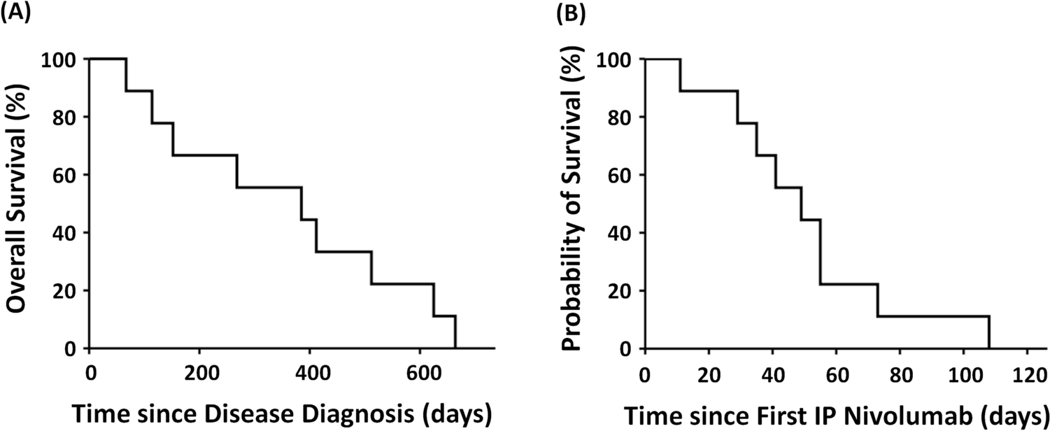
The outcome measure was the clinical response of ascites, defined as a reduction in ascitic fluid volume and a prolonged interval between paracenteses following IP nivolumab treatment. Duration of response was defined as the time interval from the initiation of IP nivolumab until it was deemed ineffective by the treating physician, upon the occurrence of unacceptable toxicity, or stopped at the patient’s request. All statistical analyses were performed using Statistical Product and Service Solutions version 26 for Windows (IBM Corporation, Armonk, NY, USA), with two-sided p-values < 0.05 considered statistically significant.

Comments (0)