Edentulous mandibular fractures account for less than 5% of all mandibular fractures; however, they present unique clinical challenges [34]. The progressive alveolar bone resorption associated with edentulism results in reduced and sclerotic bone volume, poor vascularisation, and the absence of stable anatomical landmarks [35]. Additionally, the lack of occlusal support eliminates a critical reference point for proper alignment of fracture segments and restoration of vertical dimension and the atrophic bone structure complicates the placement of rigid reconstruction plates and increases the risk of injury to the inferior alveolar nerve (IAN). These anatomical and biomechanical limitations make accurate anatomical reduction more difficult and raise the risk of local complications.
Moreover, edentulous patients with mandibular atrophy are often elderly and frequently present with multiple comorbidities, which heighten both surgical and anaesthetic risks [23, 29,30,31]. In light of these complexities, the use of virtual surgical planning (VSP) and computer-aided design/computer-aided manufacturing (CAD/CAM) technologies has emerged as a promising strategy to enhance the management of atrophic mandibular fractures [36, 37].
As early as 2010, Van Sickels and Cunningham reported favourable outcomes in mandibular fracture management using preoperatively bent plates on standard plastic mandibular models, which were subsequently adjusted intraoperatively to match patient-specific anatomy [38]. Although VSP was not employed in their study, the authors noted a reduction in operative time when plates were pre-contoured before surgery. In recent years, with the progressive development of new technologies, several authors have advocated the application of VSP and CAD/CAM-assisted approaches in the treatment of fractures of the atrophic edentulous mandible (Table 2) [22, 31].
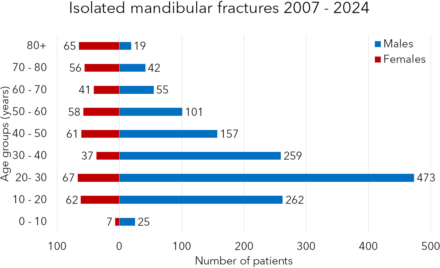
Table 2 Literature review A review of the literature confirms that these fractures occur more frequently in females and are most commonly located in the mandibular body. In all reported cases, the fractures were approached via an extra-oral route and treated using load-bearing osteosynthesis. Virtual surgical planning (VSP) was consistently employed preoperatively to achieve virtual reduction of the fracture segments and to generate an STL model of the reconstructed mandible. This model was then 3D printed and used to pre-bend a load-bearing plate prior to surgery, with the exception of five cases reported by Caruso et al. [31].
In this case series, the digital mandibular model was utilised to design patient-specific titanium plates and drilling guides with notably high precision, achieving fragment positioning errors of less than 2 mm in 88.2% of cases. The authors also reported that the use of patient-specific implants (PSIs) facilitated accurate condylar seating and provided high predictability in screw placement, thereby reducing the risk of injury to the inferior alveolar nerve (IAN). Moreover, these custom plates were described as thinner than standard stock plates while maintaining sufficient mechanical strength, thus minimising interference with future dental rehabilitation. The authors emphasised that this technique is particularly beneficial in cases of severe atrophy, comminuted fractures, or when bone grafting is required to bridge fracture gaps, as observed in several cases within the series [31]. However, the production of titanium PSIs remains both costly and time-consuming. Additionally, the design of accurate and unambiguous drilling guides can be particularly challenging in the absence of dental reference points.
On the other hand, Façanha de Carvalho et al described the use of a pre-bent plate together with fragments repositioning guides [22]. However, in cases of non-comminuted fractures and without bony gaps, we believe that the fracture can be anatomically reduced using the pre-bent plate as a reference for proper positioning. Moreover, the repositioning guide described does not incorporate drilling references, therefore its primary advantage is stabilising the fracture prior to placing the load-bearing plate; this can be achieved more efficiently and cost-effectively with the use of miniplates. In our study the use of a stock plate bent preoperatively on mandible model obtained from the virtual anatomic reduction of the fractures allowed for good reduction in all cases. Moreover, the in-house printing of the models enabled maintaining low costs, and the planning times were relatively short, did not depend on the number of fractures and decreased with the learning curve and training of dedicated personnel. Additionally, this process eliminated the lengthy intraoperative bending of thick plates. Although this study does not include an accuracy analysis, the same protocol demonstrated a very high accuracy in the case series by Abbate et al., where all the cases showed discrepancies of less than 1.5 mm between the virtual planning and postoperative CT [28].
On the other hand, in cases of comminuted fractures or those requiring bone grafts, when anatomic reduction is impossible, the use of drilling guides and custom plates as described by Caruso et al [31] could offer greater advantages, justifying the higher costs and production times of the PSIs.
The integration of new technologies in the management of atrophic mandibular fractures offers a tailored approach that addresses many challenges associated with these complex injuries, with a potential reduction in operative times in a vulnerable population and a potential improvement in the clinical outcomes.
The progressive availability of specific in-house tools for virtual surgical planning and 3D printing has allowed a simpler, quicker, and more cost-effective utilisation of these technologies. Furthermore, with a short learning curve, the in-hospital 3D laboratory could also be considered a valuable tool for learning, as the virtual model allows the junior surgeons to simulate the operation and share the decision making with the senior surgeons. The implementation of preoperative planning enhances the accessibility of the procedure for less experienced surgeons, enabling them to perform it safely and achieve favourable outcomes even in their early cases. This is especially advantageous given the condition's low epidemiological prevalence, which limits the opportunities for younger surgeons to gain practical experience [4, 22, 29, 30].
Despite the numerous advantages, the higher costs compared to conventional techniques are a significant consideration. Furthermore, the planning and fabrication process requires preparation time that can delay surgery by several days or weeks. However, for atrophic edentulous fractures, there is rarely an indication for immediate emergency treatment, making a brief delay usually not clinically significant.
Despite the limited number of patients, this case series represents the largest cohort of edentulous atrophic multiple mandibular fracture patients treated with the aid of VSP.
Beyond the already acknowledged limitation of the small sample size, this study presents additional constraints. Firstly, its retrospective nature inherently limits the strength of the conclusions that can be drawn. Secondly, in the absence of postoperative CT scans, the assessment of fracture reduction adequacy relied solely on the intraoperative judgement of the operating surgeon. This introduces a potential source of subjectivity and variability, which could be addressed in future studies through more objective accuracy assessments. Prospective, and ideally multicentre, investigations would allow for a larger and more representative cohort, particularly given the relatively low incidence of this specific pathology. In such studies, the inclusion of postoperative CT imaging would enable a more robust analysis of accuracy by allowing for a direct comparison between the 3D models of the mandible reconstructed after virtual fracture reduction and those obtained following osteosynthesis. The superimposition and comparison of these models could provide valuable insights into the actual precision of preoperative plate bending on stereolithographic replicas, and may further elucidate whether such an approach has a statistically significant impact on clinical outcomes—such as the potential reduction in the incidence of inferior alveolar nerve injuries when compared to cases treated without digital planning tools.

Comments (0)