Diagnosing pulmonary thromboembolism (PTE) on non-contrast computed tomography (NCCT) remains diagnostically challenging due to the modality’s inherently limited sensitivity. In patients experiencing cardiac arrest, PTE is associated with high mortality, ranging from 77% to 95% [11]. A substantial proportion of fatal cases remain undiagnosed antemortem, and over 90% of CT pulmonary angiograms performed in emergency settings yield negative results [12].
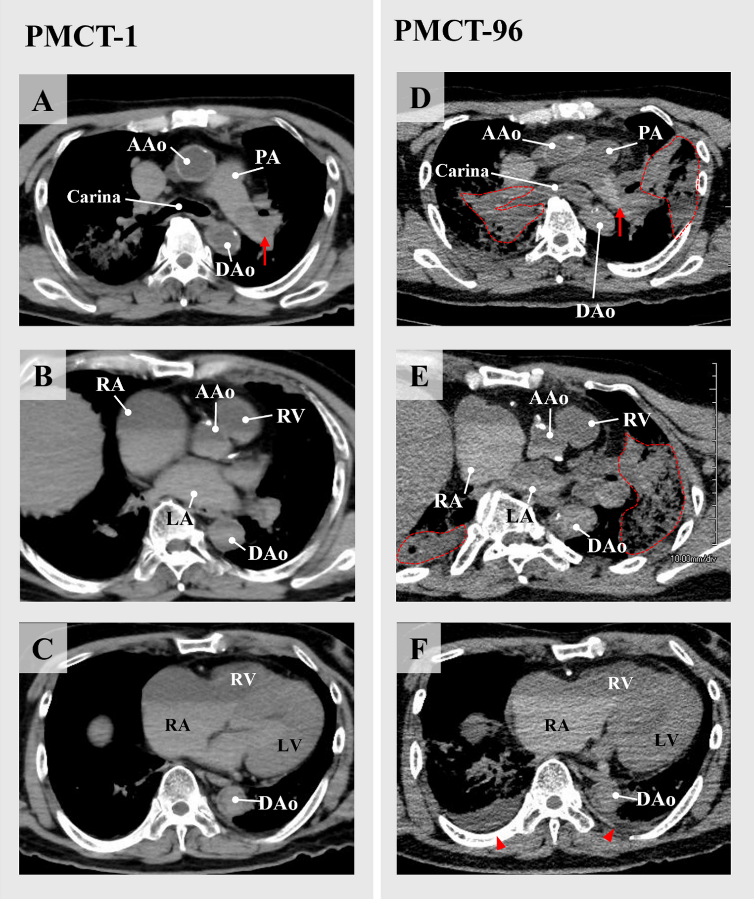
In the present case, thrombi appeared more conspicuous on the delayed-phase scan (PMCT-96), likely due to enhanced contrast resulting from time-dependent postmortem changes. While antemortem studies have reported a progressive decline in thrombus attenuation due to protein degradation [6], our findings demonstrated the opposite trend. Postmortem factors, such as ambient low temperatures and reduced enzymatic activity, may inhibit proteolysis, whereas plasma leakage and gravitational sedimentation may contribute to increased thrombus density over time.
It is noteworthy that variability in CT acquisition parameters (e.g., slice thickness, tube current) and differences in scanner types may influence the absolute attenuation values. Accordingly, relative attenuation differences between thrombi and the adjacent pulmonary arterial lumen may serve as more reliable indicators. The progressive enhancement in thrombus conspicuity observed in this case suggests that postmortem hypostasis and plasma separation enhance the visualization of thrombi over time.
Furthermore, the composition of the thrombus is likely to influence its radiologic appearance on CT imaging. Antemortem studies have shown that erythrocyte-rich (red) thrombi generally exhibit higher CT attenuation than fibrin-rich (white) thrombi [13]. However, it remains uncertain whether such compositional differences are preserved in venous thrombi or can be detected on PMCT. Layered thrombi may also exhibit heterogeneous attenuation, reflecting the variable proportions of erythrocytes and fibrin. These findings underscore that thrombus composition is a critical determinant of postmortem imaging characteristics and highlight the need for systematic investigation into its role.
PMCT has been employed in the diagnosis of fat embolism [14]; however, to our knowledge, no prior studies have documented serial alterations in thrombus attenuation or visibility. Our findings indicate that delayed-phase PMCT may enhance thrombus detectability, particularly when initial imaging yields inconclusive results.
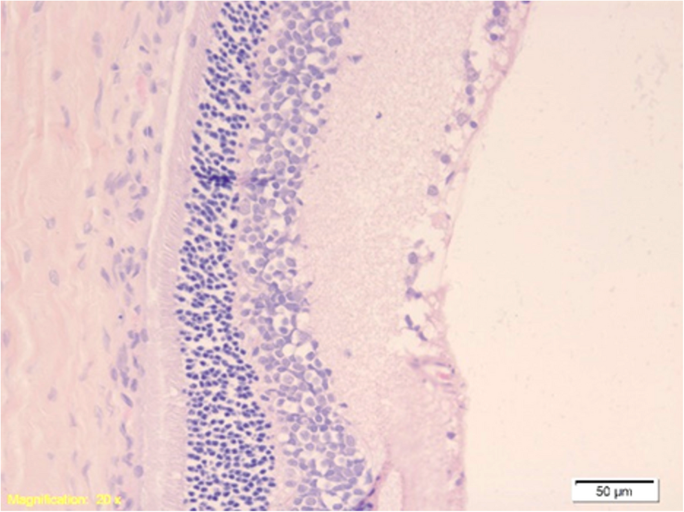
This case has several limitations. First, it represents a single case report, and attenuation assessments were performed by a single observer. Additionally, the body was stored at 2 °C, which could have influenced the rates of decomposition and coagulation. Although histological examinations of the thrombi and lung parenchyma were performed, the findings were insufficient for determining thrombus age or composition and thus were not included in this report. Consequently, the precise age of the thrombus (acute versus organized) could not be determined, which represents an additional limitation. Future studies involving larger cohorts and standardized imaging protocols are warranted to validate these preliminary observations.
From a forensic perspective, this case highlights a crucial implication: thrombi may not be readily apparent on PMCT immediately after death but can become progressively more detectable over time due to postmortem physiological processes. Awareness of these time-dependent imaging changes may enhance the diagnostic utility of PMCT and inform optimal imaging intervals in forensic investigations of sudden, unexpected death.




Comments (0)