Forensic postmortem imaging plays a crucial role in modern forensic investigations by providing non-invasive, detailed visualization of the body after death. Postmortem imaging offers several significant advantages in forensic investigations, because it enables detailed three-dimensional visualization, which improves accuracy in identifying fractures, hemorrhages, or foreign objects [14,15,16,17,18,19].
Postmortem angiography is a valuable technique for visualizing the vascular tree in forensic investigations, allowing detailed assessment of blood vessels and detection of vascular injuries. By injecting a contrast medium, it makes it possible to visualize the entire circulatory system within the body, which aids in identifying causes of death related to vascular pathology. However, the effects of the contrast medium on surrounding tissues and its potential impact on subsequent histological analysis must be carefully considered. The contrast agents may alter tissue appearance or interfere with microscopic examination, so understanding these effects is essential to accurately interpret findings and avoid misdiagnosis during the forensic evaluation [9].
It is well known that the oily contrast agent can provide an image of fatty embolism in the lungs. Since MPMCTA with oil-based contrast medium leads to these false positive histological results, it should not be used to investigate such cases unless lung tissue samples have been previously collected – and, even in the latter case, the biopsy procedure itself could cause artefacts [20]. Some authors conclude that it is not possible to assess a potential fatal fatty embolism after MPMCTA with oily contrast agent. Therefore, MPMCTA should be performed with an aqueous contrast agent if there is any suspicion of fatty embolism [9].
Histo-morphological artifacts induced by contrast media used in MPMCTA have also been observed in the kidneys, where they can present as glomerular collapse. This artifact may significantly interfere with the interpretation of tissue morphology. Capuani et al. (2014) discussed a case in which a systemic vasculitis was diagnosed. In that case, the presence of glomerular collapse prevented a clear histological diagnosis, ultimately limiting the ability to better understand the renal involvement [11].
On the other hand, MPMCTA can significantly enhance the accuracy of histological sampling in forensic investigations. As Sabatasso et al. (2018) have pointed out, MPMCTA aids in identifying myocardial regions most likely to yield diagnostically valuable tissue samples. This targeted approach increases the probability of detecting myocardial infarction, if present, and thereby improves the chances of accurately determining the cause of death. In particular, areas of the myocardium that exhibit abnormal contrast enhancement should be prioritized for sampling, as these regions are more likely to harbor ischemic or necrotic changes indicative of infarction [21].
To the best of our knowledge, little is known about these artifacts, and most existing studies have been conducted in experimental settings.
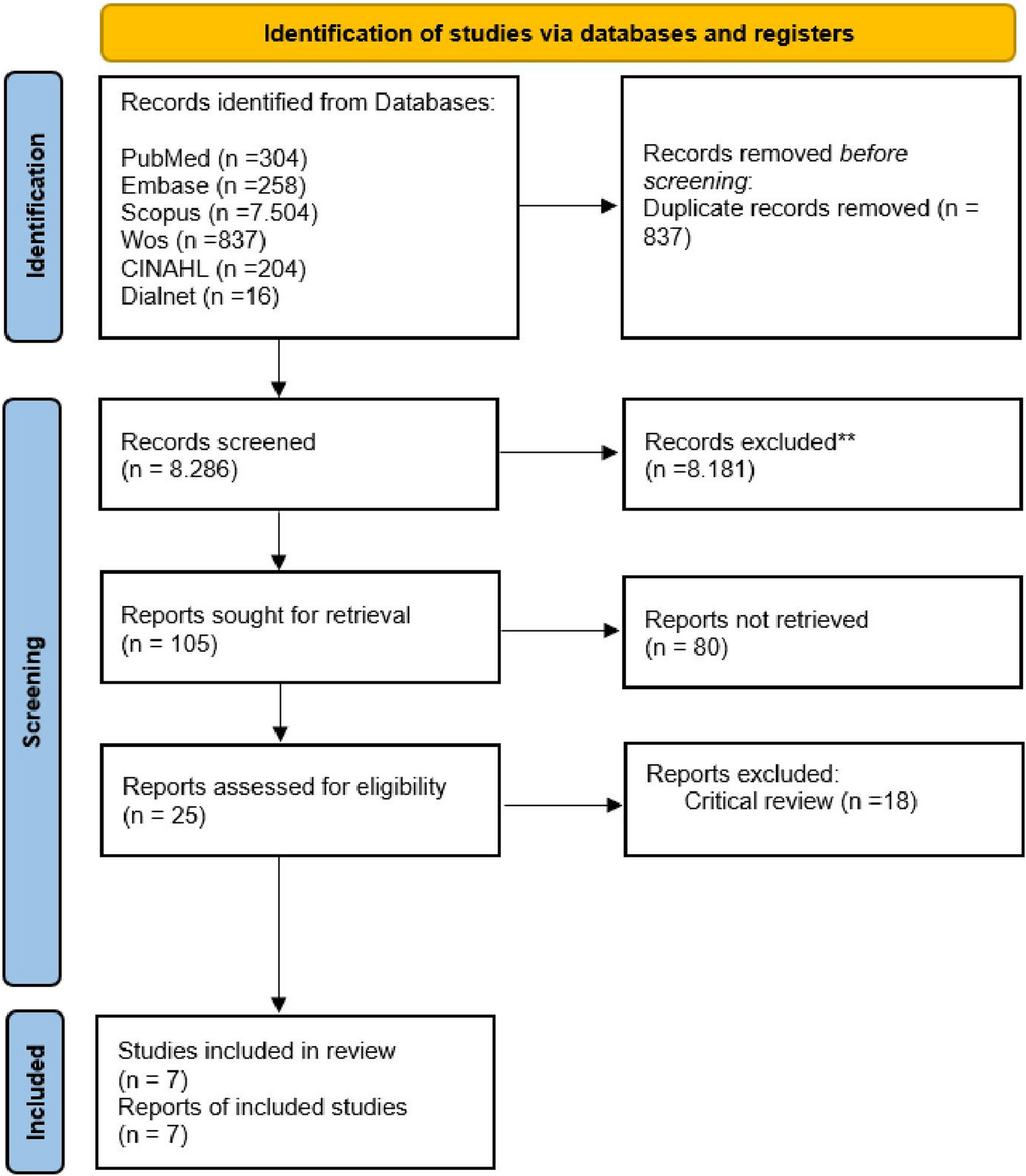
The objective of this study is to systematically investigate potential artifacts associated with the oily contrast agent used in multi-phase postmortem computed tomography angiography (MPMCTA), specifically the mixture of Angiofil® and paraffin oil. Histological samples from 37 medico-legal autopsy cases, all of which underwent MPMCTA, were examined. Through detailed histopathological analysis, this study aims to identify and characterize any tissue alterations or artifacts attributable to the contrast agent. The findings are intended to support forensic pathologists in accurately interpreting histological changes following MPMCTA, thereby enhancing the diagnostic reliability of postmortem vascular imaging.
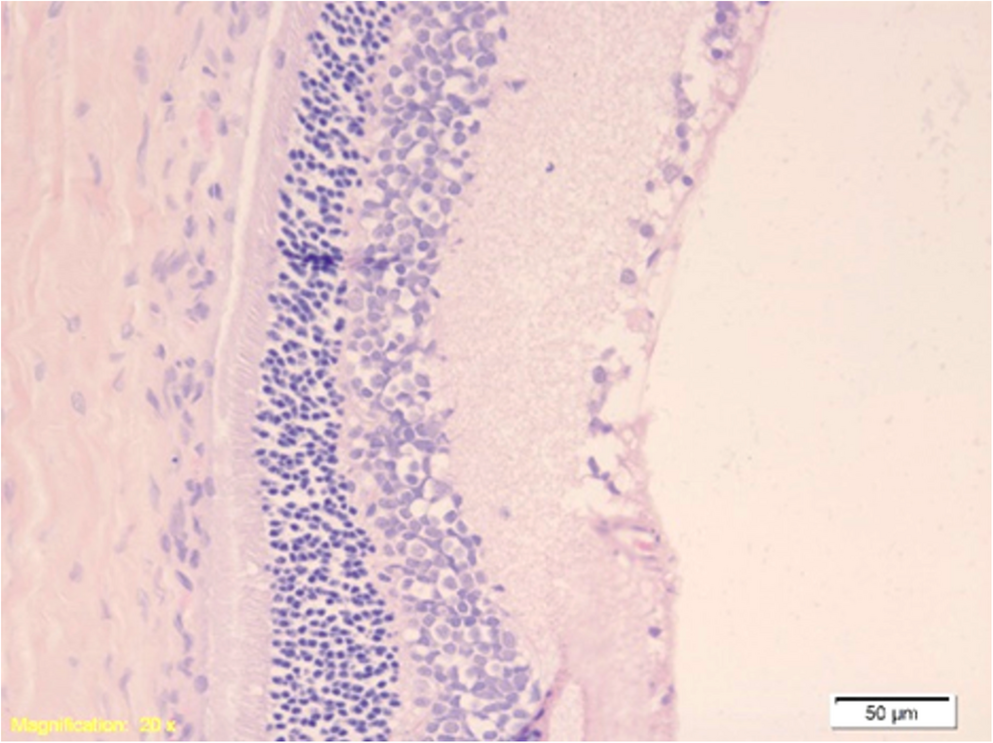
A total of 270 histological specimens were examined. In all cases, we were able to accurately identify the organ or tissue as well as the histological structures that characterize each tissue type. On the other hand, different types of artifacts were encountered, which can be grouped into four main categories. The first one refers to the OES, which result from the displacement of red blood cells within vessels by the oily medium and are the most common type of artifact, especially at the vascular level. OES often have curved/semilunar contours or a round shape, typically circumscribed by blackish material, which can be attributed to chemical residues from the contrast medium. The second one regards the compression of surrounding structures, which is common in organs with a loose vascular structure, where the distension of vessels filled with contrast medium exerts pressure on the surrounding parenchyma. The third type of artifacts relates to hyperaemia of small vessels and capillaries, which is due to the artificial propulsion of blood by the contrast solution under the pressure induced by the Virtangio®, giving the appearance of congestion. The fourth type of artifacts is associated to vessel dilation, which is also caused by the pressure induced by the Virtangio®, in which vessels filled with contrast medium appear completely empty, gaping, and with significantly distended walls.
Artifacts in the liver, particularly sinusoidal dilatation, are in differential diagnosis with the hepatic peliosis variant “parenchymal peliosis,” which consists of irregular cavities that are lined neither by sinusoidal cells nor by fibrous tissue, with the adjacent hepatic tissue occasionally showing necrosis of the hepatocytes [22]. The issue of differential diagnosis can play a role in the kidney as well, especially when collapsing glomerulopathy may be suspected, as the end-stage of focal segmental glomerulosclerosis [23]. Therefore, forensic pathologists should take this into account when examining specific autopsy samples.
Postmortem angiography is a powerful tool for visualizing the vascular system and enhancing the accuracy of forensic investigations. However, it is associated with the risk of inducing histological artifacts due to the use of contrast media. Because these artifacts can potentially interfere with microscopic examination and the interpretation of tissue samples, forensic pathologists should carefully consider, in advance, whether to perform postmortem angiography—especially in cases where detailed histological analysis is critical. This preliminary decision is essential to balance the benefits of vascular imaging against the possibility of compromising histological evaluation.
While the Atlas of Postmortem Angiography by Grabherr et al. (2016) [7] represents a landmark reference in the field by offering a descriptive and illustrative overview of angiographic findings, the present study addresses a different and complementary perspective. We provide a systematic histo-morphological assessment of artifacts induced by MPMCTA in a consecutive series of forensic autopsy cases. Unlike experimental or isolated organ studies, this approach allows the categorization of artifacts across multiple organ systems within a standardized medico-legal workflow, with quantitative data on sample inclusion and exclusion. To our knowledge, this is the first structured attempt to define the spectrum of histological artifacts in normal tissues following MPMCTA, thereby supporting forensic pathologists in differentiating imaging-related changes from genuine pathological findings during routine practice.
Our results clearly demonstrate that this method should not be performed in the presence of suspected fatty embolism, as the resulting images could be confused with artifacts. In contrast, marrow embolism remains identifiable, as does bone embolism [24]. Nevertheless, in cases where the cause of suspected death is attributed to the rupture of a major vessel, any potential artifacts do not compromise the interpretation of histological findings, since the diagnosis is primarily macroscopic, and microscopic images of the parenchyma are not relevant in such scenarios.
Further studies are needed to comprehensively characterize the artifacts induced by contrast media used in MPMCTA in pathological tissues. Pathological changes may alter tissue permeability, vascular integrity, or contrast retention, potentially resulting in misleading histological appearances. A deeper understanding of these interactions is essential to differentiate true pathological findings from imaging-related artifacts, ensuring accurate histopathological interpretation in post-mortem examinations.
Further research is needed to better identify and understand artifacts caused by contrast media in corpses at advanced stages of putrefaction. These situations may present additional challenges due to tissue degradation and altered vascular integrity, which can affect the appearance and interpretation of imaging and histological findings. Expanding studies to include such cases will help improve the accuracy and reliability of postmortem angiographic examinations across a wider range of forensic scenarios.



Comments (0)