
Remember me

Human mesentery samples were obtained from two different sources with written informed consent from each patient or their guardians. Discarded human mesentery tissues came from patients with CD that were undergoing bowel resections at Washington University School of Medicine. Control tissues came from surgical patients undergoing resection for gastrointestinal cancers or polyps. Staff affiliated with the Washington University Digestive Disease Research Core Center coordinated consent, oversaw tissue collection and managed de-identification services. Further control samples were obtained from organ donors consented for tissue use in research through Mid-America Transplant under a material transfer agreement. CD patient tissues were derived from inflamed or uninflamed regions of the ileum, collected under the supervision of a pathologist. Control ileal-draining mesentery was collected from patients with gastrointestinal cancer upstream of the tumor. Organ donors were excluded if they had previous bowel resections or a diagnosis of IBD. Participant demographics are summarized in Supplementary Table 1. Participants were not compensated. No statistical methods were used to predetermine sample sizes. All collections were conducted as approved by the Human Research Protection Office at Washington University (Institutional Review Board protocol no. 201111038) and followed the principles of the Declaration of Helsinki.
MiceMice were bred and housed at the Washington University vivarium in specific pathogen-free facilities under standard housing conditions of 12-h light/dark cycles. Food was provided ab libitum, cages were changed weekly and water was provided either through a Lixit valve or in water bottles that were changed twice a week. All experiments and procedures were approved by the Institutional Animal Care and Use Committee at Washington University (protocol nos. 20170154, 20-0032 and 22-0433). All mice were on a C57BL/6 background (Jackson Laboratory (JAX) cat. no. 000664); some were crossed to express the Ptprca CD45.1 allele (B6.SJL-PtprcaPepcb/BoyJ, JAX cat. no. 002014). Other strains obtained from JAX were μMT (B6.129S2-Ighmtm1Cgn/J, JAX cat. no. 002288)52, CD19cre (B6.129P2(C)-Cd19tm1(cre)Cgn/J, JAX cat. no. 006785)53, Lta−/− (B6;129S2-Ltatm1Dch/J, JAX cat. no. 002257)54 and Tdtomato (B6.Cg-Gt(ROSA)26Sortm9(CAG-tdTomato)Hze/J, JAX cat. no. 007909) mice. Initial TNFΔARE breeder mice were provided through a material transfer agreement by the Cleveland Digestive Disease Research Core Center (NIH P30 DK097948). TNFΔARE were kept and bred as heterozygotes, cohoused with WT littermates. M. Baldridge (Washington University School of Medicine) provided Pigr−/− mice backcrossed to C57BL/6 mice (B6.129P2-Pigrtm1Fejo/Mmmh)55,56, and Ltb−/− (B6;129-Ltbtm1Flv/J)57 and TNFfl/fl mice58 were acquired from University of Texas Health San Antonio and expanded and used at Washington University after crossing to CD19Cre/+ mice. More detailed description of mouse breeding is given in the Supplementary Methods.
BM chimerasFemale mice (7–9 weeks old) were irradiated with 8 Gy in an X-ray irradiator, corresponding to a lethal dose without reconstitution, validated for our colony and irradiator. At 6 h after irradiation, mice received 5 million donor BM cells retro-orbitally, and were then housed for ≥8 weeks before further experiments.
Anti-LTα3, anti-TNF treatment and LTβR-FcAnti-LTα3 antibody S5H3 (S5H3 LT-a.Fc-MT)33 was a gift from Genentech33. Mice were weighed and then injected intraperitoneally with 6 mg kg−1 of body weight with anti-LTα3mAb or mouse IgG2a isotype control (Clone C1.18.4; Leinco Technologies) every 2 days. Some mice were also (or instead) injected with 600 μg anti-mouse TNF (clone TN3-19.12, Leinco Technologies)59 or Armenian Hamster IgG isotype control (clone PIP, Leinco Technologies). LTβR-Fc fusion protein60 and isotype control were provided by Biogen or Genentech33. These reagents were injected intraperitoneally weekly using 200 μg.
Human and mouse tissue and single-cell processingHuman ileal mesentery samples were collected in cold saline or RPMI-1640 (GIBCO, cat. no. 11875-085) and processed within 1 h. After rinsing in HBSS, the mesentery was separated using a scalpel, minced, weighed and digested (0.2 g tissue per milliliter) in RPMI-1640 with 10% fetal bovine serum (FBS) (ThermoFisher GIBCO, cat. no. 26140-079), 1 mg ml−1 Collagenase IV (Sigma-Aldrich, C5138) and 0.2 mg ml−1 DNase I (Sigma-Aldrich, cat. no. DN25) at 37 °C for 45 min, orbital shaking at 100 rpm. Samples were vortexed, filtered (100 μm) and washed in fluorescence-activated cell sorting (FACS) buffer (2% FBS, 5 mM EDTA (Corning, cat. no. 46-034-CI), 0.2% sodium azide in PBS) then centrifuged (10 min, 500g, 4 °C). Red blood cells (RBCs) were lyzed for 5 min, washed with 10 ml FACS buffer and spun again before resuspension in 1 ml FACS buffer. Cells were counted using an automatic cell counter (Cellometer Auto T4, Nexcelom), frozen at −80 °C in 10% DMSO/90% FBS then moved to liquid nitrogen. When needed, aliquots were thawed rapidly at 37 °C.
For collecting mouse mesentery, small intestine, MLNs, spleen, mice were perfused with cold PBS (pH 7.4) after blood plasma collection. For lamina propria (LP) isolations, intestines were cut longitudinally, washed in PBS to remove feces, then cut into 1-cm pieces. Mucus and epithelial cells were removed by incubation in HBSS (ThermoFisher, cat. no. 14175-079) containing 10% FBS, 20 mM HEPES (Corning, cat. no. 25-060-CI) and 5 mM EDTA, 20 min with shaking. Tissues were vortexed, gently agitated and revortexed. Murine mesentery and processed LP were then digested similarly to human tissues, described above. Leukocytes were enriched by centrifugation on a discontinuous Percoll gradient (70%/40%, GE Healthcare, cat. no. 17-0891-01) at 850g for 25 min at room temperature. Spleens and MLNs were crushed and passed through a 70-μm strainer; spleens underwent RBC lysis using BD Pharm Lyse (BD Biosciences, cat. no. 555899) for 5 min at RT. Blood was collected in EDTA-containing tubes, centrifuged (10,000g, 10 min, 4 °C) and plasma frozen. RBC were lyzed. Single-cell suspensions were resuspended in 1 ml FACS buffer and counted using the Cellometer Auto T4.
Flow cytometrySingle-cell suspensions were incubated with either murine FcBlock (BD Biosciences, cat. no. CD16/CD32, clone 2.4G2) or human FcR blocking reagent (Miltenyi Biotec, cat. no. 130-059-901) for 15 min at 4 °C, then stained with cell surface antibodies for 30 min at 4 °C. Dead cells were excluded using a Live/Dead Aqua Fixable Cell Stain Kit (Invitrogen, cat. no. L34957), 4′,6-diamidino-2-phenylindole (DAPI) or Helix NP Green (Biolegend, cat. no. 425303). Human cells were fixed before acquisition after Live/Dead Aqua Fixable Cell Stain by incubating the cells in 2% paraformaldehyde (PFA) for 15 min at 4 °C and then washed and resuspended. For ex vivo detection of TNF protein, cells were incubated for 4 h at 4 °C in Brefeldin-A (Biolegend, cat. no. 420601), before blocking, extracellular FACS staining, staining with Live/Dead Aqua Fixable Cell Stain, fixation and permeabilization (Biolegend, cat. no. 426803), and overnight staining with an anti-TNF antibody (TNF-PE; Biolegend, cat. no. MP6-XT22, 1:200)). For transcription factor staining, cells were fixed and permeabilized (eBioscience, cat. no. 00-5523-00) before blocking and staining for transcription factors. Human cells were stained for CD45-BUV395 (BD Biosciences, clone HI30, 1:100) or CD45-PerCP/Cyanine5.5 (Biolegend, clone H130, 1:100), CD19-PE or CD19-BV605 (Biolegend, clone HIB19, 1:200), and, as an exclusionary stain, CD3-Alexa Fluor 700 (Biolegend, clone HIT3a, 1:50), CD14-Alexa Fluor 700 (Biolegend, clone HCD14, 1:25) or CD16-Alexa Fluor 700 (Biolegend, clone 3G8, 1:50). Mouse cells were stained with cell surface antibodies in different combinations: CD45.1-Alexa Fluor 700 or CD45.1-PE-Cy7 (Biolegend, clone A20, 1:200), CD45.2-Pacific Blue, CD45.2-Alexa Fluor 700 or CD45.2-APC/Cy7 (Biolegend, clone 104, 1:200) or CD45.2-eFluor 506 (eBiosciences, clone 104, 1:200) or (Biolegend, clone 104, 1:200), pan-CD45-Pacific Blue or pan-CD45-BV421 (Biolegend, clone 30-F11, 1:400), CD4-BUV496 or CD4-BB700 (BD Biosciences, clone GK1.5, 1:400), CD8a-BV785 (Biolegend, clone 53-6.7, 1:400), CD11b-BUV737 (BD Biosciences, clone M1/70, 1:600), Ly6G-BUV805 (BD Biosciences, clone 1A8, 1:400), CD44-BV605 (Biolegend, clone IM7, 1:400), CD62L-PEDazzle 594 (Biolegend, clone MEL-14, 1:400), SiglecF-Alexa Fluor 647 (BD Biosciences, clone E50-2440, 1:200), TCRβ-PerCP/Cy5.5, TCRβ-BV711 or TCRβ-FITC (Biolegend, clone H57-597, 1:400), CD19-PE/Cy7 (Biolegend, clone 6D5, 1:400), CD11c-BUV395 (BD Biosciences, clone N418, 1:200), CD64-BV605 (Biolegend, clone X54-5/7.1, 1:200), CD115-BV605 (Biolegend, clone AFS98, 1:200), IgA-PE, IgA-FITC, or IgA-APC (eBioscience, clone mA-6E1, 1:200), MHC Class II-BUV496 (BD Bioscience, clone M5/114.15.2, 1:200) or MHC Class II-AF700 (Biolegend, clone M5/114.15.2, 1:200) and Ly6C (Biolegend, clone HK1.4, 1:200). Intercellular mouse staining used either TNF-PE (Biolegend, clone MP6-XT22, 1:200) or FOXP3-FITC (Invitrogen, clone FJK-16 s, 1:1,000), T-bet-eFluor 660 (Invitrogen, clone eBio4B10 (4B10), 1:400) and RORyt-BV421 (BD Biosciences, clone Q31-378, 1:800).
Cells were acquired on a LSR Fortessa X20, a FACSymphony, or a five-laser Cytek Aurora. Human cells were sorted for scRNA-seq on an Aria II in a Bioprotector cabinet. DAPI− CD45+ KikGR+ mouse cells were sorted for scRNA-seq on a Sony Synergy HAPS cell sorter.
Photoconversion of the ileumTNFΔARE/+ or wild-type (WT) KikGR+ BM chimeras 30 weeks post-transplant were treated with anti-LTα3 mAb33 or isotype control before surgery to allow adequate systemic absorption, then anesthetized using inhaled isoflurane and placed on a heating pad. A midline incision exposed the intestines and a 365-nm fiber optic light source was used to photoconvert 15 discrete ileal regions, with each exposure for 10 s, then 30-s rest, three cycles per site. The peritoneum was closed with continuous absorbable 4-0 vicryl sutures and the skin closed with nonabsorbable nylon surgical knots. Mice recovered on a heating pad. A second dose of anti-LTα3 or isotype control (0.6 mg kg−1) was administered before tissue harvest. Mice were sacrificed in the order in which they were photoconverted. LNs were harvested, digested enzymatically as described above, and sorted on DAPI− CD45+ KikRED+ cells. Sorted cells were pooled from three mice for each experimental cohort.
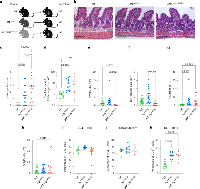
Histology on distal ileumThe distal 2 cm of the ileum was measured, flushed with PBS, cut open, pinned on wax then fixed overnight in 4% PFA. Ileal tissue was washed in PBS, embedded in 2% agar and embedded for paraffin sectioning and staining with hematoxylin and eosin before scoring61.
Whole-mount imaging and confocal microscopy of the mouse mesenteryIntact mesenteries were pinned on wax, fixed with 4% PFA overnight at 4 °C, then washed in PBS, blocked and permeabilized in 5% donkey serum, 3% BSA, and 1% Triton X-100 in PBS buffer overnight at 4 °C. Tissue were incubated with antibodies to B220 (eBioscience, 14-0452-05), Lyve-1 (Abcam, ab14917), CSF1R (R&D, AF3818) diluted in 0.6% BSA and 0.2% Triton X-100 for at least 24 h, 4 °C. Samples were washed and incubated with donkey secondary antibody conjugated to Cy2, Cy3, or Cy5/Alexa Fluor 647 for at least 12 h, 4 °C. For quantification of TLSs, the tissue was blinded before proceeding. The mesentery was pinned flat on a SYLGARD® 184-coated (Ellsworth, 4019862) plate and dehydrated in ethanol, then cleared with methyl salicylate solution (Sigma-Aldrich, M6752) for10 min. For quantification of TLS, blinded mesenteries were examined using a Leica M205FA stereoscope while pinned to the SYLGARD plates. Mesenteries were quantified before unblinding. For confocal imaging, the mesenteries were instead cleared inside a custom-designed metal chamber with 0.2 mm thick coverslip glass bottom (Washington University Machine Shop) and imaged with the tissue remaining within the chamber, using a nine-laser inverted Leica SP8 microscope with full spectral hybrid detectors. Quantification of TLS area is described in the Supplementary Methods.
Whole-mount imaging of the mouse ileum or spleen using ADAPT-3DIleum (distal 2 cm) and spleen were rinsed in ice-cold PBS containing 10 U ml−1 heparin, fixed in 4% PFA (pH 9.0, 30% sucrose) for (ileum) or 24 h (spleen). Ileal segments were pinned mucosal side up before fixation. Fixed spleen was embedded in 4% agarose and sectioned at 200 µm on a vibratome. Tissues were processed for whole mount using the ADAPT-3D protocol62 (ADAPT-3D kit, Leinco Technologies, cat. no. A630). Briefly, ileal tissue was incubated for 24 h in ADAPT-3D Decolorization/Delipidation buffer62, washed twice for 30 min each in PBS, and blocked for 24 h in ADAPT-3D blocking buffer, then incubated for 24 h with unconjugated rabbit anti-mouse IgA (NSJ Bioreagents, cat. no. R20169) and mouse anti-mouse smooth-muscle actin–Cy3 (Sigma-Aldrich, cat. no. C6198). Samples were washed in PBS + 0.2% Tween-20 (PBST) and incubated for 24 h with donkey secondary antibody conjugated to Alexa Fluor 647. Spleen sections were blocked for 6–8 h in ADAPT-3D blocking buffer62, followed by a 6–8 h in Alexa Fluor 488-conjugated anti-mouse CD3ε (BioLegend, cat. no. 152322), Alexa Fluor 647 anti-mouse IgD (BioLegend, cat. no. 405708) and rat anti-mouse CD21/CD35 (BD Pharmingen, cat. no. 553817) conjugated to Mix-n-Stain CF568 (Biotium, cat. no. 92255). Sections were washed three times (1 h each) in PBST + 0.2% Tween-20, stained with DAPI for ≥2 h, equilibrated for 30 min in 0.5× and ≥ 2 h in 1× ADAPT-3D, mounted in fresh matching medium and imaged on a Leica TCS SP8 confocal microscope with a ×20/1.0 numerical aperture (NA) oil-immersion objective. Tile stitching and three-dimensional rendering used Imaris v.10.1.1 software.
Intravital lymphatic tracer imagingIsoflurane-anesthestized mice were cut along the midline. Mesentery and intestine were splayed out onto a custom-built plate, kept moist and at physiological temperature with dripped, warm PBS at 37 °C (ref. 7). From 1 to 1.5 μl of 2,000 kDa fluorescein isothiocyanate (FITC)-Dextran (Sigma-Aldrich, cat. no. FD2000S or Invitrogen, cat. no. D7137) was injected into the most distal Peyer’s Patch of the ileum using a custom Hamilton syringe. Fluorescent signal (excitation, 488 nm; emission, 520 nm) along the exposed mesentery was recorded at 1 frame per second for 60 min (3,600 frames) or until the tracer reached the MLN7.
Metabolic cages and EchoMRIMice were weighed and body composition evaluated using an EchoMRI-100H 2n1 with a horizontal probe configuration (EchoMRI). Core body temperature was taken using rectal thermometer (RET-3 probe). Mice were housed singly in a 16-metabolic cage Comprehensive Laboratory Animal Monitoring System (CLAMS) (Columbus Instruments) and acclimated to cages for approximately 16 h, followed by 24 h of measurement occurring from 06:00 a.m. to 5:59 a.m. the next day at 22 °C, 12 h:12 h light:dark cycle, with cages positioned to distribute different experimental groups throughout in the CLAMS. Mice had ad libitum access to food and water, hung on load cells for measurement. Activity was monitored using infrared laser/detector arrays positioned at the level of the bedding on the x and y axes. Oxygen consumption and carbon dioxide production were measured by indirect calorimetry using a zirconia O2 sensor and CO2 sensor at air flow rates of 0.90 l min−1 (18-s line bleed, then a 2-s measurement per cage). Cages were measured individually in series, with CLAMS system enclosure air sampled each at 304-s intervals.
Assessment of intestinal permeabilityFollowing a 4-h fast, mice received 167 mg kg−1 4-kDa FITC–dextran (Chondrex, cat. no. 4013) by gavage, then plasma was collected 2 h later for fluorescence intensity measurement (Cytation 5; excitation, 485 nm; emission, 525 nm). Relative permeability was calculated from the mean fluorescence intensity of the BMT cohort with WT B cells.
Enzyme-linked immunosorbent assayStool collection and processing are described in the Supplementary Methods. Stool homogenates or plasma were thawed on ice and assayed for Lipocalin-2/NGAL DuoSet ELISA kit (R&D Systems, cat. no. DY1857), or for IgA by sandwich enzyme-linked immunosorbent assay (ELISA) using high-binding 96-well plates (ThermoScientific, cat. no. 442404) coated overnight at 4 °C with rat anti-mouse IgA (SouthernBiotech, cat. no. 1165-01) at 2 µg ml−1 in carbonate–bicarbonate buffer (pH 9.2). The next day, wells were washed four times with PBS + 0.05% Tween-20, then plates were blocked with 1% BSA in PBS for 2 h at 22° (room temperature). Thawed plasma or stool was diluted in 1% BSA/PBS and incubated for 2 h at room temperature. After washing, detection used goat anti-mouse IgA-HRP (SouthernBiotech, cat. no. 1040-05) at 1:4,000 in 1% BSA/PBST at 2 h at room temperature. Tetramethylbenzidine substrate (Sigma; cat. no. T0440-100) was added at room temperature for 5–10 min in the dark, then stopped with 100 µl 1 N H2SO4 and absorbance read at 450 nm. IgA concentrations were interpolated from a four-parameter logistic fit of a standard curve generated with Mouse IgA-UNLB (SouthernBiotech, cat. no. 0106-01).
Assessment of ileal cytokines using LuminexThe distal 2 cm of ileum was collected, washed and snap-frozen until use. Thawed tissues were weighed, and lysates prepared using the M-PER Mammalian Protein Extraction Reagent (ThermoScientific, cat. no. 78501). Cytokine levels were assessed using the Milliplex Mouse Th17 Premixed 25 Plex Magnetic Bead (Millipore, cat. no. MT17MAG47K-PX25) were run on a FlexMAP 3D (Luminex) at the Bursky Center, Washington University. Cytokine levels were normalized to total protein in the lysate determined by the bicinchoninic acid assay.
Single-cell RNA profilingAfter sorting, cells were placed on ice and centrifuged at 500g for 5 min, 4 °C. Supernatant was removed carefully and resuspended in 0.04% BSA in PBS at 1,000 cells μl−1. cDNA was prepared using gel beads in emulsion (GEM) generation and barcoding, followed by the GEM-reverse transcription (GEM-RT) reaction and bead cleanup steps. Purified cDNA was amplified for 11–16 cycles, then cleaned using solid phase reversible immobilization (SPRI) select beads, cDNA concentration was determined using a Bioanalyzer. For sample preparation on the 10x Genomics platform, the Chromium Next GEM Single Cell 5′ Kit v.2 was used for mouse cells and Chromium Single Cell 5′ Library Kit was used for human cells, along with the Chromium Next GEM Chip K Single Cell Kit, 48 reactions (cat. no. PN-1000286) and Dual Index Kit TT Set A. 96 reactions (cat. no. PN-1000215) were used. The concentration of each library was determined through quantitative PCR utilizing the KAPA library Quantification Kit (KAPA Biosystems/Roche) to produce cluster counts appropriate for the sequencer. For the mouse data, normalized libraries were sequenced on a NovaSeq X Plus Flow Cell, and the human single-cell data were sequenced across several Illumina NovaSeq 6000 high output runs. A median sequencing depth of 50,000 reads per cell was targeted for each Gene Expression library. Fastq files were generated using the Cell Ranger (10x Genomics) command mkfastq. For the human data, raw sequencing data were processed using the Cell Ranger multipipeline, v.7.1.0 (10x Genomics) using the human reference genome GRCh38-2020-A. For the mouse data, raw sequencing data was processed using the Cell Ranger multi pipeline, version 8.0.1 (10x Genomics) using the mouse reference genome mm10-2020-A to generate gene expression matrices. scRNA-seq data was processed using the R package Seurat63 (v.5.3.0). More detailed description of data processing is given in the Supplementary Methods.
SoftwareFlowJo (v.10.8.1, BD) was used for flow cytometry analysis. Linear regression was performed in R (v.4.4.3) using base functions and the model-based package (v.0.8.6); scatterplots were also made in R using ggplot2 (v.3.5.2), RColorBrewer (v.1.1-3), Seurat63 (5.3.0), scDblFinder64 (v.1.2.0) and patchwork (v.1.3.0). Other statistical analysis and graphing used GraphPad Prism (v.9.5.1 or v.10.4.2). Imaris (v.10.1.1) processed confocal and stereoscope images. Fiji software65 ((Fiji Is Just) ImageJ v.2.14.0/v.1.54f/Java v.1.8.0_322 (64-bit)) was used for TLS quantification. Photoshop Illustrator was used to assemble figures.
Statistical analysis and reproducibilityThe number of patients or mice per experiment are stated in figure legends for all experiments. Distribution of the data was tested for normality in pilot experiments for each assay. For the types of data that were not normally distributed or had skewed population distributions across groups, nonparametric tests were used to test for significance. P < 0.05 was considered significant. Two-tailed Student’s t test, two-sided Mann–Whitney test, one-way ANOVA with post hoc Tukey test or Holm–Šídák test, two-way ANOVA with two-stage linear step-up method of Benjamini, Krieger and Yekutieli, and Kruskal–Wallis test with multiple comparisons corrected by Dunn’s test were used. For scRNA-seq data, testing for differentially expressed genes was calculated using the two-sided Wilcoxon rank-sum test, and P values were corrected using the Bonferroni method. Statistics were calculated using either GraphPad Prism (v.10.0.3) or R (v.4.4.3).
Each human patient or each mouse was considered one independent biological replicate. Blinding for the human patient data during processing and analysis was implausible, because physical tissue features made the phenotype obvious. Similarly, the weight loss and reduction in activity in mice with ileitis made blinding mice during most experiments impossible. However, histological scoring or counting the number of TLSs in the mesentery were done blinded, with unblinding done only after analysis was completed. For BMT experiments, mice were excluded from analyses if they had to be euthanized for predetermined reasons before the experimental endpoint, that is, if a BMT mouse lost ≥20% of their initial body weight, or if a mouse had insufficient reconstitution of from donor BM, defined as >5% of blood cells of recipient congenotype. Up to ten mice were irradiated simultaneously, whereas more than ten mice required irradiation in batches, in which case mice amongst groups were distributed evenly in the different irradiation rounds. For BMT donors, one donor was used for five recipients. Donor pools were made if more than five recipients of the same group were planned. Recipient mice in each experiment were obtained from Jackson Laboratories in the same shipment and assigned randomly to receive a given donor genotype. For neutralization experiments, mice were assigned randomly to receive a given treatment using Research Randomizer (http://www.randomizer.org/). All datapoints from all human patients are included. Pilot experiments used to optimize the experimental model, such as the length of time to wait for TLS development or work to optimize staining panels are not included in the final compiled data.
Reporting summaryFurther information on research design is available in the Nature Portfolio Reporting Summary linked to this article.
Comments (0)