Mouse lines
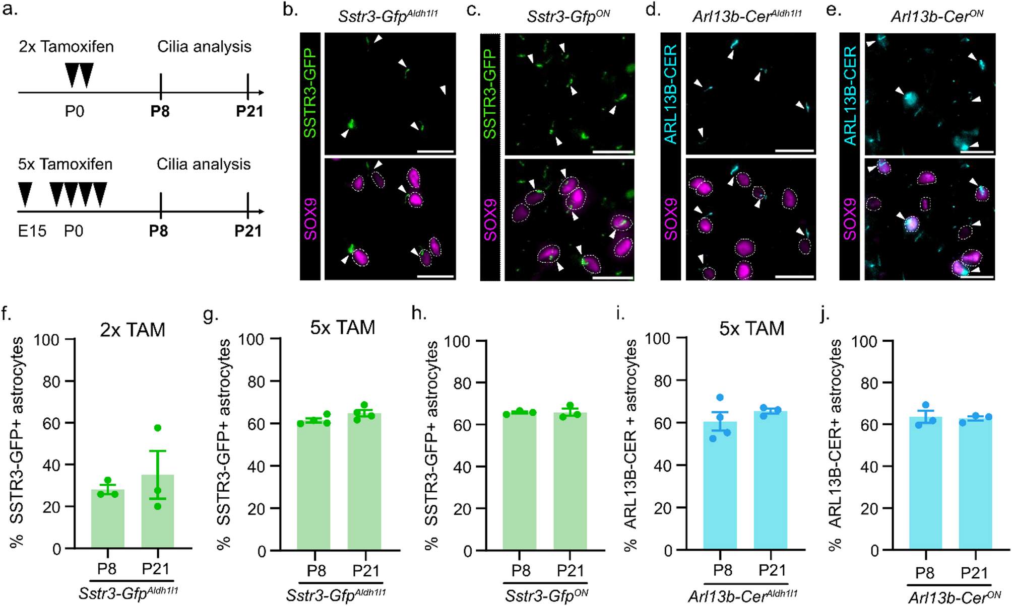
All mice were cared for in accordance with NIH guidelines and Emory University’s Institutional Animal Care and Use Committee (IACUC). Lines used were: Aldh1l1-CreERT2 obtained from The Jackson Laboratory [Tg(Aldh1l1-cre/ERT2)1Khakh, MGI:5806568, RRID: IMSR_JAX:031008] [16, 22], CMV-cre acquired from The Jackson Laboratory [Tg(CMV-cre)1Cgn/J, MGI:2176180, RRID: IMSR_JAX:006054], Sstr3-Gfp obtained from Dr. Bradley Yoder [Gt(ROSA)26Sortm1(Sstr3/GFP)Bky, MGI:5524281, RRID: MGI:5524974] [18], and Arl13b-Cerulean obtained through the European Mouse Mutant Archive [Gt(ROSA)26Sortm1(CAG−Cerulean/Arl13b,−Venus/GMNN,−Cherry/CDT1)Rmort, MGI:6193732, RRID: IMSR_EM:12168] [17]. Sstr3-GfpON and Arl13b-CerON mice were generated by crossing loxP-STOP-loxP-Sstr3-Gfp or loxP-STOP-loxP-Arl13b-Cerulean mice to the germline CMV-Cre and identifying the progeny with recombined LoxP sites eliminating the STOP so they constitutively express the cilia-reporter. Genotyping was performed on DNA extracted from ear punch via PCR (36 cycles: 95 °C 0.5 min, 60 °C 0.5 min, 72 °C 1 min) using the following primers. For Aldh1l1-Cre, forward primer GGCAAACGGACAGAAGCA and reverse primer CTTCAACAGGTGCCTTCCA; for CMV-Cre, forward primer TGACCCGGCAAAACAGGTAGTTA and reverse primer TTCCCGCAGAACCTGAAGATGTT; for Sstr3-Gfp, a common forward primer CTCGTGATCTGCAACTCCAG was multiplexed with reverse primers in the ROSA locus (to detect the wild-type allele, 223 bp product) GCTGCATAAACCCCAGATGACTCC, in the PGK-Neo region (to detect the non-recombined allele, 317 bp product) GCGCATGCTCCAGACTGCCTTG, and in the Sstr3 coding sequence (to detect the recombined allele, 433 bp product) GCGGATGTGTTCCCCAGGGTGG. For Arl13b-Cerulean, five primers were multiplexed to detect the wild-type allele (212 bp product), the non-recombined allele (374 bp product), and the recombined allele (253 bp product): in the ROSA left arm, AGGGAGCTGCAGTGGAGTAG, in the ROSA right arm, CTTTAAGCCTGCCCAGAAGA, in the Arl13b coding sequence, CGACCATCACAAGTGTCACC, in the loxP-STOP-Neo-loxP region, AAAACCTCCCACACCTCCC, and in the region between the promoter and the LoxP sites, CGTGCTGGTTATTGTGCTGT.
Tamoxifen administration
Tamoxifen (Sigma T5648) stock solution was prepared once a month at a concentration of 10 mg/ml in 100% EtOH and stored at -20 °C. Each dose of tamoxifen was freshly prepared in corn oil the day of injection and dissolved using a speed vacuum centrifuge (Eppendorf Vacufuge Plus). To induce gene expression in astrocytes, a dose of 3 mg tamoxifen/ 40 g mouse weight in 300ul (adult) or 20ul (pup) of corn oil was prepared. For 2x tamoxifen treatment, tamoxifen was administered intraperitoneally at P0 and P1 to the dam using a 1 ml syringe and a 25G 5/8-inch needle. For 5x tamoxifen treatment, tamoxifen was administered intraperitoneally at E15.5 and E19.5 to the pregnant dam using a 1 ml syringe and a 25G 5/8-inch needle followed by subcutaneous administration at P0, P1, and P2 to pups using a 1/2 ml syringe with attached 29G 1/2-inch needle.
Caesarean section and cross fostering
A Caesarean section was performed on tamoxifen-treated, timed-pregnant dams at E20.5. CD-1 mice were used as foster dams. Briefly, the experimental dams were euthanized via cervical dislocation and the pups were dissected, placed on a heating pad, and tapped gently until they could breathe on their own. Pups were transferred to a CD-1 foster dam and integrated with the foster dam’s litter.
Tissue harvesting
P21 mice were euthanized by isoflurane inhalation followed by a trans-cardiac perfusion with ice-cold 1x phosphate-buffered saline (PBS) and ice-cold 4% paraformaldehyde (PFA). P8 pups were euthanized by decapitation. Brains were harvested following perfusion or decapitation and drop-fixed in 4% PFA overnight. Fixed tissue was washed with 1x PBS and then incubated with 30% sucrose in 0.1 M phosphate buffer overnight at 4 °C until the tissue sank. Samples were washed in optimal cutting temperature (OCT) compound to remove sucrose (Tissue-Tek OCT, Sakura Finetek), embedded in OCT, frozen on dry ice, and stored at -20 °C.
Immunofluorescent (IF) staining
OCT-embedded tissues were sectioned at 40 μm using a cryostat microtome and placed directly on microscope slides (Fisherbrand Superfrost Plus). Sections were let to air-dry and either processed immediately or stored at -20 °C. Sections stored at -20 °C were brought to room temperature before starting IF. Sections were first rehydrated in 1X Tris Buffered Saline (TBS), permeabilized in 1% SDS, and blocked in antibody wash (1% heat inactivated goat serum, 0.1% Triton X-100 in 1X TBS). Sections were incubated overnight at 4 °C with primary antibodies (chicken anti-GFP, 1:8,000, Abcam ab13970; rabbit anti-SOX9, 1:500, Millipore AB5535). Sections were washed with cold antibody wash and incubated for one hour at room temperature in the dark with secondary antibodies (goat anti-chicken, AlexaFluor 488; goat anti-rabbit, AlexaFluor 647; Hoechst 33342; all 1:500 dilution, ThermoFisher). Sections were washed with cold antibody wash and then mounted with a glass coverslip using ProLong Gold (ThermoFisher) mounting media. Slides cured overnight at room temperature in the dark and were stored short term at 4 °C or long term at -20 °C. Slides were imaged at 20x on a BioTek Lionheart FX automated microscope.
Quantification of cilia
Z-stack images of brain sections stained for ciliary markers were captured at 2 μm intervals. Analysis of cilia was performed on maximum projection images using cell counting in Fiji/ImageJ.


Comments (0)