
Remember me
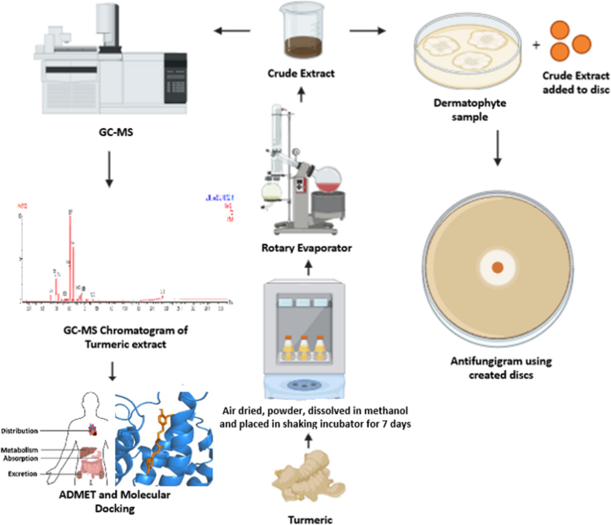


The overall workflow has been demonstrated in Fig. 1.
Fig. 1
Schematic diagram of the whole study. Initially, fresh turmeric rhizomes were collected and subjected to thorough air-drying. The dried material was next ground into a fine powder and extracted using methanol through a rotary evaporator to obtain the crude extract. The antifungal effect was tested by placing discs made from the crude extract on samples of dermatophytes. In addition, the bioactive compounds in the crude extract were identified using Gas Chromatography-mass spectrometry (GC–MS) analysis. The detected compounds were further evaluated through ADMET (Absorption, Distribution, Metabolism, Excretion, and Toxicity) profiling as well as molecular docking studies to predict their pharmacokinetic properties and possible biological interactions
In vitro antifungal activityThe Curcuma longa rhizome extract demonstrated notable in vitro antifungal activity against several species of Trichophyton, a group of dermatophytes known to cause skin infections. At concentrations of 20 mg, 30 mg, and 40 mg per disc, the methanol extract was evaluated against Trichophyton mentagrophytes (PQ394944), Trichophyton indotineae (PQ357663, PQ394963), and Trichophyton interdigitale (PQ357286) (Fig. 2 and Table 1). Results showed that the extract inhibited fungal growth, producing inhibition zones ranging from 10 to 25 mm, depending on the concentration and species tested. For example, the inhibition zone increased from 20 mm at 20 mg to 25 mm at 40 mg against Trichophyton indotineae (PQ394963), demonstrating a dose-dependent response. In contrast, at the greatest extract concentration, Trichophyton mentagrophytes (PQ394944) demonstrated a maximum inhibition of 23 mm. As a control, fluconazole, a common antifungal, produced varying inhibition zones ranging from 0 to 23 mm, demonstrating the efficacy of the C. longa extract against fluconazole-resistant strains. These findings suggest that methanol extract of Curcuma longa rhizome could serve as a potential natural alternative for treating dermatophytic infections, particularly in cases where conventional antifungal agents are less effective.
Fig. 2
In-vitro antifungal activity of methanol extract of C. longa rhizome. The antifungal susceptibility test was carried out following the Clinical and Laboratory Standards Institute (CLSI) guidelines using the disk diffusion method. Methanolic crude extracts were tested at concentrations of 20, 30, and 40 mg, with fluconazole (25 μg) used as the positive control. The assay was performed against three types of fungal strains: Trichophyton mentagrophytes (PQ394944), Trichophyton indotineae (PQ357663, PQ394963), and Trichophyton interdigitale (PQ357286)
Table 1 Inhibition zone of Curcuma longa rhizome extract against Trichophyton spp.GC–MS analysis and identification of major compoundsGC–MS analysis was used to identify the chemicals found in the methanolic extract of Curcuma longa rhizome (Fig. 3). Table 2 lists the active principles along with their concentration (%), molecular formula, molecular weight (MW), and retention time (RT). Using GC–MS, eleven components were found in the methanolic extract. The major components present in the Curcuma longa rhizome were Curlone (44.19%), 3-Isobutylidene-6,7-dimethyl-3H-isobenzofuran-1-one (17.93%), Tumerone (17.29%), 7-Epi-sesquithujene (6.63%), β-Sesquiphellandrene (4.87%), (E)-Atlantone (2.47%), 1-Bisabolone (1.80%) and numerous additional compounds were found to be at low levels.
Fig. 3
GC–MS Chromatogram of Curcuma longa rhizome methanolic extract showing peak intensity (Y-axis) versus retention time (X-axis). The major components detected in the C. longa rhizome extract were Curlone (RT:9.426),3-Isobutylidene-6,7-dimethyl-3H-isobenzofuran-1-one (RT:8.979), Tumerone (RT: 9.066). 7-Epi-sesquithujene (RT: 7.005), β-Sesquiphellandrene (RT: 7.365), (E)-Atlantone (RT: 10.172), 1-Bisabolone (RT: 9.849)
Table 2 Compounds detected from C. longa rhizome extraction and their GC-MS resultIn silico studyMolecular dockingThe study used molecular docking to assess each compound’s capacity to bind to the target protein, lanosterol 14α-demethylase. Using PyRx tools Autodock vina, all eleven compounds were docked with the protein lanosterol 14α-demethylase to assess their binding ability. Among them, four compounds α-Curcumene, 3-Isobutylidene-6,7-dimethyl-3H-isobenzofuran-1-one, Curlone, 1-Bisabolone, and Fluconazole showed better binding affinity with the corresponding values − 8.3 kcal/mol, − 8.7 kcal/mol, − 8.4 kcal/mol, and − 8.2 kcal/mol, −7 kcal/mol (Table 3). The chemicals’ binding affinities are all listed in Supplementary Table 1.
Table 3 QSAR Properties of the selected four compoundsThe docking protocol was validated by redocking the co-crystallized ligand in the lanosterol 14α-demethylase (PDB ID: 4LXJ) structure, using the same docking settings as applied to the test compounds. The redocked pose showed strong agreement with the original crystallographic conformation, yielding an RMSD of 1.219 Å, which is well within the acceptable threshold for docking validation. This result confirms the accuracy and reliability of the docking procedure employed in the study. A structural overlay of the native and redocked poses is presented in Supplementary Figure S1.
Protein–ligand interactionsProtein–ligand interactions of C. longa extracts were characterized by their van der Waals contacts, electrophoresis, hydrogen bonds, and hydrophobic interactions. The binding affinities between ligands and proteins are largely determined by these interactions. Every chemical showed a wide variety of binding and unbound interactions with several residues either within the site or outside of it. As determined, 1-Bisabolone exhibited a single conventional hydrogen contact with the residue Arg A:98, as well as seven hydrophobic interactions (p-Alkyl and Alkyl interactions) with Leu A:95, Leu A:96, Pro A:238, Phe A:241, Val A:242, Tyr A:72, and His A:381 (Fig. 4). Ten hydrophobic interactions, such as p-Alkyl and Alkyl contacts, p-p Stacked/T-Shaped interactions, and Pi-Cation and Anion interactions, were generated by α-Curcumene in place of conventional hydrogen bonding, with the following amino acid residues: Val A:242, Tyr A:72, Leu A:95, Phe A:241, Phe A:384, Ala A:125, His A:381, Leu A:96, His A:405 and Pro A:238 (Fig. 5). The compound 3-Isobutylidene-6,7-dimethyl-3H-isobenzofuran-1-one showed a single typical hydrogen contact with the residue Arg A:98. Six hydrophobic connections were also generated by it, including p-Alkyl and Alkyl contacts, as well as p-p Stacked/T-Shaped Interactions, with Leu A:95, Pro A:238, Leu A:96, Phe A:241, Ala A:125, and Phe A:384 (Fig. 6). Curlone demonstrated eight hydrophobic contacts and five Vander Waals interactions in total (p-Alkyl and Alkyl contacts) with the following amino acids: Leu A:96, Val A:242, Leu A:95, Tyr A:72, Met A:509, Phe A:384, Phe A:241, and His A:381 (Fig. 7). The positive control, Fluconazole exhibits multiple non-covalent interactions, including van der Waals forces, halogen (fluorine) bonds, carbon-hydrogen bonding, and hydrophobic Pi-alkyl and Pi-sigma interactions. Key interacting residues include ILE205, TYR229, LEU221, and ALA226. Table 4 lists the protein–ligand interaction mechanism for each of the four drugs.
Fig. 4
Protein–ligand complex interaction in 2D and 3D. In this case, 1-Bisabolone (yellow color) demonstrates the ligand–protein interaction with lanosterol 14α-demethylase following molecular docking
Fig. 5
2D and 3D interactions within the protein–ligand complex. α-Curcumene (yellow) demonstrating ligand interaction with the protein lanosterol 14α-demethylase following molecular docking
Fig. 6
Interactions in both 2D and 3D contexts involving the protein–ligand complex. In this instance, 3-Isobutylidene-6,7-dimethyl-3H-isobenzofuran-1-one (Yellow color) is presented, illustrating the interaction of the ligand with the protein lanosterol 14α-demethylase following molecular docking analysis
Fig. 7
Interaction between the protein–ligand complex in both 2D and 3D contexts. This illustration depicts Curlone (in yellow), highlighting the interaction between the ligand and the protein lanosterol 14α-demethylase following molecular docking analysis
Table 4 Results of the interactions between the 5 selected ligands and the protein lanosterol 14α-demethylaseADME and toxicity analysisFollowing the administration of the drug via any route to the human body or in an animal model, it experiences processes of absorption, distribution, metabolism, and excretion, leading to either active or passive transport to the target site. In order to meet these criteria, the analysis of in-silico pharmacokinetics provides insights on the behavior of a molecule within the body. The evaluation of the drug-likeness of the compounds was mainly conducted through Lipinski’s rule of five, which considers factors including molecular weight, topological polar surface area (TPSA), hydrogen bond acceptors, hydrogen bond donors, and the number of rotatable bonds (Nb). According to the molecular docking data, all of the drugs that underwent pharmacokinetic analysis performed exceptionally well in terms of Lipinski’s rule of five and other criteria. In terms of ADMET properties, every drug exhibited positive results for the proportion unbound in plasma, BBB permeability, MDCK permeability, CaCO2 permeability, CNS permeability, volume distribution, and human intestine absorption.
Regarding adsorption, curlone exhibited superior human intestine absorption compared to the other three chemicals. 3-Isobutylidene-6,7-dimethyl-3H-isobenzofuran-1-one exhibited superior caco2 permeability and MDCK. However, when considering the F20% value, only one chemical (3-Isobutylidene-6,7-dimethyl-3H-isobenzofuran-1-one) yields a negative result (Table 5).
Table 5 ADMET properties of the selected four compoundsIn terms of distribution, α-Curcumene had a higher volume of distribution and CNS permeability whereas curlone had better result for fraction unbound in plasma and BBB permeability (Table 5).
Human cytochrome P450 (CYP450) enzyme is crucial in phase I oxidative metabolism and is responsible for the majority of drug biotransformation [39]. Surprisingly, the curlone compounds had very few negative effects on the activity of CYP450 enzymes (Table 5). However, compounds such as 3-Isobutylidene-6,7-dimethyl-3H-isobenzofuran-1-one, alpha curcumene, and 1 bisabolone showed significantly higher inhibition against CYP450 enzymes (specifically CYP1A2 and CYP2C9). The clearance and half-life (t1/2) parameter demonstrated favorable outcomes, with curlone exhibiting a superiority compared to the other chemicals (Table 5).
The skin permeability, as indicated by the logarithm of the skin permeability coefficient (Log Kp), showed considerable variation among the compounds studied. α-Curcumene exhibits the highest predicted skin permeability, while 3-Isobutylidene-6,7-dimethyl-3H-isobenzofuran-1-one shows the lowest. Solubility assessments revealed that all compounds except α-curcumene exhibited good solubility; α-curcumene showed only moderate solubility. Notably, none of the compounds in this study were categorized as insoluble. Transdermal absorption depends on the polar surface area (PSA), with a lower PSA indicating greater skin permeability. α-Curcumene, possessing a PSA of 0.00 Å2, aligns with its high permeability profile. In contrast, 3-Isobutylidene-6,7-dimethyl-3H-isobenzofuran-1-one has a significantly higher PSA of 26.30 Å2, supporting its lower permeability. Curlone and 1-Bisabolone had middle-range PSA values of 17.07 Å2, showing that they can moderately pass through the skin, which is better than 3-Isobutylidene-6,7-dimethyl-3H-isobenzofuran-1-one but not as good as α-curcumene. All four compounds demonstrated potential for poor skin sensitization (Table 5).
Fluconazole was used as a control in this study for comparison with the compounds detected in our results. In terms of absorption, fluconazole demonstrated similar results to human intestinal absorption, showing poor Caco-2 permeability and a negative result for F 20%, similar to curlone. In terms of distribution, fluconazole exhibits poor brain barrier permeability, lower volume of distribution (Vd) (L/kg), and inability to penetrate the CNS. In terms of metabolism, fluconazole performs well and similarly to Curlone for CYP2C9-sub, but is not as effective as α-Curcumene and 1-Bisabolone. Fluconazole displays hepatotoxicity, which is not present in the other four compounds. Furthermore, fluconazole demonstrated reduced skin permeability, indicated by a more negative Log Kp value and relatively high topological polar surface area (TPSA) of 81.65 Å2 (Table 5).
Comments (0)