Leiomyosarcomas comprise fewer than 2% of all malignant vaginal neoplasms. The most frequently observed vaginal tumors are squamous cell carcinomas (75–90%), followed by adenocarcinoma (5–10%), melanoma (3%) and sarcoma (2%) [1]. Amongst vaginal sarcoma, leiomyosarcoma is the most common [2] It peaks in fifth decade of life, with a range extending from 21 to 86 years [2]. They tend to occur in older age than SCC of vagina. The etiology of leiomyosarcomas remains unknown and majority occurs de novo, except for a history of pelvic irradiation in three patients reported by Malkasian et al. [3].
Vaginal LMS presents most commonly with an asymptomatic vaginal mass as was seen in our case. Patient complaints can range from vaginal, rectal pain, vaginal discharge or bleeding, difficulty in micturition to rarely, dyspareunia. Majority (45%) arises from the posterior vaginal wall, followed by 34% from the lateral vaginal wall and 21% from the anterior vaginal wall [4]. Although in our case it was situated anteriorly, mostly submucosal, they can arise from any part of vagina [4].
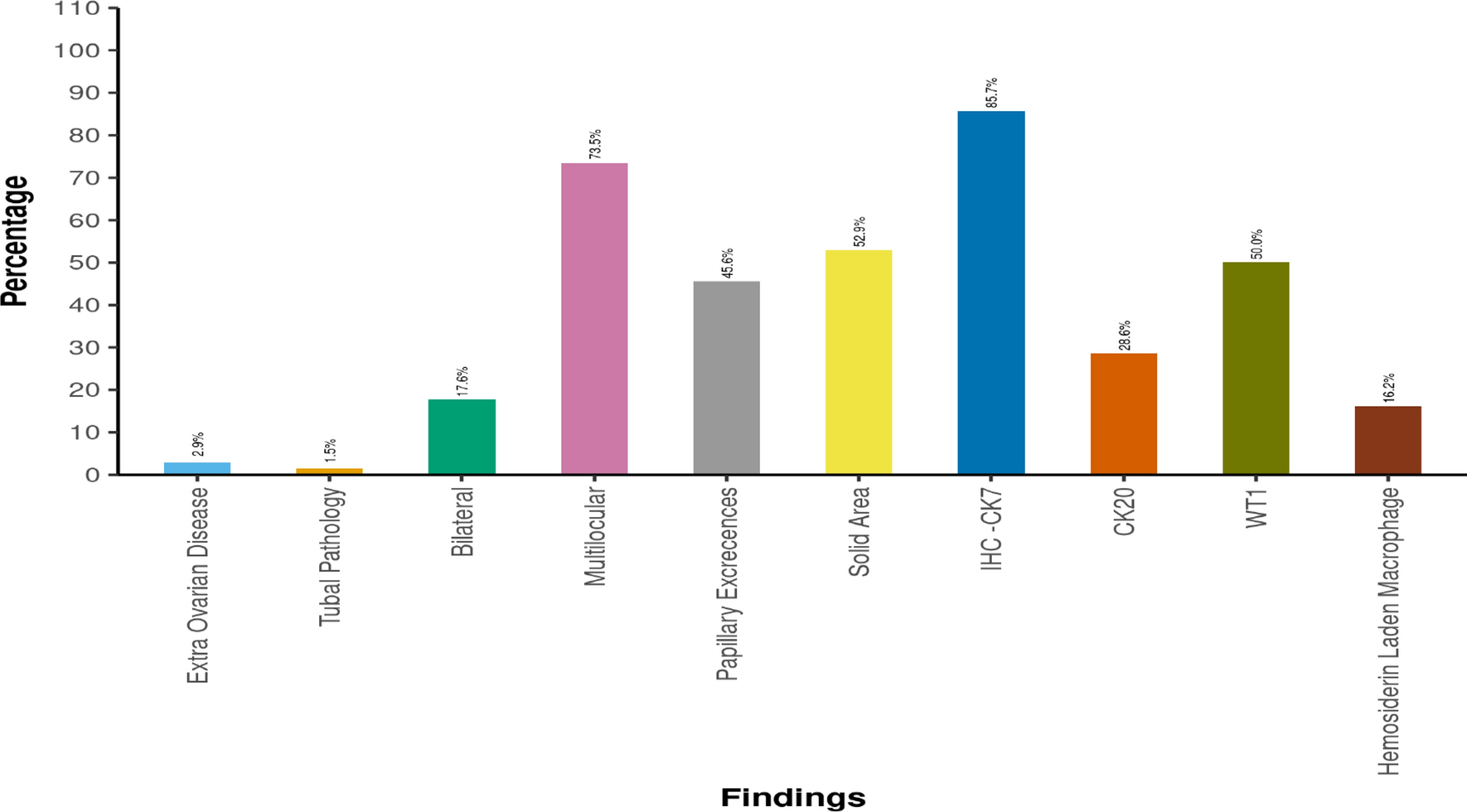
Preoperative biopsy is highly advocated both for definitive diagnosis and as an important aid in planning optimal surgical strategy [4]. The criteria between benign leiomyoma and leiomyosarcoma include high mitotic index and histologic evidence of cellular dysplasia. The diagnostic criteria established by Tavassoli and Norris for smooth muscle tumors of the vagina suggest that any tumor with greater than 10 mitoses per 10 HPF or a tumor with a mitotic count between 5 and 9 and at least moderate atypia should be considered a leiomyosarcoma [5].
Leiomyosarcomas have a better outcome than other sarcomas, as suggested by some authors [6,7,8]. Differential diagnoses include Gartner’s duct cyst, granuloma, vaginal leiomyoma, fibroepithelial polyp, capillary hemangioma, neurofibroma rhabdomyosarcoma, and other soft tissue sarcoma. Metastasis from gestational trophoblastic disease and endometrial adenocarcinoma, or direct extension from cervix, rectum, and bladder should be ruled out.
Complete surgical resection remains the mainstay of treatment, with adjuvant radiotherapy considered for high-grade tumors. Although Peters et al. and Ciaravino et al. have shown pelvic exenteration to increase OS, few studies concluded that extent of resection may not be related to clinical course [7, 9, 10].
A meta-analysis encompassing 14 controlled trials of adriamycin-based chemotherapy for soft tissue sarcomas demonstrated significant improvement in delaying both local and distant recurrence and enhancing overall survival. This study did not elucidate the impact of chemotherapy on vaginal leiomyosarcoma; however, it combined data from many different types of sarcomas [11].
LMS are notorious for recurrences. within 2 years of treatment. It spreads locally or hematogenous metastasis. Pulmonary metastasis is common [12]. Pelvis is the earliest, most common site of recurrence and only site of recurrence in half of the reported cases [7]. The risk factors include positive resection margin, large size, high mitotic index, and age. The prognosis depends on factors such as tumor size, mitotic index, and margin status, stage. Close and life-long follow-up is essential due to the high risk of recurrence and metastasis, especially if the initial treatment includes radiotherapy as second primaries are more common in this group.


Comments (0)