
Remember me
Family Ochodaeidae Streubel, 1846
Subfamily Ochodaeinae Streubel, 1846
Tribe Ochodaeini Streubel, 1846
Genus Atlantochodaeus n. gen.
Atlantochodaeus Costa-Silva, Sousa, Fuhrmann, Grossi and Vaz-de-Mello, n. gen.
(Figs. 1, 2a–g, 3e–l, 4, 5, 6, 7, 8, 9, 10, 11, 12, 13).
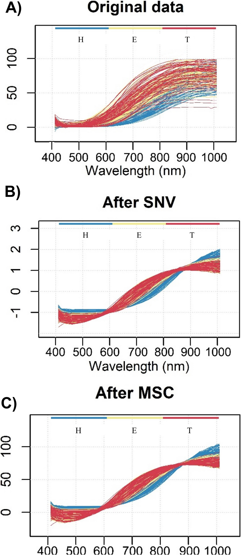
Fig. 1
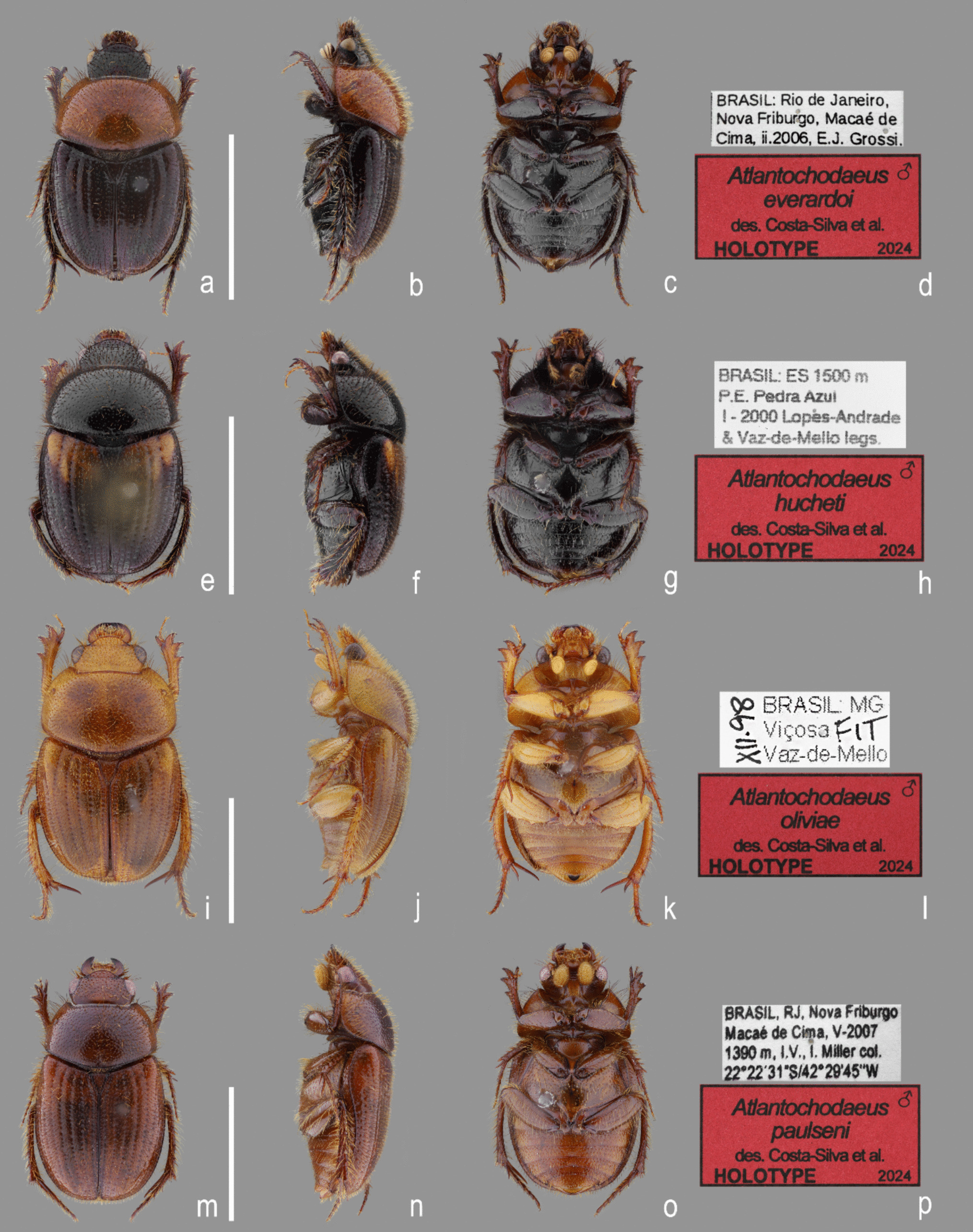
Atlantochodaeus new genus, holotypes (dorsal, lateral, ventral, labels). a–d, A. everardoi new species. e–h, A. hucheti new species. i–l, A. oliviae new species. m–p, A. paulseni new species. Scale = 5 mmss
Fig. 2
Ochodaeidae morphology. a–g, Atlantochodaeus everardoi new genus and species, male. h–n, Parochodaeus pectoralis (LeConte, 1868), male. a, h, head, dorsal view. b, i, head, lateral view. c, j, head, ventral view. d, k, eye posterolateral margin. e, l, mesocoxae (arrow pointing coxae separation). f, m, detail of propygidium-elytra interlocking apparatus. g, n, protibia
Fig. 3
Head (dorsal, frontal, ventral, lateral). a–d, Parochodaeus pectoralis (LeConte, 1868). e–h, Atlantochodaeus everardoi new species. i–l, A. oliviae new species. Scale = 1 mm
Fig. 4
Atlantochodaeus new genus, mouthparts. a–j, A. everardoi new species. k–t, A. oliviae new species. a–c, k–m, left mandible (dorsal, inner, ventral). d–f, n–p, right mandible (ventral, inner, dorsal). g, q, labium (ventral). h, r, Detail of anterior area of prementum. i, s, labrum (dorsal). j, t, maxilla (ventral). Scale = 0.5 mm; details without scale
Fig. 5
Atlantochodaeus everardoi new genus and species, mouthparts. a, labrum (dorsal). b, epipharynx (ventral, with detail of asperites). c, labium and hypopharynx (dorsal). d, labium (ventral, with detail of setae). pgl, paraglossa; gls, glossa. Scale = 0.5 mm
Fig. 6
Atlantochodaeus everardoi new genus and species, mouthparts. a, b, right mandible (ventral with details of internal morphology, dorsal with detail of small fovea). c, d, maxilla (dorsal, ventral, respectively). bst, basistipe; gal, galea; lac, lacinia; lst, laterostipe; pst, parastipe; vst, ventrostipe. Scale = 0.5 mm
Fig. 7
Atlantochodaeus new genus, prothorax and legs. a–f, h, A. everardoi new species. g, i, A. oliviae new species. a, prothorax, frontal (left side with cervix); b, prothorax, posterior (left side with intersegmentar membrane); c, prothorax, ventral (left side with arm of hypomeron dissected); d, detail of ornaments of posterior arm of hypomeron; e–g, protibia, outer view (male, female, male); h, i, metatibia posterior view. Scale = 1 mm
Fig. 8
Atlantochodaeus everardoi new genus and species, pterothorax. a, anterior (right side with mesoalinotum omitted). b, posterior. c, dorsal, internal (right side with epimesterna and epimera ommited). d, ventral, internal (left side with mesopostnoum elements omitted). e, f, lateral (external, internal with mesoalinotum setarate). Acronyms suffix 2 and 3 indicating mesothorax and metathorax elements, respectively (except to fragma: ph1–3); apn, apodeme of postnotum; awp, anterior notal wing process; ba2–3, basalare (* apodeme); em2–3, epimeron; es2–3, episternum; fu2–3, furca; lp2–3, lateral arm of postnotum; ls2–3, lateral arm of scutellum; msb, mesendosternite bridge; mwp, medial notal wing process; pe2–3, prealare; ph1–3, first, second and third phragma; pla, pleural arm; pwp, posterior notal wing process; sc2–3, scutum; su3, subalare (* apodeme); st2–3, scutellum; vt2–3, meso- and metaventrite; wp2–3, pleural wing process. Scale = 1 mm
Fig. 9
Atlantochodaeus everardoi new genus and species, pterothorax. a, dorsal (right side with mesoalinotum omitted). b, mesoalinotum, internal, ventral. c, d, pleurites (external, internal with margins of apodemes of basalare and suralare in red). e, right posterior wing. f, detail of posterior wing basis. g–i, first axillary sclerite (head, dorsal, ventral, respectively). j, k, second axillary sclerite (dorsal with radial fulcalare associated, ventral). l, third axillary sclerite, dorsal. Acronyms suffix 2 and 3 to thorax indicating mesothorax and metathorax elements, respectively (except to axillary sclerites and phragma, Ax1–3, ph1). awp, anterior notal wing process; ba2–3, basalare; em2–3, epimeron; es2–3, episternum; lp2–3, lateral arm of postnotum; ls2–3, lateral arm of scutellum; mts, metathoracic spiracle; pe2–3, prealare; ph1, first phragma; pla, pleural arm; su3, subalare; wp2–3, pleural wing process. Veins (anterior to posterior): C, costa; Sc, subcosta; RA, radius anterior; RP, radius posterior; MP, medial posterior; CuA, cubital anterior; AA, anal anterior; AP, anal posterior. Folds (dotted lines; anterior to posterior): dl1, dl2, longitudinal fold 1 and 2; mf, medial fold; tf, transversal fold; af, anal fold; jf, jugal fold. Scale: A–F = 1 mm; G–L = 0.5 mm
Fig. 10
Ochodaeidae, abdomen. a–c, e–j, Atlantochodaeus everardoi new species. d, Parochodaeus pectoralis (LeConte, 1868). k, l, Atlantochodaeus hucheti new species. m, n, A. oliviae new species. o, p, A. paulseni new species. a–d, abdomen (dorsal, ventral, lateral, and lateral view, respectively). e–g, genital ring. h, k, m, o, aedeagus, dorsal. i, l, n, p, aedeagus, lateral. j, aedeagus, posterior. as1–7, abdominal spiracle I–VII; cs3, lateral concavity of sternite III; ft9, ventral fold of tergite IX; ps2, medial process of sternite II; pyg, pygidium; s2–9, sternite II–IX; sp, sternite stridulatory peg; t1–10, tergite I–X. Scale = 1 mm
Fig. 11
Atlantochodaeus new genus, female terminalia. a–i, k, o, A. everardoi new species. l, p, A. hucheti new species. j, m, q, A. oliviae new species. n, r, A. paulseni new species. a–d, terminalia (dorsal, ventral, posterior, lateral). e, internal genitalia, ventral. f, left paraprocts (tergite 9). g, internal structure of puncture of paraprocts and proximal gonocoxite. h, left proximal gonocoxite. i, j, left medial gonocoxite. k–n, left distal gonocoxite and gonostyle. o–r, spermatheca. bcx, bursa copulatrix; dpg, distal gonocoxite; gst, gland of spermatheca; ovd, oviduct; mpg, medial gonocoxite; ppg, proximal gonocoxite; spt, spermatheca; stl, gonostyle; t9, paraproct or abdominal tergite IX; t10, proctiger or abdominal tergite 10. Scale: a–f, h–r = 0.5 mm; g = 0.1 mm
Fig. 12
Atlantochodaeus everardoi new genus and species, colour variation: (a) orange pronotum and black elytra; (b) yellow pronotum and black elytra; (c) orange pronotum and black elytra with a yellow spot in the humeral region; and (d) yellow pronotum and black elytra with yellow-milk colour band in the humeral region. Colour variation non-sex related
Fig. 13
Atlantochodaeus new genus, metafemur. a, A. everardoi new species. b, A. hucheti new species. c, A. oliviae new species. d, A. paulseni new species
(urn:lsid:zoobank.org:act:F551 FDA9-1B76-433 C-A3 AF-C4BCC1E19D3B).
Generic differential diagnosisThe Atlantochodaeus can be separate from the genus Parochodaeus by the following characters (Parochodaeus pectoralis morphology between square brackets): latero-posterior border of eyes round (Fig. 2d) [emarginated, Fig. 2k]; ventral surface of eye small (Figs. 2c, 3f–h, j–l) [large, Figs. 3b–d]; gula not or slightly prominent posteriorly (Fig. 3h, l) [greatly prominent posteriorly, Fig. 3d]; mesocoxae widely separate (Fig. 2e) [subcontiguous, Fig. 2l]; apex of elytra slightly prominent (Fig. 2f) [strongly prominent, Fig. 2m]; longest metatibial spur as long as or longer than metatarsomere I [shorter]; metatarsomere I straight [usually sinuous] (see Table 1).
Table 1 Comparative table among Parochodaeus pectoralis and the species of the Atlantochodaeus n. gen DescriptionBody light or dark yellowish brown (A. olivae and A. paulseni), or black with pronotum black (A. hucheti), orange or yellow (A. everardoi), humerus with or without light macula. Head (Figs. 2a–c, 3e–l): Surface flat, without tubercles or carina, densely punctate and setose, fronto-clypeal suture distinct, complete or indistinct medially, slightly curved (Fig. 3a, i). Gula slightly prominent posteriorly (Fig. 3h, l). Eyes (Fig. 3e–l) prominent, small in dorsal view (head 1.4–1.6 wider than minor dorsal interocular distance; Figs. 2a, 3e, i), small in ventral view (head 1.4–1.6 wider than minor ventral interocular distance; Figs. 2c, 3g, k); posterior margin round (Figs. 2d, 3h, l). Clypeus wider than long, flat; anterior margin rounded, thickened with row of long yellow setae. Labrum (Figs. 4i, s, 5a) slightly sinuous, with strong transversal carina delimiting the anterior and posterior area. Epipharynx (Fig. 5b) with distal medial area with row of about five large sensilla, medial area with about 20 small sensilla, posterolateral areas and posteromedial area with dense comb of cuticular setae-like projections and some asperites. Mandibles (Figs. 4a–f, k–p, 6a–b) slightly asymmetric; outer margin continuously rounded; two large incisor teeth present; mola almost flat and smooth; prostheca densely setose; dorsal proximal area of incisivus with two large punctures. Maxillae (Figs. 4j, t, 6c, d) with stipe divided in four free plates: basistipes (bst), ventrostipes (vst), laterostipe (lst) and parastipe (pst); laterostipes with longitudinal row of minute spines and some long setae; lacinia (lac) falciform and articulated with parastipes; galea (gal) somewhat trapezoid with some thick bent setae; palpus with four palpomeres. Labium (Figs. 4g, q, 5c, d) with submentum trapezoid, fused with gula, deflected and forming angle with the basis of mentum. Mentum slightly convex, deeply depressed anteriorly, dorsolateral fold with large fovea, disc with about four lateral long setae and with one transverse strong keel. Prementum membranous; palpiger prominent and with ventroproximal asperites; glossa (gls) club-like and with five distal setae; paraglossa (pgl) blade-like with outer setae-like cuticular projections; palp with three palpomeres. Hypopharynx (Fig. 5c) covered with dense setae-like cuticular projections, anterior area with a transverse row of about 15 large sensilla. Antenna with ten antennomeres; scape as wide as pedicel, and slightly wider than funicle antennomeres; antennal club with proximal flagellomere longer than medial one, and medial flagellomere longer than distal one. Prothorax (Figs. 7a–d): Pronotum wider than long, entirely beaded, marginal bead with long yellow or testaceous setae, surface slightly convex, sparsely punctate and setose, interpuncture smooth and shiny; pronotal scar as a shallow concavity; anterior angles acute, anteriorly prominent; posterior angles rounded. Hypomeron with posterior arm with two carinate articular process (Fig. 7d). Prosternum with basisternum anteriorly prominent and medially with longitudinal carina; posterior sternal process slightly acuminate. Cervix (Fig. 7a) with three sclerites, two ventrolateral and one minor dorsolateral. Pterothrorax (Figs. 8, 9a–d): Mesothoracic spiracle about 2.5 times longer than wide (Fig. 7b), metathoracic spiracle about 3.3 times longer than wide (Fig. 9d). Mesoscutellum (Figs. 9a, b) triangular, laterals slightly arcuate, surface punctate and setose as pronotum, separate from scutum by a rounded deflection. Mesepisternum (Figs. 9c, d) with anterior ventral angle prominent, metepisternum (Fig. 8e) separate from mesocoxal cavity by the mesepimeron-metaventrite contact. Mesoventrite with posterior area separate mesocoxae cavity, deflected and forming an angle of 120º with the anterior half in lateral view (Fig. 8e). Metaventrite longer at the middle, punctate and setose, posterior margin sinuous, medial posterior area pointed backward. Metendosternite (Fig. 8b, c, f) with large basis and anterior area prominent. Elytra: Outer, distal and inner margins beaded; interstriae granulated and setose; nine punctate stria present, I–V at disc, inner to humerus and separate from each other by about 3 punctures diameter, VI–IX at lateral and outer to humerus and separate from each other by less than one puncture diameter; inner posterolateral surface with longitudinal carina; posterior angle slightly prominent. Posterior wings (Fig. 9e–l): Anterior margin fused with radial cel forming club-like area formed by costa (C), subcosta (Sc) and anterior sector of radius (RA); basis of fourth anterior radial vein (RA4) and basis of first posterior radial vein (RP1) indistinct; third and fourth posterior medial veins (MP3 and MP4) distinct; distal part of anterior cubital vein (CuA) distally bent; radial fulcalare distinct as slightly sclerotized band basally in contact with second axillary sclerite (Fig. 9f, j); head of first axillary sclerite narrow (Fig. 9g–i); dorsal distal ridge of second axillary sclerite short (Fig. 9j, k); caudal area of third axillary sclerite slightly shorter than cranial anterior area (Fig. 9l). Legs: Profemur with inner carina between trochanter articulation and posterior femur-tibia articulation. Protibiae (Fig. 2g, 7e–g) with three outer teeth; inner distal angle not prominent, rounded; males with posterior distal area with two tubercles each with one thin seta (Fig. 7e, g), females with tubercles inconspicuous and bearing one thick seta (Fig. 7f), posterior proximal and medial area with a row of four to eight acute teeth. Mesocoxae oblique, posteriorly convergent; widely separate from each other (Fig. 2e). Anterior side of meso- and metafemora with four longitudinal rows of long setae. Metafemora inner margin crenulate or with one small tooth. Mesotibiae shorter than mesotarsomeres I–V combined; surface covered by spines and setae. Metatibiae with three longitudinal rows of spine-like setae. Meso- and metatarsomere I slightly dilated; longer than tarsomeres II–IV combined. Longest mesotibial spur spiny serrate. Abdomen (Figs. 10, 11): Sternites III–VIII ventrally exposed; II thin, with medial lamellate process (ps2), dorsal fold small and trapezoid. Surface of sternites III–VIII smooth, covered by yellow setae; dorsal fold of sternite III (cs3) with anterior cavity as long as the length of fold (Fig. 10c); dorsal fold of sternite VII with small club-like stridulatory peg (sp; Fig. 10a). Propygidium with spiracles at anterior angles (as7); elytral interlocking apparatus formed by two posterior teeth, medial area without process or carina (Figs. 2f, 10a). Pygidium (pyg) densely punctate and setose; broad triangular, anterior angles with spiracles; anterior margin with transversal shallow sulcus (Fig. 10c). Male terminalia (Figs. 10e–g) flat and tubular; tergite IX (t9) medially interrupted, posterolateral lobes setose, and with each anterolateral area ventrally bent, fold as a thin bar anterior to sternite IX; sternite IX long, with posterior area setose. Aedeagus (Fig. 10h–p) with short and almost symmetrical parameres. Female terminalia (Fig. 11a–c) with paraprocts (t9; Fig. 11f) long, with posterior inner membranous punctate area; each side of external genitalia with shield-like punctate proximal gonocoxite (ppg; Fig. 11h); small medial gonocoxite (mpg) bearing two setae (long and minute; Fig. 11i, j); distal club-like setose gonocoxite (dpg; Figs. 11k–n); and small distal gonostyle (stl) bearing three distal setae. Internal genitalia (Fig. 11d–e) with long bursa copulatrix (bcx) connected to left side of duct, with some long thick straight setae at basis; spermatheca somewhat oblong (spt; Fig. 11o–r); gland of spermatheca (gst) small, connected to basis of spermatheca; accessory glands indistinct.
Sexual dimorphismMales and females are easily separated from each other specially by the ornaments of protibia. Males of Atlantochodaeus present protibial spur strongly bent (Figs. 2g, 7e), while it is slightly curved in females (Fig. 7f). Males with posterior side of protibia with two distal tubercle-like process, each bearing one thin seta; otherwise, females have inconspicuous processes, and the setae associated with each process are thick. Also, mentum presents the truncated transverse keel (Fig. 4g, q) that is relatively larger in males than in females. Metafemora ornaments (Fig. 13) smaller in females than in males, sometimes indistinct.
RemarksFemale terminalia of Atlantochodaeus is similar to that found in other Ochodaeinae, but the medial gonocoxite is smaller (Fig. 11i, j) than in Ochodaeus species (Dupuis 2005).
Type-speciesAtlantochodeus everardoi n. sp., here designated.
Geographic distributionAtlantic Forest of Southeast Brazil (Fig. 14).
EtymologyThe name is derived from “Atlant-” (referring to the Atlantic Forest), the geographical origin of the species comprising the new genus, combined with the generic name “Ochodaeus”; gender masculine.
Atlantochodaeus everardoi n. sp.
(Figs. 1a–d, 2a–g, 3e–h, 4a–j, 5, 6, 7a–f, 7h, 8, 9, 10a–c, 10e–j, 11a–i, k, o, 12, 13a, 14).
Fig. 14
Occurrence of Atlantochodaeus species from the Brazilian Atlantic Forest: A. everardoi n. sp. (grey green circle), A. hucheti n. sp. (black red circle), A. oliviae n. sp. (black rhombusblue circle), and A. paulseni n. sp. (white black circle). Legend of Brazilian States: ES = Espírito Santo; MG = Minas Gerais; and RJ = Rio de Janeiro; and SP = São Paulo
(urn:lsid:zoobank.org:act:32D27 C46-A50D-413B-87 A8-A7 C929DB623 F).
Type materialHolotype, male (Fig. 1a–c). Labels (Fig. 1d), first label [white, printed]: “BRASIL: Rio de Janeiro,/Nova Friburgo, Macaé de/Cima, ii.2006, E.J. Grossi.”. Second label [red with black frame, printed]: “♂/Atlantochodaeus/everardoi/des. Costa-Silva et al./HOLOTYPE/2024” (CERPE). Type locality. Macaé de Cima, Nova Friburgo municipality, Rio de Janeiro State, Brazil.
Paratypes (40 specimens)First label [white, printed]: “BRASIL: Rio de Janeiro,/Nova Friburgo, Macaé de/Cima, ii.2006, E.J. Grossi.”. Second label [yellow with black frame]: “Atlantochodaeus/everardoi/des. Costa-Silva et al./PARATYPE/2024” (1 female [MZSP 22217], MZSP). First label [white, printed]: “BRASIL: RJ/Nova Friburgo/III-1998 FIT/P. and E. Grossi”. Second label [yellow with black frame]: “Atlantochodaeus/everardoi/des. Costa-Silva et al./PARATYPE/2024” (1 male [MZSP 22216], MZSP). First label [white, printed]: "BRASIL, RJ, Nova Friburgo/Macaé de Cima, RPPN/Bacchus, iii.2007, 1500 m/E. and P. Grossi leg". Second label [yellow with black border, printed]: “Atlantochodaeus/everardoi/des. Costa-Silva et al./PARATYPE/2024” (1 female, CERPE). First label [white, printed]: "Coleção E./and P. Grossi". Second label [white, printed]: “BRASIL, RJ, Nova/Friburgo, Macaé de/Cima, 1300 m, I.2010 E.J. Grossi.”. Third label [yellow with black frame]: “Atlantochodaeus/everardoi/des. Costa-Silva et al./PARATYPE/2024” (1 male, CEMT; 1 male and 2 females, CERPE). First label [white, printed]: “BRASIL, Rio de Janeiro, Nova/Friburgo, Macaé de cima,/1500 m, xii.2009, FIT,/P.J. Grossi leg”. Second label [white, unknown’s handwriting] “M.C. XII.09/FIT”. Third label [yellow with black frame]: “Atlantochodaeus/everardoi/des. Costa-Silva et al./PARATYPE/2024” (1 male, CERPE). First label [white, printed]: “BRASIL: Rio de Janeiro, Nova Friburgo, Macaé de/Cima, ii.2006, E.J. Grossi.” Second label [yellow with black frame]: “Atlantochodaeus/everardoi/des. Costa-Silva et al./PARATYPE/2024” (2 males and 2 females, CEMT; 2 males and 2 females, CERPE). First label [white, printed]: “BRASIL: Rio de/Janeiro, Nova Fribur/go, Macaé de Cima,/x.2000, P. Grossi”. Second label [yellow with black frame]: “Atlantochodaeus/everardoi/des. Costa-Silva et al./PARATYPE/2024” (1 female, CEMT). First label [white, printed]: “BRASIL: Rio de Janeiro/Nova Friburgo, Macaé/de Cima I-1999 PGrossi.”. Second label [yellow with black frame]: “Atlantochodaeus/everardoi/des. Costa-Silva et al./PARATYPE/2024” (2 males and 2 females, CEMT, 2 females, CERPE). First label [white, unknown’s handwriting]: “BRASIL: RJ/Nova Friburgo/Macaé de Cima/IV-2000/P. and E. Grossi”. Second label [yellow with black frame]: “Atlantochodaeus/everardoi/des. Costa-Silva et al./PARATYPE/2024” (2 males, CEMT; 2 males, CERPE). First label [white, printed]: “BRASIL: RJ, Nova Friburgo/Macaé de Cima XII-2000/P and E Grossi legs”. Second label [yellow with black frame]: “Atlantochodaeus/everardoi/des. Costa-Silva et al./PARATYPE/2024” (1 male, CEMT; 1 male CERPE). First label [white, printed]: “BRASIL, RJ, Nova Friburgo/Macaé de Cima, III-2000/1390 m, I.V. [Interception Trap], I. Miller col./22º22′31”S/42º29′45”W”. Second label [white, printed]: “COLEÇÃO/E., and P. Grossi”. Third label [yellow with black frame]: “Atlantochodaeus/everardoi/des. Costa-Silva et al./PARATYPE/2024” (1 male and 1 female, CEMT; 2 males and 1 female, CERPE). First label [white, printed]: “BRASIL: RJ Nova Friburgo/Macaé de Cima 1500 m/III-2000 Lopes-Andrade, Gumier and Vaz-de-Mello”. Second label [yellow with black frame]: “Atlantochodaeus/everardoi/des. Costa-Silva et al./PARATYPE/2024” (1 male, CEMT). First label [white, printed]: “Macaé de Cima/Nova Friburgo/RJ—BRASIL/I-2006/Leg: B.Miller”. Second label [yellow with black frame]: “Atlantochodaeus/everardoi/des. Costa-Silva et al./PARATYPE/2024″ (1 male, CEMT). First label [white]: “Macaé de Cima/Nova Friburgo/RJ—Brasil/I—2006/Leg: B. Miller”. Second label [yellow with black frame]: “Atlantochodaeus/everardoi/des. Costa-Silva et al./PARATYPE/2024″ (3 males and 1 female, AMBC).
Differential diagnosisAtlantochodaeus everardoi is similar to A. hucheti and both share the body mainly black, metafemur with inner area crenulate (Fig. 13a–b), and male protibial spur abruptly bent (as in Figs. 2g and 7e). Atlantochodaeus everardoi can be separated by following characteristics (A. hucheti morphology between square brackets): pronotum reddish or yellowish brown (Figs. 1a, 12) [pronotum dark reddish brown, almost black, Fig. 1e]; metafemur inner area weakly crenulate (Fig. 13a) [strongly, Fig. 13b]; female spermatheca somewhat ovoid (Fig. 11o) [oblong, Fig. 11Q].
Holotype(Figs. 1a–c) length: 6.8 mm, width: 3.9 mm, body dark reddish brown, almost black; pronotum reddish brown; setae yellow or black. Head. Eyes very small in ventral view (head 1.4 wider than minor ventral interocular distance; Fig. 3g). Fronto-clypeal suture complete. Mentum with curved keel, U-shaped in ventral view (similar to Fig. 4g). Prementum with anterior area lobed (Fig. 4h). Thorax. Pronotum length: 2.1 mm, width: 3.4 mm. Elytra: Length: 3.7 mm, width: 3.9 mm. Legs: Protibiae with posterior proximal and medial areas with row of six acute teeth (Fig. 7e). Metafemur narrow with inner area slightly crenulate (Fig. 13a). Metatibia inner side arched, without emargination (Fig. 7h). Aedeagus with anterior half of parameres abruptly bent in lateral view (Figs. 10h–j).
VariationsThis species presents a great variability in length and colour (Fig.
Comments (0)