
Remember me
The dMEGA-PRESS pulse sequence was implemented on two different instruments. Initial in vitro experiments were performed on phantom solutions using a 11.7 T Bruker Ascend 500 MHz vertical wide bore spectrometer. These experiments were used to validate the performance of the pulse sequence, both when it comes to spectral selective refocusing and diffusion-weighting. The method was then implemented on a 7.0 T Bruker Pharmascan 70/16 horizontal small animal MRI scanner. Due to an abrupt out-phasing of the animal scanner, in vivo experiments were prioritised, with very limited time for systematic experiments on phantom solutions, which is therefore not included. In both in vitro and in vivo experiments the total echo time was kept constant at 68 ms, which is optimal for MEGA-PRESS editing of GABA. To achieve full refocusing the time delays in Fig. 1 were: t2 = t3 = t4 = t1 + t5 = TE/4 = 17 ms [13, 15]. The bandwidth of the 180\(^\) slice selective pulses were 3.5 kHz (7.0 ppm) and 2.5 kHz (8.3 ppm) respectively for 11.7 T and 7.0 T. Shinnar–LeRoux pulses with a duration of 10 ms and bandwidth of 170 Hz, corresponding respectively to 0.34 ppm and 0.56 ppm at 11.7 T and 7.0 T, were used for spectral selective refocusing. Spoiler gradients were rectangular pulses with a duration of 3 ms and with an amplitude of 62 mT/m. Other time intervals were: \(\delta = 3\) ms, \(\tau = 1.5\) ms, \(\Delta = 27\) ms, and \(\Delta _s = 31.5\) ms. Due to the relatively large voxel size in the in vivo experiments the effects from anisotropy is expected to average out, and this, combined with the restriction of a 1 h time limit for animal scanning, diffusion gradients were applied only in the z-direction. Outer volume suppression was enabled using the built-in subroutines of the Paravision software. Local iterative shimming on the voxel was performed to the second-order, both in the in vitro and in vivo experiments. The shimming quality was determined by evaluating the peak of water or NAA at 2.0 ppm. Shimming gradients were adjusted until a full width at half maximum of less than 3 Hz for in vitro experiments and less than 12 Hz for in vivo experiments was achieved, ensuring optimal magnetic field homogeneity. A built-in algorithm was used for online evaluation of eddy currents, where a water reference scan was recorded before each single experiment for the purpose of eddy current correction.
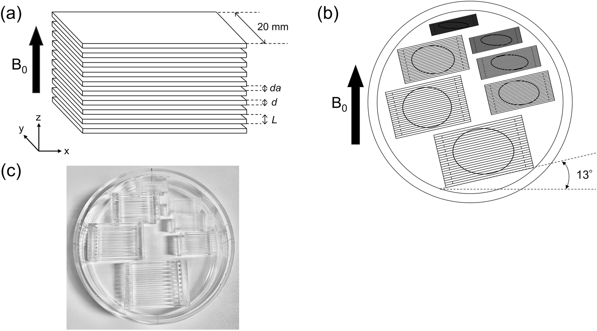
In vitro phantom studyIn vitro experiments were performed on aqueous solutions of selected metabolites. All solutions were prepared using distilled water where the pH was adjusted to 7.4 using a phosphate buffer. Each solution was added to a 10 mm NMR tube. To examine the spectral editing performance of the dMEGA-PRESS pulse sequence, experiments were performed on a solution containing only GABA (5.0 mM) and NAA (20.0 mM). The pulse sequence was then applied to a series of solutions with varying concentration of GABA (2.5, 4.2, 5.9, 7.9 and 10.3 mM), and where the concentrations of the other metabolites were kept constant (in mM): Glu: 12.5, Cho 3.0, Cr 10.0, NAA 12.5, Lac 5.0, and Ins 7.5. This series of solutions are referred to as ‘metabolite phantoms’. The reliability of the diffusion-weighting was tested on the metabolite phantom containing 5.9 mM GABA.
All in vitro experiments were conducted at 25 \(^\)C on a 11.7 T Bruker Ascend 500 MHz vertical wide bore spectrometer equipped with a commercial Bruker MicWB40 micro imaging probe, using ParaVision 6.0.1 software (Bruker BioSpin, Billerica, MA, USA). The maximum available gradient strength was 1.4 T/m. For placement of the voxel, the Paravision multi-slice localiser was utilised. The shimming was done automatically based on an acquired \(B_0\)-map. Water suppression was manually adjusted for each sample using VAPOR. A \(4\times 4\times 4~\text ^3\) voxel was placed in the middle of the 10 mm NMR tube. All spectra were acquired using a regular PRESS sequence or the dMEGA-PRESS sequence shown in Fig. 1, with respectively TE/TR = 16/3000 ms and TE/TR = 68/3000 ms. 4096 data points were acquired with 16 averages. Diffusion weighting was performed with a total of 9 b values in the range 0.01–4.19 \(\text /\upmu \text ^2\). Two datasets were acquired for each b value, one ‘ON’ spectrum, where the spectral selective 180\(^\) RF-pulses were placed at 1.9 ppm, causing refocusing of the signals from GABA at 2.3 and 3.0 ppm (GABA-editing) [13], and the signals from Glu at 2.4 and 3.75 ppm (co-editing) [16]. In the ‘OFF’ spectrum, these pulses were placed at 7.5 ppm to acquire a spectrum without selective refocusing.
In vivo animal studyIn vivo experiments were performed on adult Wistar rats (N = 10, 5 male and 5 female). Animal experiments were conducted according to ethical guidelines at University of Bergen. The animals were anaesthetised with isoflurane (2.5 vol% added to 1:2 oxygen:nitrogen). Respiration was monitored and a physiological temperature (37.0 ± 0.2 \(^\)C) was maintained by a heater module (SA instruments Inc, NY, USA). After positioning the animal in the scanner, isoflurane was reduced to maintain a respiration level of 60–70 breaths min\(^\) (1.6–2.0 vol%). Two animals were excluded (one male, one female) due to problems with water suppression during scans. All animal experiments were performed on a 7.0 T Bruker Pharmascan 70/16 horizontal small animal MRI scanner with software ParaVision 6.0.1 (BrukerBioSpin, Billerica, MA, USA) and a 55 mm/23 mm tx/rx quad head coil (Bruker Corporation, Model No: MT0206). The maximum available gradient strength was 0.7 T/m. The animal was fixed using a standard Bruker animal cradle.
Fig. 2
Placement of a \(7\times 4\times 7~\text ^3\) voxel in the central region of a rat brain, based on the multi-slice localiser image
Localisation was performed based on a multi-slice localiser image with FOV = \(50\times 50\) mm\(^2\). An \(8\times 5\times 8\) mm\(^3\) voxel was chosen for first, second, and third order shimming. The shimming was done automatically based on an acquired \(B_0\)-map. For localised \(^1\)H spectroscopy a \(7\times 4\times 7\) mm\(^3\) voxel, shown in Fig. 2, was placed in the middle of a heterogenous region of the brain, including mid brain, cortex, thalamus and hippocampus, averaging out regional concentrations and diffusion properties of metabolites [17]. As described above, local iterative shimming on the voxel was performed until a full width at half maximum for water and/or NAA of less than 12 Hz was achieved, which is within the recommended limit for animal MRS in the rodent brain [18]. The voxel was placed at a safe distance from the edges of the skull to avoid any susceptibility artefacts.
MR spectra were acquired using the dMEGA-PRESS sequence with TE/TR = 68 ms/3000 ms. 4096 data points were acquired with 32 averages. Water suppression was achieved using VAPOR, with manually optimised RF attenuations. ‘ON’ and ‘OFF’ diffusion-weighted spectra were acquired using the same settings for the spectral selective 180\(^\) RF-pulses as described for the in vitro experiments. Diffusion weighting was performed with a total of 5 b values in the range 0.03–4.38 \(\text /\upmu \text ^2\), and the gradient pulses were applied along the z-direction in all experiments. A navigator was used for online frequency drift, which can be more pronounced when performing diffusion experiments.
ProcessingEddy current correction of in vitro and in vivo data were performed using ParaVision 6.0.1 software (Bruker BioSpin, Billerica, MA, USA), where the procedure is based on an accumulated signal from a water reference scan, resulting in a signal which is demodulated based on the temporal phase evolution of the strongest spectral component of this reference scan. Averaging, combination of spectra, and frequency drift correction, was performed online in ParaVision. Further processing was performed using an in-house script made in MatLab 2019b (MathWorks inc., Massachusetts), where the raw data were first combined and averaged after correcting for frequency drifts that might have occurred during the experiment. The data were then zero filled to \(2^\) to achieve a Hz/pt resolution that follows the standard in the GANNET pipeline [19] used in the subsequent processing. Global phase correction was performed using the ‘Automated phase Correction based on Minimization of Entropy’ (ACME) algorithm [20]. Phase correction was performed in two steps, first using the water peak, then using the chosen region of interest corresponding to the metabolite being investigated. This approach resulted in a more robust phase correction. Edited spectra, which enables isolation and quantification of molecules with refocused signals, were obtained for each b value by subtracting the ‘OFF’ from the ‘ON’ spectrum.
Spectral fitting and deconvolution [6] is often performed using a basis set representing linear combinations of individual metabolite spectra at a given experimental condition [21, 22]. A basis spectrum for a specific metabolite can be generated from separate experimental data or through simulations based on density matrix theory and input of experimental conditions [6]. When analysing edited spectra a simpler approach with lineshape peak fitting and integration of each individual signal is common, where the use of GANNET [19] on data from GABA-editing is an example. An analysis based on lineshape peak fitting and integration was applied in our study, where each individual peak of interest were curve fitted using in-house algorithms inspired by the GANNET pipeline [19] and LWFIT [23]. Signal intensities of the different metabolites at varying diffusion-weightings were then obtained from integration of the curve fitted peaks. This corresponds to a “no prior knowledge” approach to spectral analysis, allowing for larger flexibility compared to methods where the spectral data are fitted using basis sets obtained from simulations or separate experiments [23]. This approach allows for the use of the same analytical method for the ‘OFF’, ‘ON’ and edited diffusion-weighted data, enabling a direct and simple comparison of the improved quality of the obtained ADC values for specific metabolites. Fit errors for the spectral analysis of each metabolite was calculated according to the procedure used in GANNET pipeline [19].
Details regarding hardware, acquisition, data analysis, and data quality are given in Supporting Information, Table S1.
Comments (0)