
Remember me

Among the 647 patients with HR + MBC in the ctDNA study cohort, 609 had HR +/HER2− MBC. Among the latter, there were 69 patients (11%) in whom ctDNA analysis identified a non-synonymous GATA3 mutation. GATA3 mutations were observed in a statistically significant larger percentage of patients with documented visceral disease (76.8% vs. 62.0%; p = 0.016) and de novo metastatic disease (33.3% vs. 20.7%; p = 0.018) compared to GATA3WT; no other distinguishing clinical characteristics, including age at metastatic diagnosis, number of prior therapies, and progesterone receptor status, were observed (see Table 1 for clinical characteristics & mutational status of the cohort). Of note, when comparing only patients with frameshift or nonsense GATA3 mutations (annotated as likely oncogenic or known oncogenic via the OncoKB tool, n = 55), observations and significance were similar to the above (Supplemental Table 1). A separate analysis showed no correlation with HER2 status and GATA3 mutations, but given the more aggressive phenotype and different treatment approach in HER2 + MBC, this subtype was otherwise excluded from this analysis. Among these 69 patients with HR +/HER2− GATA3mut MBC, 49 (71%) patients identified as White and 14 (20%) as Black. Patients had received a median of 2 prior lines of therapy in the metastatic setting (range 0–9).
Table 1 Patient characteristics in patients with GATA3WT and GATA3mut HR +/HER2− MBCNext, we aimed to characterize GATA3 mutations and their location within the gene. The GATA3 protein is encoded by 444 amino acids, which encompass 6 exons, 5 of which are coding; the gene also contains two transactivating domains and two Zinc finger (ZnFn) motifs. [13] ZnFn 1 stabilizes DNA binding, while ZnFn 2 binds DNA at the GATA3 motif. [45] Prior tissue-based analyses have found most GATA3 mutations to occur at the splice sites between exons 4/5 and 5/6, as well as within exon 5 (including Zn finger 2) and exon 6, the majority of which were frameshift mutations and annotated as likely oncogenic per the OncoKB tool [14, 25, 32, 33, 43, 46].
In our ctDNA analysis, among these 69 GATA.mut patients we found 73 GATA3 mutations: 18 (26%) occurred in exon 5, all but three of which were in the second zinc finger, 8 (11%) were c.925-3_925-2 del splice site variants, and 46 (67%) were in exon 6. In terms of mutation type, 43 (62%) were frameshift mutations, 14 (20%) were missense mutations, and 5 (7%) were nonsense mutations (Fig. 1B). Comparison to breast cancer variants in the Catalog Of Somatic Mutations In Cancer (COSMIC) database, a tissue-based cancer mutation repository, also showed the majority of variants being frameshift mutations (Supplemental Fig. 1). Median variant allele fraction (VAF) of the GATA3 mutations was 1.0% (IQR 0.34–5.5%). To approximate clonality, we calculated the fraction of the alteration of interest relative to the alteration in the ctDNA sample with the highest VAF; we used 30% as a cutoff. Four patients had two GATA3 mutations with comparable VAFs (listed in parentheses), consistent with them being two clonal hits (based on above criteria to estimate clonality as ratio of VAF over highest VAF in the sample): F392L (6.40%)/P409fs (6.20%), 336* (1.29%)/435* (0.71%), W329R (0.8%)/A395 V (0.1%), and A442fs (2.9%)/R353 T (2.4%). While the Guardant360 test is not intended to report germline mutations, we set a VAF cutoff of 40% to identify potential germline GATA3 alterations, which result in HDR syndrome (hypoparathyroidism, deafness, renal dysfunction) [47, 48]. By the above criterion, no GATA3 alterations appeared to be germline (i.e., all VAFs were under 40%). Fifty-five patients (80%) were determined to have likely oncogenic variants via OncoKB, which in this dataset included all frameshift, nonsense, and splice site variants (Fig. 1B) [43].
Fig. 1
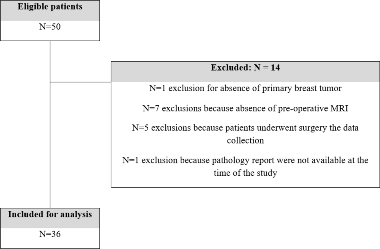
GATA3 gene mutation overview—A Flowchart of patient ctDNA cohort harboring GATA3 gene mutations; B Mutational distribution within the GATA3 gene
GATA3 mutational landscapeIn 3 of the 69 patients with GATA3.mut HR +/HER2− MBC, GATA3 was the only detectable ctDNA alteration, while remaining patients had concomitant co-mutations with a median number of 4 variants (range 1–44) (Fig. 2). The most frequent co-mutations were in PIK3CA (n = 30), ESR1 (n = 26), and TP53 (n = 23), and the most frequent amplification was in CCND1 (n = 14). There were no statistically significant associations (positive or negative) between GATA3 and TP53 (Fisher exact p = 0.30), and GATA3 and PIK3CA (p = 0.52). These findings are distinct from the co-mutational landscapes based on tissue analyses of primary breast cancers, where GATA3 mutations appeared to be mutually exclusive from both TP53 and PIK3CA mutations [14, 25, 33].
Fig. 2
CoMut plot of GATA3 and other gene mutations in our study population
In the ctDNA-detected GATA3mut population, TP53 co-mutations (n = 23) were found with a median VAF of 1.0% (range 0.03–30.5%). Of note, plasma-based cell-free DNA assays cannot definitively distinguish tumor-derived somatic mutations from clonal hematopoiesis (CH), in which TP53 is a commonly mutated gene [49]. 55 of the 69 patients (80%) had predicted clonal GATA3 mutations (based on above criteria), and 12/23 (52%) TP53 co-mutations appeared to be clonal. Among the clonal TP53 variants, 8 (67%) had a VAF > 1%. 5/12 patients had both TP53 and GATA3 as suspected clonal variants, only 2 of whom also had a VAF > 1% (Supplemental Fig. 2).
Within the GATA3mut population, GATA3 was the mutation with the highest VAF in 23 patients (34% of patients). Among patients where GATA3 was not the highest VAF, the other highest mutations included PIK3CA (n = 19), ESR1 (n = 7), TP53 (n = 8), BRCA1 (n = 2), BRCA2 (n = 2), and SMAD4 (n = 2).
Interestingly, there were 3 patients with germline mutations in BRCA1/2. As mentioned above, the Guardant 360 assay does not report germline mutations, but suspicion is raised when allele fraction is above 40% (and much higher than other mutations). By this criterion, 3 of the 7 (43%) patients with non-synonymous BRCA1/2 mutations were noted to have findings suspicious for germline variants, which was later confirmed on retrieval of documented genetic testing. This appears to be a new finding compared to an earlier study of GATA3 mutations in familial breast cancer, where GATA3 mutations were found in 22% (7/32) of patients without BRCA1/1 mutations and not identified in patients with germline BRCA1 or BRCA2 mutations (n = 0/23) [50].
Among the 69 patients with GATA3mut HR +/HER2− MBC, there were 20 patients with serial sampling data. Among these, in 5 patients, GATA3 mutation(s) had not been identified on initial ctDNA sampling but rather were identified on subsequent draws. New detection on serial sampling may be due to one of several factors: emergence of a new variant, or else the variant existing in the first sample but below the limit of detection. While the numbers are relatively small, many of the GATA3 frameshift mutations appeared to increase in serial sampling in synchrony with known driver alterations such as PIK3CA or ESR1, as well as ARID1A and ATM, though disparate growth patterns were also observed in 3 patients. (Supplemental Fig. 3).
Fig. 3
GATA3 proteomic profiling using CPTAC database—A GATA3 mutation and variant distribution; B Proteomic and phosphoproteomic expression changes between GATA3WT vs GATA3mut samples
GATA3 mutations and proteomic correlatesTo understand the functional implications of GATA3 mutations, the National Cancer Institute's tissue-based Clinical Proteomic Tumor Analysis Consortium (CPTAC) database was interrogated for HR +/HER2− breast cancers harboring GATA3 mutations, and the expression and phosphorylation of GATA3 and associated proteins were analyzed. Among 121 patients with breast cancer, 10 patients had a single GATA3 mutation. We identified 6 luminal A, 3 luminal B, and 1 HER2-enriched cases. The latter was excluded given the focus of this analysis on HR +/HER2− breast cancer. Among the remaining 9 GATA3-mutated, there were 4 frameshift, 3 missense, and 2 splice site variants. We did not detect differences in GATA3 protein abundance when comparing GATA3-mutated to HR +/HER2− unmutated samples (Fig. 3). The frameshift mutations were located in the C-terminal domain and have not been demonstrated to impact protein levels of function. [51] Similarly, there was no difference in protein abundance of breast cancer-associated genes, including ESR1, PIK3CA, FOXA1, FOXO3, or RB1 (0.2 < FDR < 0.9). Notably, GATA3 mutations were associated with a significant increase in abundance of the deubiquitinating enzyme USP48 (LogFC = 0.76, FDR = 1.7 × 10⁻⁵), which stabilizes MDM2, and thus enhances p53 ubiquitination and degradation [52]. All above samples with GATA3 mutations had wildtype TP53.
Global differential expression analysis of phosphorylation sites revealed a significant increase of RPS6 KA3 phosphorylation levels on sites S369 and S715 (FDR = 0.014 & 0.006 respectively), which both reside on the highly conserved catalytic domain of the S6 kinase, a component of the RAS/ERK signaling pathway. Similarly, we saw increased phosphorylation on FOXO3 (Sites S7 and S12, FDR = 0.048), both suggesting increased downstream signaling, potentially via ERK. KRT18 showed significantly reduced phosphorylation at multiple sites (such as S31, FDR = 0.001); this protein is linked with the epithelial-mesenchymal transition and was explored given the association of GATA3 mutations and metastasis. In addition, the histone chaperone ANP32E, which has been shown to be inversely correlated with tumor progression and relaxation of chromatin at FOXA1 binding sites, was found to be higher in GATA3mut cancers, which is in keeping with published literature (LogFC = 1.09, FDR = 2.2 × 10⁻⁴) [53]. Activation of these pathways may lead to estrogen receptor signaling independence.
GATA3 mutations and clinical outcomesFinally, we evaluated the association of ctDNA-detected GATA3 mutations with clinical outcomes in the HR +/HER2− MBC setting, stratified by type of therapy. Among patients who received endocrine monotherapy (ET; GATA3WT, n = 74, GATA3mut, n = 6), GATA3mut were associated with worse progression-free survival (PFS; p = 0.061) and worse overall survival (OS; p = 0.004); sample sizes were not powered to detect statistically significant differences. There was no statistically significant difference in PFS or OS between GATA3mut (n = 25) and GATA3WT (n = 188) subgroups that received chemotherapy and those that received ET + CDK4/6 inhibitor treatment.
In two contrasting index patient cases, we describe this clinical story at the individual level along with ctDNA clonal pattern. One patient with known lung metastases treated with palbociclib in combination with fulvestrant, remained on this therapy for 14 months, during which GATA3 T329fs and ESR1 D538G emerged, likely both in a resistant dominant clone (Fig. 4a). The disease remained stable for over a year of therapy, after which the patient developed a new lung metastasis. In contrast, in Fig. 4d, a patient with known liver metastases was treated with a novel oral selective estrogen receptor degrader (SERD) monotherapy. The liver metastases increased in size and number after only 3 months. During this time, there was a steady increase in the allele fractions in 3 mutations: PIK3CA E542 K, ESR1 Y537 N, and GATA3 K358f in a manner suggesting the PIK3CA mutation was clonal, and the ESR1 and GATA3 variants were subclonal. No amplifications in PIK3CA were detected.
Fig. 4
Survival analyses of GATA3 mut patients treated with ET + CDK4/6 and ET alone
Comments (0)