
Remember me
Small bowel obstruction (SBO) is a common indication for diagnostic imaging, hospital admission, and surgical consultation. At our institution, patients with SBO identified on an initial CT scan without enteric contrast often underwent a second CT with enteric contrast. A new protocol was implemented in a collaboration between the department of surgery to eliminate this second CT, instead utilizing enteric water-soluble contrast (WSC) and serial abdominal radiographs for further assessment of SBO in clinically stable patients. This study aims to assess the impact of this protocol on radiation exposure and resource utilization.
MethodsA retrospective cohort study was conducted on patients with SBO diagnosed on initial abdominopelvic CT for whom the general surgery service was consulted. The control group included patients prior to protocol implementation who underwent two abdominopelvic CT scans within 24 h–one with and one without enteric contrast. The experimental group included patients managed under the new protocol. Ionizing radiation exposure, contrast media utilization, and CT technologist time were recorded for both groups.
ResultsEighteen patients were included in the experimental group and 38 patients were included in the control group. Total effective dose (mSv) and CT technologist time were significantly less with the new protocol (p = 0.02 and p < 0.001, respectively). Although the use of intravenous contrast was lower in the experimental group, this did not reach statistical significance (p = 0.06).
ConclusionThe implementation of a collaborative SBO imaging and care algorithm between general surgery and radiology resulted in reduced radiation exposure to patients and decreased CT technologist time. This highlights the value of multidisciplinary approaches in improving the efficiency of imaging strategies for SBO.
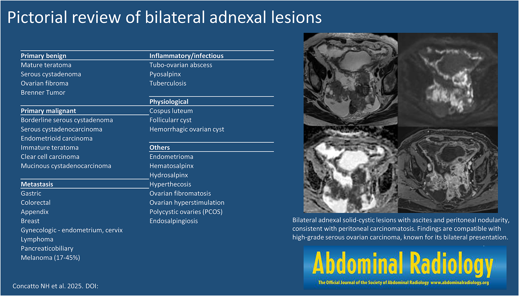
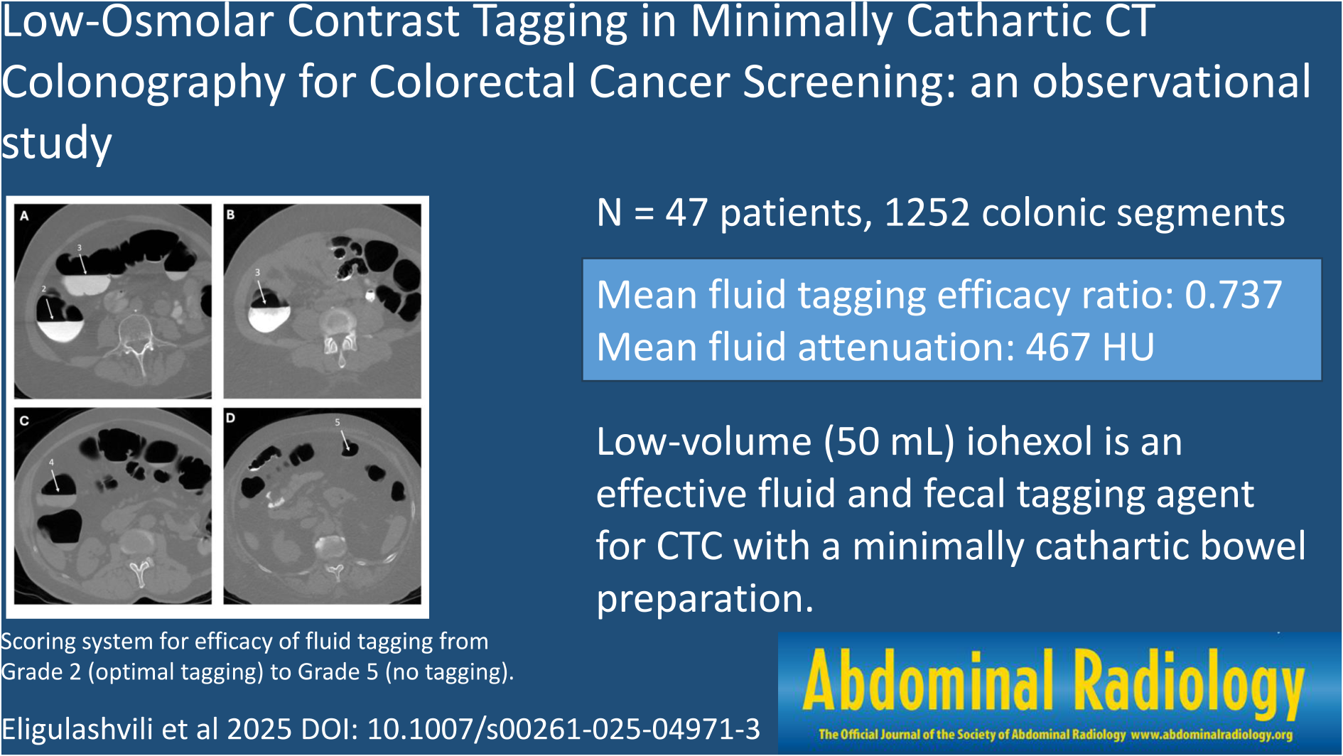
Graphical abstract
Comments (0)