
Remember me


Wild-type (WT) mice (C57BL/6J) were purchased from the Jackson Laboratory (Bar Harbor, ME, USA). A mix of male and female mice were used for all studies. The mice were 11 weeks old at the start of the study and housed under controlled standard housing conditions (dark/light cycle of 12 h at a range of 20–23 °C). All procedures involving mice were performed in accordance with Office of Laboratory Animal Welfare and National Institutes of Health guidelines and the approval of The Roskamp Institute Institutional Animal Care and Use committee (AAALAC certified).
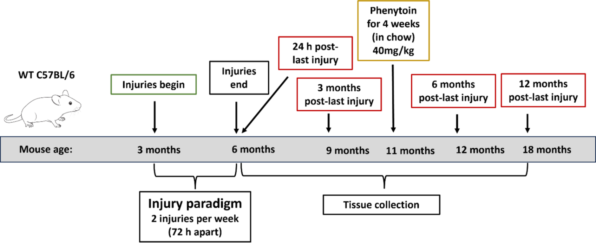
Traumatic brain injury protocolA mouse model of closed head injury was used to investigate the effects repetitive mild traumatic brain injury (r-mTBI) as previously described [1, 32]. Mice were shaved at the injury site and then anesthetized with 1.5 L/min if oxygen and 3% isoflurane for 3 min. Animals were positioned on a heating pad to maintain their body temperature at 37 °C and the head was placed in a stereotaxic frame (Stereotaxic Instrument, Stoelting, Wood Dale, Illinois) connected to an impact device (Impact One Stereotaxic Motorized Impactor, Richmond, Illinois). The impact was performed using a 5 mm blunt metal impactor tip which was then retracted to a strike depth of 1 mm. The impact was delivered at 5 m/s with a force of 72 N. To prevent possible hypothermia, mice were allowed to recover on a heating pad set at 37 °C until they became ambulant again. Mice in the r-mTBI group received 2 impacts per week (approximately every 72 h) for 3 months. The r-sham group was exposed to isoflurane for the same frequency and time as the r-mTBI mice, but they did not receive an injury, to control for the effects of repeated anesthesia. For the pericyte isolation studies, mice were euthanized, and brains collected after 6 months post-injury. For the PDGF-BB disposition studies, mice were euthanized at post-injury time points of 24 h, 3 months, 6 months and 12 months. Additional information on the injury timeline and tissue collections can be found in Fig. 1.
Fig. 1
Timeline of the brain injury paradigm and tissue collection for the mouse studies. C57BL/6 mice at 3 months of age received 2 injuries per week for 3 months. Mice were euthanized and tissue was collected at 24 h, 3 months, 6 months and 12 months after the final brain injury or anesthesia exposure for r-sham control mice. Moreover, for pericyte isolation studies, mice were fed with phenytoin for 4 weeks before euthanasia
Animal treatmentBeginning on the fifth month post-injury, mouse chow incorporating phenytoin (PHT) (40 mg/kg, Inotiv) was administered to r-sham and r-mTBI mice ad libitum for a total of 4 weeks. The PHT dosage administered here was based on prior reporting [9]. The total cohort was split in 4 groups which included r-sham and r-mTBI fed with normal chow (control mice) and r-sham and r-mTBI fed with PHT chow (PHT-administered mice). On a weekly basis, we monitored the body weight and food intake of the PHT-administered mice for comparison to the control mice.
Human and primary mouse cerebrovessel isolationParenchyma and cerebrovasculature from brains were isolated using a protocol as previous described [32]. Freshly extracted mouse brains were collected (minus the cerebellum), the meninges and other outer vessels were removed with a dry cotton swab, and then minced with a blade in a Dounce homogenizer on ice. HBSS was added in fivefold excess of the brain volume, then the minced brain material was stirred with the spatula to break up larger material. After 6–8 passes with a Teflon pestle, a sample of the brain homogenate was collected as a representation of the whole brain. This homogenate fraction was stored in an equal volume of lysis buffer consisting of M-PER (Pierce Biotechnology, Rockford, IL, ISA) supplemented with phenylmethanesulfonylfluoride fluoride (PMSF) (1 mM) and Halt protease and phosphatase inhibitor cocktail (Thermo Scientific, Waltham, MA, USA). An equal volume of 40% dextran solution was added to the brain homogenate for a final concentration of 20% dextran, and subsequently centrifuged at 6000 x g for 15 min at 4 °C. This results in a compact mass at the top of the solution (parenchyma) and a pellet at the bottom of the container (cerebrovasculature) separated by a clear dextran interface (non-cell associated fraction). The parenchyma and dextran were collected and added to an equal volume of HBSS, then centrifuged at 6000 x g for 10 min at 4 °C. The parenchyma was resuspended in HBSS and centrifuged for 5 min at 4 °C to remove any residual dextran, and parenchymal pellet was collected in lysis buffer. After the parenchyma and dextran were removed, the remaining cerebrovascular pellet was washed with ice cold HBSS and collected in lysis buffer. All samples were stored at −80 °C until further analysis.
Human brain specimens were obtained from Dr. Thomas Beach, Director of the Brain and Body Donation Program at the Sun Health Research Institute (Sun City, AZ). Frozen cortical samples (500 mg) from the inferior frontal gyrus were collected from autopsied brains, representing non-demented control subjects (no history of TBI), and TBI. For the TBI group, donors reported one or two brain injuries accompanied by loss of consciousness, each lasting less than 30 min. Given our primary focus on the chronic phase post-injury, samples with a longer duration since the last injury were prioritized, with an average of approximately 40 years post-injury. A summary of the human brain specimens is provided in Table 1.
Table 1 Specifications of human brain specimensPrimary brain vascular mouse pericyte isolation by magnetic cell sortingPrimary mouse pericytes were prepared from C57BL/6 mice at 6 months post-injury (r-mTBI) and r-sham. Entire brains were collected using stringent aseptic conditions, minced, dissociated using an enzyme-based brain dissociation kit (Cat No.130-107-667, MACS, Miltenyi Biotec), and passed through a sterile 70 μm nylon cell strainer (Cat No. 130-098-462, MACS, Miltenyi Biotec). After resuspending the cell pellets with 5 mL of cold PBS, cold debris removal solution was applied to the cell suspensions. Cells were centrifuged and resuspended with a primary FITC-conjugated CD13 + antibody (Cat No. ) for 10 min in the dark at 4˚C. After centrifugating the cells to remove the unbound primary antibody, pellets were resuspended with anti-FITC Microbeads (Cat No. 130-048-701) for 15 min at 4˚C. Cells were applied onto the LS columns (Cat No. 130-042-401) and placed in the magnetic field of a suitable MACS separator and unlabeled cells were washed out 3 times with 3 mL of Dulbecco’s Phosphate Buffered Saline (DPBS, Cat No. 20012-027, Gibco™). After removing the columns from the magnetic separator and placing them in new collection tubes, magnetically labeled cells were firmly flushed out by applying the plunger (Supplementary files: Figure S1). Cells were centrifuged and resuspended in culture media (complete pericyte medium). Cells were allowed to grow at 37 °C in a humidified chamber with 5% CO2 and cultured for 3–5 days before collecting conditioned media.
Mouse brain vascular pericyte cell culturesMouse brain vascular pericytes (MBVP) (Cat No. M1200-57) were purchased from ScienCell Research Laboratories. Cells were resuspended in poly-L-lysine-coated T-75 flasks, (PLL, Cat No. A005C, MilliporeSigma™), with 15 mL PM-medium (ScienCell) composed of DMEM (490 ml), 10 mL of fetal bovine serum (FBS, Cat No. 0010), 5 ml of growth supplement-mouse (PGS, Cat No. 1282), and 5 ml of penicillin/streptomycin solution (P/S, Cat No. 0503) and incubated at 37 °C with 5% CO2. Fresh medium was replaced every 2–3 days. When forming a confluent layer, cells were rinsed with DPBS followed by 2 ml of Trypsin/EDTA (0.25%), phenol red (Cat. No 25–200-072, Gibco™) and incubated at 37 °C until the cells were fully detached. Next, cells were counted and centrifuged for 5 min at 1000 rpm, resuspended in fresh culture medium, and finally plated at 50,000 cells/cm2. MBVP were used withing a narrow passage range P2-P6 for the experiments.
Cytokines and PDGF-BB exposure to mouse brain vascular pericytesMBVP were treated with either TNFα, IL1β, or IFN-γ alone (100 ng/mL) (resuspended in sterile PBS) or the combination of all three for either 2–24 h for the cell viability studies. PDGF-BB or vehicle was used at 1 ng/mL and 10 ng/mL for 1, 2–3 h to test PDGFRβ internalization. PDGF-BB was used either 24 h prior, simultaneously, or 24 h following cytokine stimulation at a concentration of 10 ng/mL (Table 2).
Table 2 Specifications for cytokines and growth factors usedHuman brain microvascular endothelial cell culturesHuman brain microvascular endothelial cells (hBMEC) (Cat No. 1000) were purchased from ScienCell. Cells were then resuspended in fibronectin-coated T-75 flasks, with 20 mL complete endothelial cell medium (ECM) (ScienCell, Cat No. 1001) and incubated at 37 °C with 5% CO2. Fresh medium was replaced every 2–3 days. When forming a confluent layer, cells were rinsed with Dulbecco’s Phosphate Buffered Saline (DPBS, Cat No. 20012-027, Gibco™) followed by 2 ml of Trypsin/EDTA (0.25%), phenol red (Cat. No 25–200-072, Gibco™) and incubated at 37 °C until the cells were fully detached. Next, cells were counted and centrifuged for 5 min at 1000 rpm resuspended in fresh culture medium, and finally plated at 50,000 cells/cm2.
In vitro PDGF-BB stimulation via phenytoin treatment in human brain microvascular endothelial cellsHMBEC were treated with either 0, 0.2, 2, 20 µg/mL of PHT for 48 h. HBMEC lysates were collected and probed for PDGF-BB using an ELISA.
Pericyte viability assayMTT (3-(4,5-dimethylthiazol-2-yl)−2,5-diphenyltetrazolium bromide) (cat No. M5655, Sigma-Aldrich) was dissolved in water to 5 mg/mL. Mouse brain vascular pericytes were seeded in 96-well plates containing a final volume of 100 µL/well and incubated for 2 and 24 h. MTT solution (10 µL) was added per well to achieve a final concentration of 0.45 mg/mL and plates were incubated for 2 h at 37 °C. Next, MTT solution was aspirated off and 100 µL solubilization solution (DMSO, cat No. D45, Sigma-Aldrich) was added to each well to dissolve the formazan crystals for 10 min on an orbital shaker. Plates were read at 570 nm with a plate reader.
Pericyte edu proliferation assayMBVP proliferation was measured by an Edu proliferation Kit (iFluor 488) (cat No. ab219801, Abcam). Edu solution was added to wells for 2–4 h at 37 °C with 5% CO2 prior to completion of experiment. Next, cells were fixed with 4% paraformaldehyde for 15–20 min followed by three washes in a 3% BSA -PBS solution. Reaction buffer was added to the plate for 30 min at RT protected from light. After three PBS washes, mounting medium with DAPI (Cat No. ab104139, Abcam) was used to counterstain cell nuclei and to mount the rounded coverslips on slides. Images were captured with a ZEISS LSM 800 confocal microscope.
Conventional western blottingCell lysates were diluted in 4X Laemmli buffer (with 20x β-mercaptoethanol; Cat No. 1610747, BioRad) separated by 4–15% precast gels SDS-PAGE (BioRad) and transferred onto PVDF membranes (Cat No. 1620239, BioRad). Membranes were blocked with 5% non-fat milk in 0.05% Tween 20 Tris-buffered Saline (TBST) for 1 h at RT, followed by an incubation with primary antibodies (Table 3) at 4 °C overnight. Membranes were exposed to horseradish peroxidase-conjugated secondary antibodies (secondary antibodies were obtained from Cell Signaling and used in 1:500 dilution) for 1 h at RT Then, membranes were washed in TBST and deionized water and developed using the enhanced chemiluminescence (ECL) system (Cat No. A38554,Thermofisher). β-actin was used as the housekeeping protein to normalize the protein levels. Band quantification was performed with ImageLab software.
Table 3 Details of antibodies used for these studiesCharacterization of PDGFRβ and total MMP-9 expression by ELISAMBVP lysates were collected from adherent cells by first removing the media and washing the cells with ice-cold PBS. Lysis buffer consisting of M-PER (cat No. 78501, ThermoFisher Scientific) supplemented with phenylmethanesulfonylfluoride fluoride (1 mM) and Halt™ protease and phosphatase inhibitor cocktail (Cat No. 78440, ThermoFisher Scientific) was added to the plate, the cells were scraped off, and the solution was transferred to new tubes and stored at −80 °C. After thawing, cell lysates were centrifuged for 5 min at 5000 x g, 2–8 °C. Quantitative determination of mouse beta-type platelet-derived growth factor receptor (PDGFRβ) and total MMP-9 concentrations were evaluated using ELISA kits (respectively Cat No. MBS919047, MyBioSource Cat No. MMPT90, R&D Systems). Intra-assay precision: CV% <8%. Normalization to total protein was measured using the bicinchoninic acid (BCA) protein assay (cat No. 23225, ThermoFisher Scientific).
Characterization of PDGF-BB expression levels by ELISAHuman and mouse isolated cerebrovessels lysates, in addition to HBMEC lysates were probed to determine PDGF-BB levels. Quantitative determination of PDGF-BB was evaluated using an ELISA kit (Cat No. ab224879, Abcam). Normalization to total protein was measured using the bicinchoninic acid (BCA) protein assay (cat No. 23225, ThermoFisher Scientific).
Immunofluorescence assayCells were fixed with 4% paraformaldehyde (PFA, Cat No. 158127, Sigma-Aldrich) for 15–20 min followed by three PBS washes. Firstly, cells were permeabilized with a solution containing 0.1% Triton X-100 (Cat No. 93443, Sigma-Aldrich) in PBS for 15 min at RT. Then, cells were blocked with a solution of 5% donkey serum (Cat No. D9663, Sigma-Aldrich) and 0.1% Triton X-100 in PBS for 1 h at RT. After that, cells were incubated with a primary antibody (Table 3) solution containing 5% donkey serum and 0.1% Triton X- 100 overnight at 4 °C, followed by PBS washing to remove the excess primary antibody solution. Next, cells were incubated with a secondary antibody solution containing 5% of donkey serum 0.1% Triton X-100 and Alexa Fluor-647 conjugated donkey anti-rabbit (Cat No. A31573, Invitrogen) used at a dilution of 1:500 for 1.5 h at RT, protected from light. After three washes with PBS, mounting medium with DAPI (Cat No. ab104139, Abcam) was used to counterstain cell nuclei and to mount the rounded coverslips on slides. Images were captured with a ZEISS LSM 800 confocal microscope.
Confocal imaging analysisQuantitative analysis of intensity and positively stained cells was performed using ImageJ software. Quantification of intercellular (both cytoplasmic and nuclear) pAkt, pPDGFRβ and VCAM1 was performed by setting a threshold level equal to all samples and measuring the corrected total cell fluorescence (CTCF) using the formula: CTCF = integrated density– (Area of selected cell X Mean fluorescence of background readings). Quantification of nuclear translocation of NF-kB and STAT1 was performed by setting a level equal to all samples and measuring the corrected total nuclear fluorescence (CTNF) using the same formula mentioned above.
Collection of conditioned media and cytokines measurement (secretome analysis) by MSD assayCytokine levels in the cell media of treated MBVP with cytokines and/or PDGF-BB and primary isolated pericytes were analyzed through electrochemiluminescence detection using the Meso Scale Discovery (MSD) 96-well MULTI-SPOT® Ultra-sensitive V-PLEX Proinflammatory Panel 1 Mouse Kit on an MSD Sector Imager™ 6000 with Discovery Workbench software (version 3.0.18) (MSD®, Gaithersburg). This assay measured ten cytokines as follows: IFN-γ, IL-1β, IL-2, IL-4, IL-5, IL-6, KC/GRO, IL-10, IL-12p70, TNF-α. All assays were performed according to manufacturer’s instructions in triplicate. Cell supernatants (50 µL) were added to the plate wells which contained the capture antibody immobilized on a working electrode.
Statistical analysisData were analyzed by ordinary one-way analysis of variance (ANOVA) with Tukey’s multiple comparisons statistical tests and t-test using GraphPad Prism 9.4.0. Significant statistical difference between groups was considered * p < 0.05; ** p < 0.01; *** p < 0.001, **** p < 0.0001. Data were reported as mean ± standard error of the mean (SEM).
Comments (0)