In this report, we present a case of melanoma metastasizing into an intracranial meningioma.
The phenomenon of tumor-to-tumor metastasis (TTM) presents a unique and complex challenge in oncology, particularly when examining the rare instances of melanoma metastasizing to meningiomas. This case adheres to the rigorous criteria established by Campbell et al. [1, 11], which stipulate that for a “true” tumor-to-tumor metastasis to be recognized, several conditions must be met: the existence of at least two primary tumors, the classification of the recipient tumor as a true neoplasm, established growth of the metastatic neoplasm within the recipient tumor (not attributable to contiguous growth or tumor emboli), and exclusion of cases where lymph node metastasis occurs in the context of existing lymphoreticular malignancies.
Meningiomas are the most reported recipient tumors in TTM. The primary sources of these metastases are breast carcinoma (33.6%), lung carcinoma (28.2%), and, less frequently, melanoma (2%) [9, 12]. Meningiomas account for 36.4% of central nervous system tumors, with an incidence of 4 per 100,000 individuals and a female-to-male ratio of 2.5:1, predominantly affecting individuals in their 50s and 60s [6, 8]. Several pathophysiological mechanisms have been associated with tumor-to-tumor metastasis (TTM). For TTM to establish in the recipient tumor, three conditions are considered essential [9]:
1)
The tumor must be hypervascular, making it susceptible to hematogenous metastasis.
2)
It must be well-nourished to support the growth of donor tumor cells.
3)
It should exhibit slow growth.
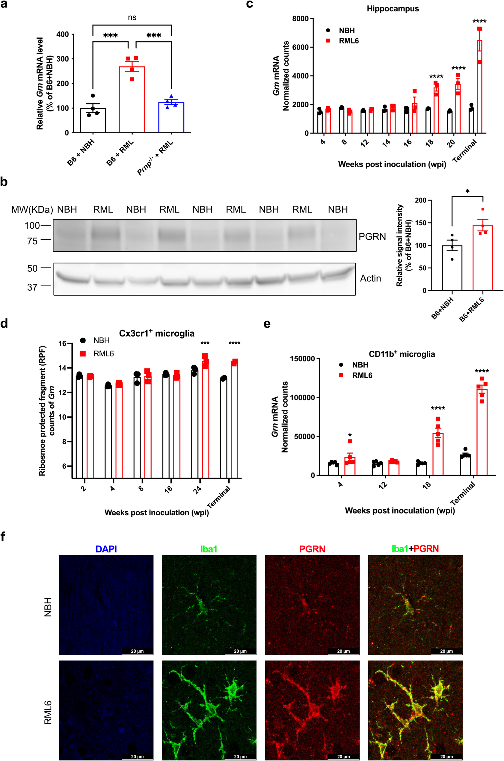
Also, ICAM expression is common in meningiomas and may also facilitate the adhesion of metastases to meningioma blood vessels [3]. However, the mechanism of tumor-to-tumor metastasis is still unknown. It is not known why this frequency is lower in melanoma than that of lung and breast cancers. The majority of brain metastases originate from primary cancers in the lung (40–50%) or breast (15–25%), or from melanoma (5–20%) [2]. There might be an actual numerical difference, but the possibility of publication bias cannot be ruled out. Collision tumors are rare, and their features are not well understood. This case is only the fifth documented instance of melanoma metastasizing to a meningioma. Notably, it is the first case to include genetic analysis of the melanoma, which revealed an NRAS mutation, although the genetic details of the meningioma were not examined.
The most recent similar case, reported in 2010, lacked molecular data, emphasizing the importance of future cases including such analyses. Understanding the genetic factors that might make melanoma more likely to metastasize to meningiomas is essential. Advances in genetic analysis may help identify markers that explain this phenomenon. Additionally, certain histologic and molecular features of a meningioma may create a more favorable environment for melanoma metastasis. Further molecular analysis of these tumors could lead to improved diagnostic and management strategies for these rare cases. A molecular model to predict response to radiotherapy for meningioma was recently developed. Using a similar approach, it may be possible to develop a predictive model for TTM in the future [13].
Cases have been reported where malignant tumors metastasize to meningiomas, leading to rapid growth of the affected meningioma [12]. Clinicians should consider the possibility of TTM in patients with a history of cancer who present with a previously stable meningioma that has recently begun to enlarge. In cases where melanoma metastasizes to a meningioma, hemorrhage, a common feature of melanoma metastases may contribute to this accelerated growth. Our case involved hemorrhage, consistent with two previously reported instances of melanoma-meningioma collision tumors. Given the frequent association of hemorrhage with CNS melanoma metastases [4], its presence in melanoma collision tumors involving meningiomas is unsurprising.
In cases of tumor-to-meningioma metastasis, most recipient meningiomas are found in the convexity (39.8%), parasagittal region (26.6%), anterior skull base (18.58%), posterior fossa or tentorium (9.73%), and spine (5.26%) [12]. The epidemiology of primary meningioma locations differs somewhat: convexity (lateral hemisphere) (20–37%), parasagittal (medial area of hemispheres) (13–22%), including falcine meningiomas (5%), spine (7–12%), and skull base (43–51%) [5].
Over the past 15 years, melanoma treatment strategies have undergone significant transformation, beginning with the approval of ipilimumab. These advancements have greatly extended the life expectancy of melanoma patients. The risk of collision tumors involving stable benign meningiomas may emerge as an important consideration in patient management. Evaluating prophylactic interventions, such as resecting benign meningiomas, or more likely to be adopted, more frequent CNS imaging in patients with melanoma and known meningioma, could help mitigate the potential risk of metastatic seeding by melanoma and/or allow for earlier intervention. However, careful assessment of the risks and benefits of such approaches is necessary.
Key questions remain, including whether the prognosis of collision tumors differs from typical metastatic brain tumors, whether non-surgical treatment options might be effective, and whether alternative methods for detecting metastasis beyond monitoring for rapid tumor growth can be developed. Addressing these questions will be critical to optimizing the management of melanoma patients with stable benign meningiomas.


Comments (0)