Artificial intelligence (AI) and machine learning are emerging technologies that can be used to create algorithms capable of decision making [6]. The whole medical scientific community has been fascinated by this new opportunity. Researchers and clinicians dedicated to rare conditions are foreseeing tools which would overcome the scarcity of numerosity of existing series to reach robust and supported diagnostic and therapeutic processes. Furthermore, the specific field of diagnosis through images both from radiology exams and pathology specimens would clearly receive huge support from AI-based assessments [7, 8]. Digital pathology, coupled with advanced digital slide scanning technology, has opened numerous possibilities for identifying various tissue types and specific target elements [9, 10]. Through the application of machine and deep learning techniques, it is now feasible to train a “computer pathologist” to recognise diverse structures, depending on their unique characteristics. However, one current limitation of fully automated pathology lies in the need for pathologist-guided delineation of specific regions within digitised slides. To achieve a diagnostically conclusive result, it has become increasingly important to blend both manual-adapted detection and automated cellular analysis through deep learning methods. Wang et al. have highlighted the advantages of combining these approaches to mitigate issues arising from the vast amount of data or a lack of inherent understanding of histological structures [12]. Deep learning relies on extensive datasets to train neural network algorithms [11]. As the number of slides/images increases, the algorithm's capability for unsupervised cellular analysis improves, enabling it to recognise disease-specific features and patterns through learned associations [12]. Until recently, AI and machine learning technologies were predominantly applied in the field of oncology [13,14,15]. However, more recently, these systems have been introduced into the diagnostic process of rare paediatric diseases, holding great promise [16,17,18,19]. Hirschsprung disease is a rare condition, belonging to the anomalies of the enteric nervous system. The condition is diffused all over the world with an incidence of 1 out 5000 newborns [20]. Although several aspects of the disease have been deeply studied, aetiology as well as variability in the phenotype and prognosis are still challenging the specialists who treat it. Guidelines for diagnosing and treating these cases are emerging from the editorial effort of medical societies and supranational institutions with methodology conditioned by poor level of evidence [21, 22]. In Hirschsprung’s disease and allied disorders, the expert’s involvement in crucial phases is rewarded as the possible guarantee of a correct approach. However, expertise definition is currently vague and volume of treated cases seems the only reliable parameter.
In this study, we have described the development and technical validation of a novel, supervised AI model for the evaluation of histopathologic features in the spectrum of Hirschsprung diagnosis. The primary goal of this study was to establish a “proof-of-principle” model in the setting of HD-AI system, showing its potential as a semi-automated tool in the field of anatomic pathology, providing accurate, reproducible, quantitative assessment of various microscopic features of interest (identifying ganglionic cells, hypertrophic nerves, normal nerves), increasing both efficiency and reporting standardisation in this specific context. Considering the rare nature of Hirschsprung's disease, there have been limited attempts to harness AI for its diagnosis. Schilling et al. made an endeavour in this direction, utilising AI to diagnose HD with the aid of histological slides stained for calretinin, microtubule-associated protein 2, Glucose transporter isoform 1, and S100. Their study involved 93 tissue blocks from 31 specimens of 27 patients. In their training set, they reported a sensitivity of 87.5% and specificity of 80%, while in the development set, they achieved 95% sensitivity and 90.4% specificity [18]. Our study diverges in both objectives and methodologies. First, mirroring a recent study by Greenberg et al., we exclusively employed H&E-stained slides, abstaining from the use of immunohistochemistry [23]. Second, through the diligent application of data augmentation and segmentation techniques, our dataset surpassed 1000 slides in volume, distinguishing it in terms of scale and potential.
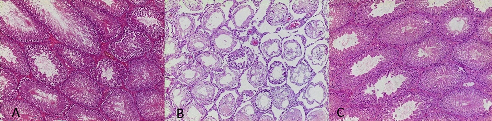
The algorithm developed in this study demonstrates an accuracy rate of 92.3% for detecting ganglionic cells and 91.5% for identifying hypertrophic nerves, respectively. In the realm of Hirschsprung’s disease diagnosis, a common trend in the literature is the consistently high reported specificity, typically exceeding 90%, which translates to a rarity of false-positive results. However, the incidence of false-negative results displays a wider spectrum, ranging from 0 to 40% [24]. In this context, the potential incorporation of immunohistochemistry could further enhance diagnostic accuracy. Nevertheless, it is noteworthy that at our centre, our experienced pathologist achieved a 100% detection rate for pathological markers (including hypertrophic nerves and the absence of ganglionic cells) exclusively through the examination of H&E-stained slides, without any instances of false positives. Consequently, the utilisation of this algorithm may simplify the diagnostic process and empower less-experienced pathologists to perform effectively. To the best of our knowledge, this study represents the pioneering effort to employ two AI models for the histological diagnosis of Hirschsprung’s disease, encompassing both ganglionic cells and hypertrophic nerves. This innovation is significant because the combined use of these models enables the AI system to identify the transition zone, the area situated between the aganglionic and ganglionic zones. Notably, our research group has previously demonstrated that the length of this transitional area serves as a predictive factor for post-HAEC development (these findings are yet to be published). One of the most significant challenges encountered in this model is the accurate detection of ganglion cells. The machine learning algorithm may occasionally misclassify immature ganglion cells as mature ganglion cells, especially when they are not in proximity to the expected context. It is essential to note that ganglion cells are exclusively located within the submucosa or muscularis propria layers. Any cell or finding identified in any other layer, regardless of how similar it may appear, is highly unlikely to represent a genuine ganglion cell. However, in the absence of this contextual information, some findings can mimic ganglion cells, particularly immature ones, leading to potential misclassification.
To address this issue, our future applications of the algorithm will include tracking the origin of each image within its respective slide. This additional contextual information will significantly enhance the algorithm’s ability to provide a more accurate assessment by considering the specific histological layer in which the cells are located.
This study has several limitations which merit mention. As stated, the available dataset was limited and significantly smaller than that of similar studies on the use of AI in pathology [25, 26]. Large data sets are considered necessary to properly represent the wide variability present in clinical samples. Smaller datasets therefore suffer both from a statistical standpoint and from excessive uniformity. Our use of data augmentation techniques somewhat circumvents this problem. Nevertheless, additional data, including data generated by other institutions, considering the rarity of the disease would allow for further validation which could improve upon the algorithm. Furthermore, from a technical point of view, there are various challenges that will have to be overcome. For instance, artefacts can be mistaken as ganglionic cells if many tissue layers overlap and create a “brown-like colouration”. In addition, in a machine learning approach for histological purposes, the hierarchical analysis of specific structures such as nerves and ganglion cells within the tissue slide is a fundamental aspect that significantly contributes to the accuracy and effectiveness of the AI system.


Comments (0)