
Remember me


Pneumoperitoneum refers to intra-abdominal and extraluminal air that most often develops from perforation of hollow viscus.1 Other etiologies of pneumoperitoneum include postprocedural (eg, upper endoscopy), intrathoracic (eg, diaphragmatic defects and high mechanical ventilation peak pressures), and gynecologic through patent oviducts (eg, sexual intercourse and intentional vaginal insufflation).2 Patients with pneumoperitoneum that cannot be classified by aforementioned etiologies are diagnosed with idiopathic spontaneous pneumoperitoneum (ISP). Diagnosis of ISP is exceedingly rare with literature describing patients with ISP being limited to case reports.3,4 We report a case of a healthy, 42-year-old woman without significant risk factors who presented with worsening abdominal pain and found to have moderate-volume ISP.
CASE REPORTThe patient is a 42-year-old woman with history significant for cesarean section who presented to the emergency department with left shoulder pain, nonfocal abdominal pain, distention, and nausea. She was afebrile, hemodynamically stable, and without signs of peritonitis on presentation. Cross-sectional imaging with computed tomography (CT) with intravenous (IV) contrast demonstrated moderate-volume pneumoperitoneum with anterior gastric wall thickening and adjacent perigastric free fluid concerning for perforated gastric ulcer (Figure 1). An oral contrasted study was not pursued because institution policy is to perform CT scans with IV contrast only in surgically naive patients and whose clinical picture is not suspicious for an enteric leak. Laboratory workup including complete blood count, basic metabolic panel, electrocardiogram, and troponins was within normal limits. Additional history was obtained from the patient—she denied recent trauma and endoscopy, nonsteroidal anti-inflammatory drug use, previous Helicobacter pylori infections, a personal and family history of colorectal cancers and inflammatory bowel disease, and smoking. The patient did report occasional alcohol use but denied additional drug use. The patient also reported snorkeling 3 weeks before presentation; however, no scuba diving was reported, and the patient denied vigorous sexual activity or intentional vaginal insufflation that would have suggested possible gynecologic etiologies of pneumoperitoneum.
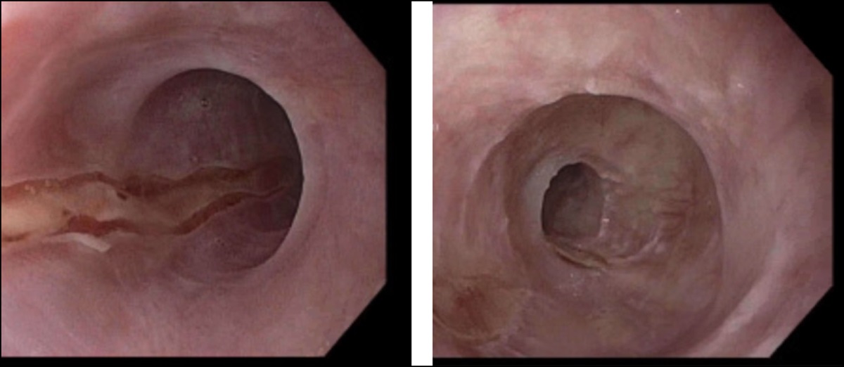
 Figure 1.:
 Representative images preoperatively and postoperatively of a patient with idiopathic spontaneous pneumoperitoneum. Preoperative axial (A) and coronal (B) cross-sectional CT imaging with IV contrast demonstrating moderate-volume pneumoperitoneum and evidence of gastric wall thickening with small-volume perigastric fluid (C and D). Postoperative axial (E) and coronal (F) cross-sectional CT imaging with IV and PO contrast demonstrating absence of PO contrast extravasation along stomach with thickened wall. CT, computed tomography; IV, intravenous; PO, per os.
Given the volume of pneumoperitoneum in an otherwise hemodynamically stable and nonperitonitic patient, we favored pursuing operative investigation through a minimally invasive approach to bridge between CT with oral contrast that may have delayed patient care and maximally invasive exploratory laparotomy. Intraoperatively, a 3-incision exploratory laparoscopy was performed with evacuation of the pneumoperitoneum. Findings were significant for healthy-appearing solid organs, stomach, small bowel, appendix, colon, rectum, and gynecologic structures including uterus and adnexa. The esophageal hiatus was intact without evidence of hiatal hernia. The gallbladder demonstrated chronic-appearing adhesions that were taken down with an energy device. The gastrocolic ligament was dissected with electrocautery, and the lesser sac was entered to examine the posterior stomach. Mobilization of the stomach failed to demonstrate wall injury or enteric contents. Given absence of findings on exploratory laparoscopy, an esophagogastroduodenoscopy (EGD) was performed intraoperatively, which demonstrated healthy-appearing esophageal, gastric, and duodenal mucosa without evidence of esophagitis, gastritis, duodenitis, and ulceration.
The patient was admitted postoperatively, and a repeat CT with oral and IV contrast was performed on postoperative day 1 to re-examine for possible missed etiologies of pneumoperitoneum and to ensure pneumoperitoneum was stable to improved. This also demonstrated mild focal gastric anterior wall and rugal fold thickening and small-volume pneumoperitoneum. Specifically, no oral contrast extravasation was identified (Figure 1). Concomitant CT of the chest was performed to rule out intrathoracic etiologies, and this study did not show evidence of pneumomediastinum nor pneumothorax and normal-appearing trachea and primary bronchi. The patient's diet was advanced as tolerated, and abdominal distention improved. Gastroenterology was consulted who recommended laboratory workup to rule out H. pylori infection and celiac disease. H. pylori antibody, immunoglobulin A, and tissue transglutaminase studies testing were nonreactive. The patient was ultimately discharged on postoperative day 3. Subsequent outpatient endoscopy evaluation demonstrated healthy-appearing esophageal, gastric, and duodenal mucosa on EGD with biopsies failing to demonstrate intraepithelial lymphocytosis and villous blunting otherwise consistent with celiac disease; rectum, colon, and terminal ileum mucosa appeared healthy on colonoscopy.
DISCUSSIONISP is a rare phenomenon that has been observed in patients with diverse demographics and comorbidities and can have variable presentations. Most patients present with nonspecific gastrointestinal symptoms such as nausea, vomiting, and abdominal pain, leukocytosis, and elevated inflammatory markers; furthermore, patients with ISP are also often nonperitonitic and hemodynamically stable.1,4,5 The workup and treatment of patients with ISP has included medical management with making patients nil per os and prescribing IV antibiotics or surgically through diagnostic laparoscopy and/or exploratory laparotomy with bowel resections.2 In this patient case, initial CT imaging demonstrated gastric wall thickening and perigastric fluid consistent with gastric perforation; however, mucosal inflammation nor adherent omentum was identified on EGD and diagnostic laparoscopy, respectively. Furthermore, given the degree of pneumoperitoneum (Figure 1) in an otherwise stable and nonperitonitic patient, minimally invasive surgical exploration was deemed appropriate. In the immediate postoperative setting, gastrointestinal medicine was involved early who recommended testing for celiac disease as possible etiology for ISP, given the patient's age and lack of other significant risk factors. Previous case reports described gas-filled pockets of submucosa and serosa, pneumatosis intestinalis, as a complication of celiac disease, leading to pneumoperitoneum.6 However, in our patient case, immunoglobulin A, tissue transglutaminase, and duodenal biopsy failed to demonstrate evidence of celiac disease. Although many other feasible etiologies of pneumoperitoneum have been explored, it seems that this patient maintains a diagnosis of ISP. Recurrence of ISP is possible, and when encountered, it is important to know the patient's ISP history in conjunction with current presentation to guide the surgeon between clinical observation vs surgical exploration.4 It is also important to consider resources available, timeliness to diagnosis and treatment based on the patient's clinical severity, and healthcare and financial expenditure in the context of this clinically challenging scenario. Given relatively low incidence of ISP, variable workup and treatment approaches, and multiple case reports of ISP, patients with ISP would benefit from a multi-institutional collaborative database to improve our understanding of ISP and contribute to creation of diagnostic and treatment guidelines in the patient who presents with pneumoperitoneum of unknown etiology.
DISCLOSURESAuthor contributions: A. Awe: conception and design, analysis and interpretation of the data, and drafting of the article. S. Campbell: analysis and interpretation of the data and critical revision of the article. L. Burke, M. Long, B. Natarajan, and D. Overby: final approval of the article. S. Campbell is the article guarantor.
Financial disclosure: None to report.
Informed consent was obtained for this case report.
REFERENCES 1. Tanaka R, Kameyama H, Nagahashi M, et al. Conservative treatment of idiopathic spontaneous pneumoperitoneum in a bedridden patient: A case report. Surg Case Rep. 2015;1(1):69. 2. Karaman A, Demirbilek S, Akin M, Gürünlüoğlu K, Irşi C. Does pneumoperitoneum always require laparotomy? Report of six cases and review of the literature. Pediatr Surg Int. 2005;21(10):819–24. 3. Pitiakoudis M, Zezos P, Oikonomou A, Kirmanidis M, Kouklakis G, Simopoulos C. Spontaneous idiopathic pneumoperitoneum presenting as an acute abdomen: A case report. J Med Case Rep. 2011;5:86. 4. Velez DR. Recurrent idiopathic spontaneous pneumoperitoneum: A case report. Cureus. 2022;14(6):e26471. 5. İflazoğlu N, Gökçe ON, Kıvrak MM, Kocamer B. Spontaneous idiopathic pneumoperitoneum with acute abdomen. Ulus Cerrahi Derg. 2013;31(2):110–2. 6. Terzic A, Holzinger F, Klaiber C. Pneumatosis cystoides intestinalis as a complication of celiac disease. Surg Endosc. 2001;15(11):1360–1.
Comments (0)