Hydatid disease/Echinococcus cyst is a parasitic disease caused by an infection with the larvae of the tapeworm Echinococcus granulosus [1]. The most frequently involved site in the heart is the myocardium, while pericardial involvement occurs in 2–10% of cardiac echinococcosis cases and 0.5–2% of all hydatidosis cases [2,3,4,5,6,7]. Pericardial involvement of hydatid cysts may be caused by systemic circulation, which results from fissuring of hydatid cysts from the liver or lung, transdiaphragmatic dissemination or lymphatic circulation [2,3,4,5].
Patients with pericardial hydatid cysts may remain asymptomatic until echinococcal cysts cause pressure mass effects on surrounding structures. Presenting symptoms of uncomplicated pericardial hydatid cysts include chest pain due to stretching of the pericardium and/or compression of coronary vessels and dyspnea [2,3,4,5].
The diagnosis of hydatidosis is based on immunodiagnostic methods along with radiological and ultrasound examinations [1,2,3,4,5,6,7,8].
In our case, the antibody titer test was negative. False negative results in human hydatidosis may range from 3 to 5% of hydatid patients up to 35–40% in hyperendemic areas, as in the case of our country [8].
Although pericardial hydatid cysts can be detected by transthoracic echocardiography, cross-sectional imaging studies such as computed tomography (CT) and/or MRI are usually necessary to identify before the surgery the cardiac anatomy, disease location and cyst size [2,3,4,5,6,7].
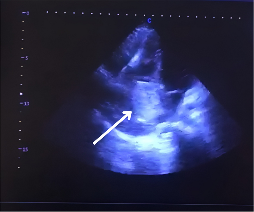
CT is a better imaging technique for revealing small calcifications, which may be a helpful imaging finding in the diagnosis of a hydatid cyst. Our case had no calcifications and no other cysts in the liver or in the other structures. The relationship of hydatid cysts with surrounding structures can be seen by MRI [2,3,4]. Hydatid cysts present as a hypointense mass on T1-weighted MRI scans (T1-weighted) images and hyperintense mass on T2-weighted MRI scans (T2-weighted) images. A hypointense rim around the mass on T2-weighted scans represents the pericyst. Daughter cysts are multiple cystic structures attached to the internal wall of the cyst. In our case, CT showed a hypodense mass characteristic of hydatid cysts with multiple daughter cysts inside. On MRI, a multicameral cystic mass was seen with no cardiac involvement/penetration infiltration after contrast medium, with differential diagnosis of a cavernous lymphangioma vs hydatid cyst. On transesophageal ultrasound, a multi-lobulated extracardiac mass adjacent to the left atrium was seen, with mass effect and probably thrombus in the left pulmonary vein (Gharbi type II) [9].
Treatment of pericardial hydatid cysts can be accomplished with surgical excision of the cystic lesion. Medical treatment (e.g., albendazole and mebendazole) is complimentary for disseminated cases and for prophylaxis [2,3,4,5,6,7].
Research has shown that 73–75% of patients respond to medical management to some extent, but the reported cure rates are only 25–30%, and this strategy is a long and tedious process involving considerable risks [1].
In our case, considering the rare position of the cyst (retrocardiac) and the fact that the cyst did not enter the intracardiac site, the surgery was performed through left anterolateral thoracotomy by thoracic surgeons after 7 days of premedication with albendazole. It was necessary to release the Botalli duct after opening the pericardium to better expose the cystic lesion and all the structures adjacent to it. The surgery was performed in cardiac operation suite/room theatre with the availability of cardiac surgeons for any intraoperative complications.
There are a lot of cardiac surgeons that would prefer to perform the surgery in cases like that through the sternotomy, but in our case the cardiac surgeons were not familiar with the surgery in the area that the cystic mass was localized.
Regarding the premedication with albendazole prior to surgery there have been recent studies that emphasise the role of albendazole in cases of rupture of large cysts of the lungs [10]. In our case the location of the pathology was intrapericardial, it was not a big cyst but a cystic mass full of daughter cysts (hydatidosis) and the premedication was necessary to prevent the dissemination.


Comments (0)