
Remember me

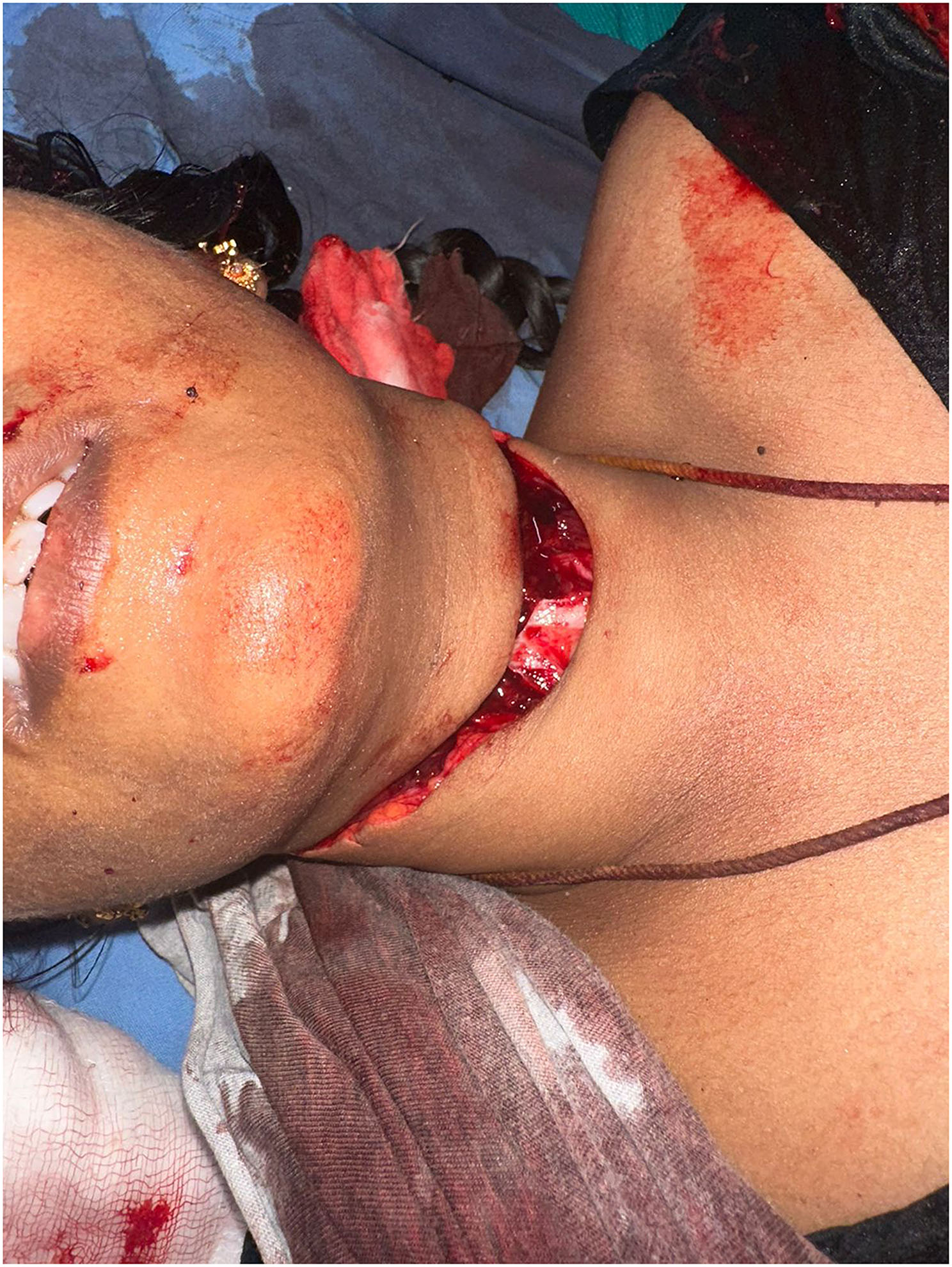

The patient is a 45-year-old female with no prior significant medical history, presenting to the emergency department after sustaining a homicidal assault (Fig. 1). The injury was inflicted with a knife, resulting in a deep penetrating neck injury. Upon arrival, the patient exhibited signs of severe hypovolemic shock, with a rapid heart rate and significantly low blood pressure, necessitating immediate fluid resuscitation and airway management according to ATLS protocol. Aggressive fluid resuscitation, TXA administration, and PRBC transfusion were initiated to control bleeding and restore hemodynamic stability.
Fig. 1
Penetrating neck injury following a knife assault, highlighting its severity on presentation
Emergency interventionDue to the complete transection of the trachea, standard airway management techniques were ineffective. An endotracheal tube was strategically inserted through the transected tracheal segment to secure the airway (Fig. 2). This manoeuvre provided temporary stabilization, allowing for safe transport to the operating theatre.
Fig. 2
Endotracheal intubation through the transected trachea for emergency airway management
Surgical explorationIn the operating room, a detailed examination of the neck injury was conducted under anaesthesia. The intra op findings were Carotid Sheath had Partial transection, with potential risk to the underlying vascular structures. Meticulous dissection was performed to assess the integrity of the carotid artery and internal jugular vein. Trachea was Completely transected was at the mid-tracheal level. The distal and proximal ends were identified, and care was taken to preserve surrounding tissues to facilitate reconstruction (Fig. 3). Esophagus had a clean transection, necessitating precise approximation to restore the continuity of the gastrointestinal tract.
Fig. 3
Intraoperative view of tracheal and esophageal transections with carotid sheath injury
Surgical Procedures included Carotid Sheath Repair: Given the partial injury, the carotid sheath was carefully sutured using 5 − 0 polypropylene sutures, ensuring the protection of the carotid artery and jugular vein. Followed by Esophageal Repair: An end-to-end anastomosis was performed using absorbable 3 − 0 polygalactin sutures over a nasogastric tube to maintain patency. Adequate care was taken to achieve tension-free closure and prevent postoperative leaks. Finally Tracheal Reconstruction: Initial attempts to approximate the transected tracheal ends proved inadequate. Subsequently, a posterior tracheal wall repair was accomplished using 3 − 0 polypropylene sutures. Given the complexity of the injury, a size 7 tracheostomy tube was inserted and securely positioned to maintain airway stability. (Fig. 4). The platysma and overlying skin were meticulously approximated in layers. A suction drain was positioned to manage postoperative fluids, and the wound was closed in a tension-free manner.
Fig. 4
Postoperative reconstruction with tracheostomy tube, esophageal repair, and carotid sheath repair
Postoperative managementThe patient was monitored in the intensive care unit (ICU) for potential complications, including airway obstruction, infection, and anastomotic dehiscence. A tracheostomy care regimen was instituted, with gradual downgrading and eventual removal of the tracheostomy tube by the 20th postoperative day. The patient received nutritional support via enteral feeding until esophageal healing was confirmed.
Discharge and follow-upThe patient showed remarkable recovery with no signs of infection or fistula formation. Psychiatric evaluation and therapy were integral to her holistic recovery, addressing the psychological aftermath of the traumatic event. She was discharged on the 25th day, with recommendations for outpatient follow-up and continued psychiatric care.
Comments (0)