
Remember me


In our study,188 patients accepting surgery in 2019 were included as the training group, and 53 patients accepting surgery from January 2022 to June 2022 were classified as the validation group. Lung adenocarcinoma patients who underwent lobectomy, wedge resection, segmentectomy and pneumonectomy with en bloc mediastinal lymph node dissection were included in this study. Patients were excluded for the following reasons: (1) adenocarcinoma in situ or minimally invasive adenocarcinoma on postoperative pathology; (2) histopathological diagnosis of invasive mucinous adenocarcinoma; (3) Treatment with neoadjuvant therapy before surgery; (4) Presence of other primary malignancies; (5) Patients with incomplete medical records. The flow chart for patient selection is shown in Fig. 1. The study was approved by the ethics committee of Huadong Hospital, and no informed consent was obtained from patients due to the retrospective nature of our study.
Fig. 1
Flowchart for patient selection
MethodsPathologic STAS evaluationHematoxylin and eosin histological staining was conducted to assess STAS status in resected adenocarcinoma tissues. Briefly, the tissues were embedded in paraffin and fixed with formalin. Tumor STAS implies that tumor cells are found in the normal pulmonary tissues outside the boundary of the main tumor, according to the literature [6, 9, 10]. It is generally accepted that the micropapillary subtype, solid nets subtype and single cancer cell subtype are predominantly found in STAS [11]. To differentiate “blade contamination” from real STAS, the following cases were excluded: (1) scattered tumor cells caused by the blade during specimen processing; (2) individual isolated tumor cells detected far away from the main tumor without disseminating in a continuous form [4, 5, 12]. Two senior pathologists independently reviewed all the slides, and a consensus-based decision was made for points of disagreement. The representative pathological images of STAS are listed in Fig. 2.
Fig. 2
Representative pathological images of STAS(Red arrow indicated the tumor nest)
Immunohistochemistry (IHC)Immunohistochemistry was used to detect PD-L1 and PD-1 expression levels in adenocarcinoma tissues. Briefly, the tissues were embedded in paraffin and then fixed with formalin. Then the tissues (4–5 μm-thick) were incubated overnight at 4 °C with rabbit anti-PD-L1 monoclonal antibody (E1L3N, 1:100, CST) and rabbit anti-PD-1 monoclonal antibody (MX033,1:100, MaiXin). Immunostaining was performed using the EnVision + System-HRP (AEC) (K4005, Dako, Glostrup, Denmark). Positive PD-L1 and PD1 expression were observed in the membrane. The expression level was classified into two groups: < 1 was grouped as negative, while ≥ 1% was grouped as positive [13].
Gene mutational analysisThe mutational status of targetable oncogenic driver gene EGFR and ALK was assessed through the standard polymerase chain reaction based method [14]. Then ALK fusion was confirmed by the fluorescence in-situ hybridization method (FISH). Rare mutation sites and common mutation sites of EGFR were all recorded in our cohort.
Other clinicopathological indexesAge, gender, location of the lesions, surgical approach, tumor differentiation grade, tumor margin, pleural invasion, tumor size, pTNM stage, Ki67, intravascular cancer embolus, NLR (neutrophil to lymphocyte ratio), PLR (platelet to lymphocyte ratio) and several preoperative blood indexes were all collected from electronic medical records. The pTNM stage was stratified based on the 8th edition of New TNM staging in lung cancer [15]. Positive pleural invasion was defined as tumor invasion beyond the elastic lamina of the pulmonary tissue. The histological types of lung adenocarcinoma mainly included lepidic, papillary, acinar, micropapillary, and solid types. The threshold for predominant histological type was set to 20%. According to the published literature, poorly differentiated types included micropapillary or solid type ≥ 20%; moderately differentiated types included acinar or papillary type, and well-differentiated types included the lepidic type without micropapillary or solid type ≥ 20% [16].
Statistical methodsAll patient databases were established, and data analysis was performed using SPSS 22.0 statistical software and R version 4.0.2. The binary logistic regression model was used for univariate analysis in the training group. Indexes with P < 0.05 were selected for multivariate logistic regression analysis. The independent predictive factors with P < 0.05 were then selected to establish a nomogram.
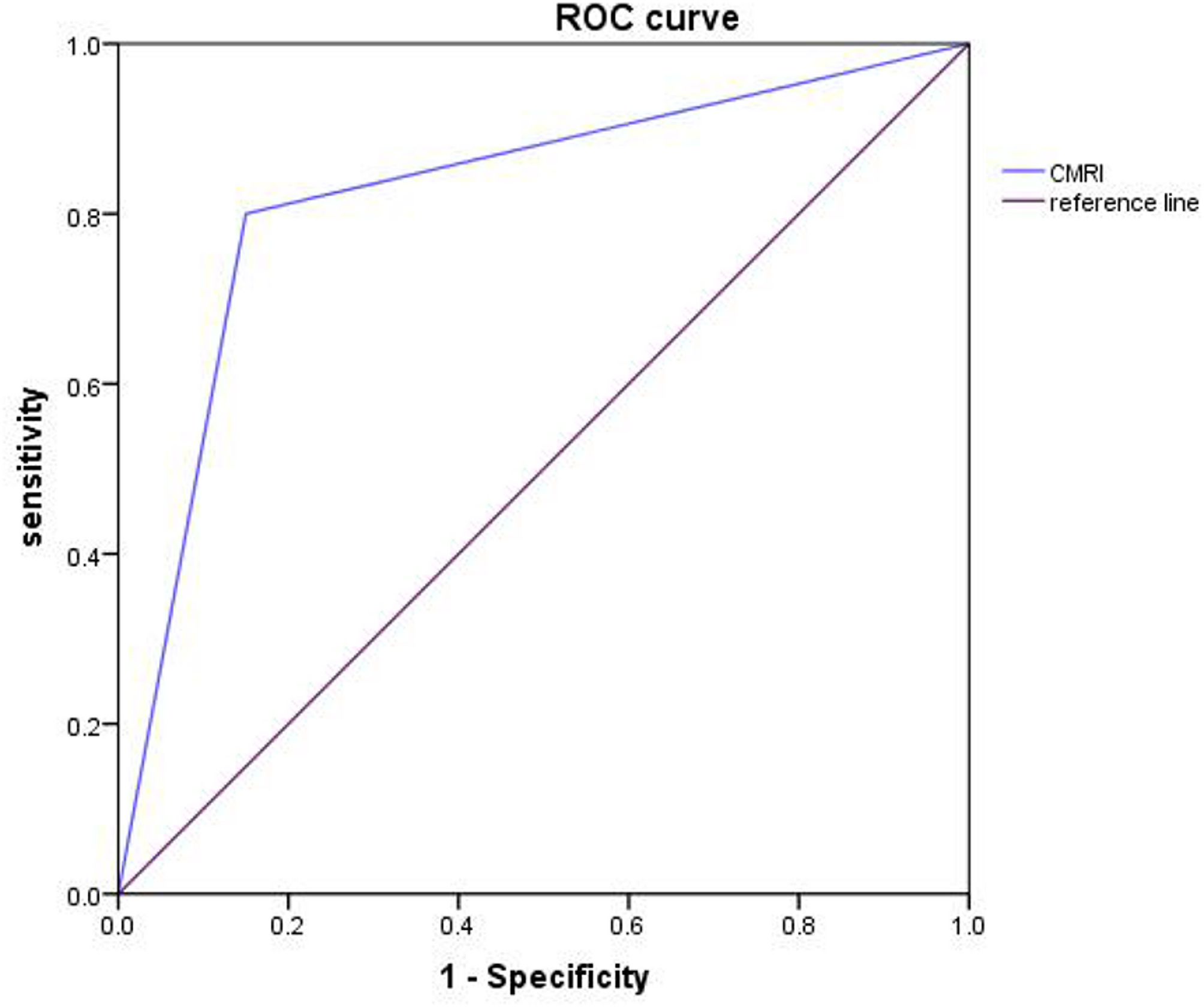
ROC and calibration curves were used to evaluate nomogram performance in both training and validation groups. In addition, a DCA analysis was conducted to assess the clinical usefulness of the nomogram.
Comments (0)