
Remember me


Hepatocyte nuclear factor-1 beta (HNF-1β/B) is a transcription factor (TCF) of the homeodomain-containing family that is expressed in various organs including the gut, liver, pancreas, kidney, and urogenital tract.1–3 Maturity-onset diabetes of the young (MODY) is a variant of autosomal dominant monogenic diabetes characterized by early onset (before age 25 years) and progressive dysfunction of pancreatic beta cells.3 MODY is difficult to differentiate from diabetes mellitus type 1 and requires genetic analysis to be accurately diagnosed.
CASE REPORTA 11-year-old boy presented with elevated liver enzymes. Workup was negative for chronic liver disorders including autoimmune liver disease, alpha-1 antitrypsin deficiency, chronic viral hepatitis, and celiac disease. Subsequently, he was diagnosed with maturity-onset diabetes of the young type 5 (MODY 5) because of an identified pathogenic mutation in HNF1B. Alanine aminotransferase (ALT) levels ranged from 144 to 603 U/L (normal < 35). Aspartate aminotransferase levels ranged from 50 to 296 U/L (normal < 40) with normal bilirubin, gamma- glutamyl transferase (GGT), and alkaline phosphatase levels. His lipid profile was slightly abnormal with a triglyceride level of 268 (normal < 120 mg/dL) and low-density lipoprotein cholesterol 112 (normal < 100 mg/dL). HbA1C levels ranged from 6.3% to 8.0% (normal 4.2%–5.6%). His body mass index ranged from 76 to 90th percentile for age and sex. Abdominal ultrasound showed no anomalies. Transient elastography of the liver revealed F0 fibrosis and <33% steatosis on 2 occasions a year apart. Needle liver biopsy revealed mild nonspecific findings including minimal to mild portal and lobular predominantly lymphocytic inflammation with occasional admixed eosinophils and neutrophils. No significant steatosis was observed.
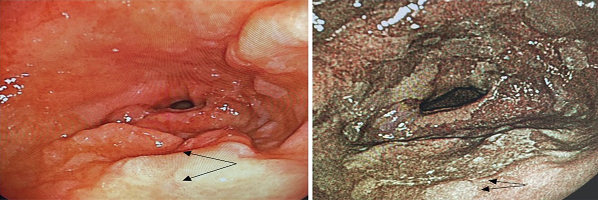
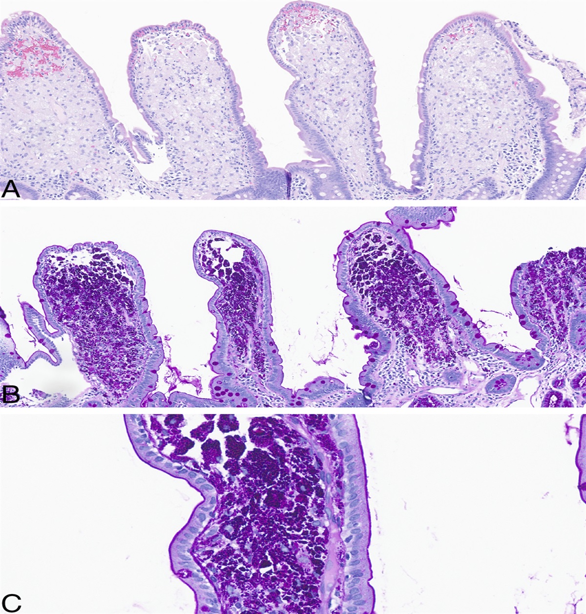
Trichrome stain revealed focal sinusoidal stage 0-1 fibrosis (Figure 1). Iron stain showed no stainable iron, and periodic acid- schiff (PAS) and PAS with diastase (PAS-D) stains were negative for intracytoplasmic globules.
 Figure 1.:
 Trichrome stain -Stage-1 perisinusoidal fibrosis.
An 8-year-old girl was evaluated for elevated liver enzymes in the setting of refractory Henoch-Schönlein purpura requiring oral steroids and methotrexate. Elevated transaminases persisted after medications were discontinued. ALT levels ranged from 77 to 211 U/L, and aspartate aminotransferase levels ranged from 35 to 137 U/L with normal bilirubin, GGT, and alkaline phosphatase. Further workup was negative for chronic liver disorders and inflammatory bowel disease. The patient subsequently developed diabetes and was diagnosed with MODY 5. HbA1c levels ranged from 5.5% to 9.4%. Body mass index was at 30th percentile for age and sex. Ultrasound of the abdomen showed no anomalies. Needle liver biopsy revealed patchy mild portal and lobular mixed inflammation including lymphocytes and neutrophils. There was no steatosis, and trichrome stain demonstrated sinusoidal stage 1 fibrosis. Iron stain as well as PAS and PAS-D stains for intracytoplasmic globules was negative (Figure 2).
 Figure 2.:
 Mild portal and lobular mixed inflammation.
DISCUSSIONHeterozygous mutations in the HNF1B gene located on chromosome 17 Q21.3 encoding for the TCF HNF-1β results in MODY 5.4 HNF-1β, also known as TCF-2, plays a key role in organogenesis and is widely distributed in kidneys, liver, pancreas, and female reproductive organs. HNF-1β is also responsible for regulating the expression of liver enzymes, and mutation in the gene may result in abnormal biochemical liver function tests.5
MODY 5 was initially described in a Japanese family as the association of early onset diabetes and nephropathy.6 Pathogenic variants in HNF1B were first reported in association with an autosomal dominant form of MODY with renal cysts also known as a renal cysts and diabetes syndrome. Pathogenic variants in HNF1B have now been described in a wider spectrum of renal and extrarenal phenotypes.7 Wide interfamilial and intrafamilial clinical variability has been reported in renal disease including renal cysts, single kidney, horseshoe kidney, irregular collecting systems, glomerular tufts, and hyperuricemic nephropathy. However, for some individuals harboring a pathogenic variant in the HNF1B gene, renal cysts may be the only disease manifestation.1 Affected individuals may also have reduced pancreatic size and abnormalities of the genital tract.8 Approximately 50% of pathogenic variants are de novo, and about 50% are gene deletions or larger deletions at 17 q 12.7 The p.Arg235Trp (R235W) mutation has been identified in unrelated patients with MODY that have kidney and liver complications.9,10 Abnormalities of the hepatobiliary tract present as neonatal cholestasis, hepatomegaly, steatosis, and increased ALT and/or GGT levels.3 Although there have been many cases of renal pathology associated with HNF-1βassociated disease, there are few reported cases of MODY with abnormal hepatobiliary findings.11
We present 2 siblings with persistent elevated liver enzymes with maternal family history for insulin-dependent diabetes. Workup was negative for chronic liver disorders including autoimmune liver disease, alpha-1 antitrypsin deficiency, chronic viral hepatitis, and celiac disease in both patients. Subsequently, both siblings were diagnosed with MODY 5 with an identified pathogenic mutation in HNF1B.
Liver biopsies performed during the evaluation of cholestasis or persistent elevation in liver enzymes in patients with HNF-1β defects show varied histological findings.3 In these cases, needle liver biopsy revealed mild nonspecific findings, indicating no significant inflammation or steatosis.
Neither of the patients had renal anomalies but will be monitored closely as they are at risk of developing renal cysts with their mutation variant.
MODY 5 is a genetic condition, and the patients are siblings with a strong maternal family history of insulin-dependent diabetes mellitus in their mother, uncle, grandfather, and great grandfather. Mother has the same point mutation for MODY 5 and has insulin-dependent diabetes diagnosed at age 38 years. She also had abnormal LFTs noted in her early 20s, similar to her father and grandfather and had a negative workup despite laboratory tests, magnetic resonance imaging of the abdomen, and a liver biopsy. Maternal grandfather also has a history of kidney cancer, another organ commonly affected by a mutation in the HFN1B gene.1
In conclusion, HNF1B disease should be considered in the differential diagnosis for young patients with idiopathic liver enzyme elevation in the setting of early onset autoantibody negative insulin-dependent diabetes.
DISCLOSURESAuthor contributions: S. Veerareddy and S. Reddy wrote the manuscript and did literature review. M. Barreto and N. Vedherey helped with literature review and editing; VV Gopalareddy wrote the case report, provided the pathology slides, and submitted the manuscript. VV Gopalareddy is the article guarantor.
Acknowledgments: We thank Michael Duncan in the Pediatric Research Department at Levine Children's Hospital for editing the manuscript.
Financial disclosure: None to report.
Informed consent was obtained for this case report.
REFERENCES 1. Edghill EL, Bingham C, Ellard S, Hattersley AT. Mutations in hepatocyte nuclear factor-1beta and their related phenotypes. J Med Genet. 2006;43(1):84–90. 2. Kotalova R, Dusatkova P, Cinek O, et al. Hepatic phenotypes of HNF1B gene mutations: A case of neonatal cholestasis requiring portoenterostomy and literature review. World J Gastroenterol. 2015;21(8):2550–7. 3. Camacho SM, Nowicki MJ. Hepatocyte nuclear factor 1-β gene mutation: Brief report and review of hepatic involvement. Ann Case Rep Images. 2018;1(1):2. 4. Fajans SS, Bell GI, Polonsky KS. Molecular mechanisms and clinical pathophysiology of maturity-onset diabetes of the young. N Engl J Med. 2001;345(13):971–80. 5. Bala V, Anania FA. Increased liver enzyme levels and HNF-1beta gene mutation. Clin Gastroenterol Hepatol. 2008;6(12):A26. 6. Horikawa Y, Iwasaki N, Hara M, et al. Mutation in hepatocyte nuclear factor-1 beta gene (TCF2) associated with MODY. Nat Genet. 1997;17(4):384–5. 7. Bockenhauer D, Jaureguiberry G. HNF1B-associated clinical phenotypes: The kidney and beyond. Pediatr Nephrol. 2016;31(5):707–14. 8. Bellanné-Chantelot C, Chauveau D, Gautier J-F, et al. Clinical spectrum associated with hepatocyte nuclear factor-1β mutations. Ann Intern Med. 2004;140(7):510–7. 9. Dusatkova P, Fang M, Pruhova S, et al. Lessons from whole-exome sequencing in MODYX families. Diabetes Res Clin Pract. 2014;104(3):e72-4. 10. Johnson SR, Ellis JJ, Leo PJ, et al. Comprehensive genetic screening: The prevalence of maturity-onset diabetes of the young gene variants in a population-based childhood diabetes cohort. Pediatr Diabetes. 2019;20(1):57–64. 11. Derencius A, Crow K, Patel S, Loo N. S2635 Abnormal liver enzymes attributed to HNF-1b associated disease. Am J Gastroenterol. 2021;116:S110.
Comments (0)