
Remember me


Hepatocellular carcinoma (HCC) is the sixth most common malignancy globally.1 Ectopic HCC is defined as HCC arising from hepatic parenchymal tissues found in organs other than the liver.2 The most common sites include the gall bladder, omentum, retroperitoneum, and thorax.1 Diagnosis is made on the histopathology.2
A 62-year-old man with diabetes treated for hepatitis C, with Child-Pugh class A5 and The Model for End-Stage Liver Disease 9, was referred for pain in his left loin region. General physical examination revealed tenderness in the left lumbar area, and straight leg test was positive. Serum alpha-fetoprotein was raised (1,560,000 ng/mL). In consultation with a neurologist, magnetic resonance imaging of the lumbosacral spine with contrast (Figure 1) was performed and revealed a soft-tissue lobulated mass in the left lumbosacral paravertebral region involving and scalloping the L4, L5, and S1 vertebrae and left anterior sacroiliac joint. This mass was inseparable with left iliacus and posterior paraspinal muscle.
It was decided to perform an ultrasound-guided TruCut mass biopsy, which showed HCC (ie, poorly differentiated carcinoma). An abdominal and pelvic computed tomography scan showed features of chronic liver disease and portal hypertension (Figures 2 and 3).
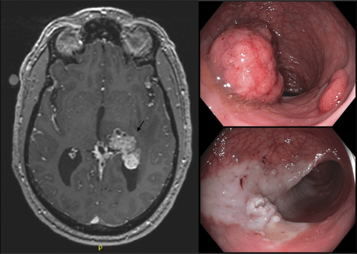
 Figure 1.:
 MRI lumbosacral spine revealed soft tissue lobulated mass in left lumbosacral paravertebral region involving and scalloping L4, L5, S1 vertebra and left anterior sacroiliac joint.
 Figure 2.:
 No convincing evidence of HCC on arterial portovenous and delayed phase.
 Figure 3.:
 Arterial enhancing soft tissue lobualted mass in left lumbosacral area with local invasion.
The final diagnosis was primary ectopic HCC. He was advised for radiation therapy for pain relief and systemic therapy (sorafenib).
The incidence rate for ectopic HCC is between 0.24% and 0.47%.3 Ectopic HCC eventhough has no connections with the native liver, but behaves like one in histology and morphology.4 Diagnosis during the early stage is difficult. Treatment option is mainly surgical resection. Other possible treatment options include sorafenib.5
Our case highlights the diagnosis and management of ectopic HCC and reflects upon the fact how this rare tumor if diagnosed early can be managed by surgery.
DISCLOSURESAuthor contributions: N. Akbar and M. Adeel managed the patient and wrote the first draft. AA Tasneem edited and wrote the final draft. SR Sanjani managed the patient and assisted writing the final draft. SA Khan managed the patient. Z. Majid edited the manuscript. N. Luck conceptualized the paper and is the article guarantor.
Financial disclosure: None to report.
Informed consent was obtained for this case report.
REFERENCES 1. Takahashi K, Putchakayala KG, Safwan M, Kim DY. Extrahepatic metastasis of hepatocellular carcinoma to the paravertebral muscle: A case report. World J Hepatol 2017;9(22):973–8. 2. Adachi Y, Hayashi H, Yusa T, et al. Ectopic hepatocellular carcinoma mimicking a retroperitoneal tumor: A case report. World J Gastroenterol 2020;26(18):2268–75. 3. George NE, Raghavapuram S, Banerjee D, Al-Shoha M, Fedda F, Tharian B. Ectopic hepatocellular carcinoma within a choledochal cyst diagnosed using single-operator digital cholangioscopy. Am J Gastroenterol 2017;112:1347–8. 4. Martinez CA, de Resende HC Jr., Rodrigues MR, Sato DT, Brunialti CV, et al. Gallbladder-associated ectopic liver: A rare finding during a laparoscopic cholecystectomy. Int J Surg Case Rep 2013;4:312–5. 5. Ko YL, Takata K, Tanaka T, et al. Unresectable ectopic hepatocellular carcinoma treated with sorafenib. Case Rep Gastroenterol 2020;14(1):226–33.
Comments (0)