
Remember me


Friday C, MS1; Krock N1; Schaefer N1; Levy H MD2; Clancy M, RDH, MPA.1
1Cure HHT, Monkton, Maryland, USA; 2University of Wisconsin School of Medicine and Public Health, Madison, Wisconsin, USA
Objective: As a grantee of the Chan Zuckerberg Initiative’s Rare as One Project, Cure HHT created a research network (CHRN) to develop a research roadmap for the next 3–5 years. CHRN is led by HHT patients working in collaboration with researchers and clinicians to prioritize research initiatives by order of feasibility, impact, importance, and logical sequence.
Methods: Comprehensive surveys were sent to the HHT patient and professional community to identify perceived gaps in research and treatments. 1,204 patients answered 49 questions, 42 HHT scientists answered 17 questions, and 96 HHT clinicians answered 25 questions. Based on the analyzed results of patient needs, broad-topic work streams were established which included patients, clinicians, and researchers. Work streams performed a comprehensive literature review on their respective topic, discussed gaps in research, and proposed recommendations to fill each knowledge gap. Finally, a 2-day convening was held with all working groups to present recommendations and vote on a consensus for a final research roadmap to meet patient needs over the next 3–5 years.
Results: 8 work streams identified 31 recommendations to address patient needs. Recommendations were selected by anonymous vote after their presentation to the group and ranked by feasibility, impact, and importance. Additionally, resources and tools were identified to achieve all recommendations.
Conclusion: CHRN is poised with specific, actionable goals to address patient priorities. The research roadmap will serve as a guide for next steps, research funding opportunities, and collaboration between patients, clinicians, and researchers at an unprecedented level for the HHT community.
O2 Efficacy and safety of bevacizumab on severe bleedings associated with hemorrhagic hereditary telangiectasia: a national, randomized multicenter phase III studyDupuis-Girod S1, 2; Rivière S3; Lavigne C4; Fargeton A.E.1; Gilbert-Dussardier B5; Grobost V6; Leguy-Seguin V7; Maillard H8; Mohamed S9; Decullier E10,11; Roux A10,11; Bernard L10; Saurin J.C.11,12; Saroul N13; Faure F14; Cartier C15; Altwegg R16; Laccourreye L117; Oberti F18; Beaudoin M1; Dhelens C19; Desvignes C20,21; Azzopardi N22; Paintaud G21,23; Hermann R24; Chinet T.25
1Hospices Civils de Lyon, Hôpital Femme-Mère-Enfants, Service de Génétique et centre de référence de la maladie de Rendu-Osler, Bron F-69677, France; 2Univ. Grenoble Alpes, Inserm, CEA, Laboratory Biology of Cancer and Infection, F-38000 Grenoble, France; 3Service de Médecine Interne A, Centre Hospitalier Universitaire, F-34000 Montpellier, France; 4Service de médecine interne-Immunologie clinique, CHU d’Angers, F-49933 Angers cedex 09, France; 5Service de génétique médicale, CHU La Milétrie, F-86021 Poitiers, France; 6Service de Médecine Interne CHU Estaing, Clermont-Ferrand University Hospital, F-63100 Clermont-Ferrand; 7Service de Médecine Interne 1, CHU F.MITTERRAND, F-21000 DIJON, France; 8Service de médecine interne, Université Lille 2, CHRU de Lille, F-59037 Lille, France; 9Département de Médecine interne et Immunologie Clinique, CHRU BRABOIS, F-54500 Vandoeuvre-lès-Nancy; 10Hospices Civils de Lyon, Pôle Santé Publique, Lyon, F-69003; 11Faculté de médecine, Université Lyon 1, F-69008; 12Hospices Civils de Lyon, Service d’Hépato-gastroentérologie, Hôpital E. Herriot, Lyon, F-69437; 13CHU Clermont Ferrand, Service d’ORL F-63100 Clermont-Ferrand, France; 14Hospices Civils de Lyon, Hôpital E. Herriot, Service d’ORL, Lyon, F-69437, France; 15Service d’ORL Centre Hospitalier Universitaire, F-34000 Montpellier, France; 16Service Hépatogastroentérologie CHU St Eloi, F-34000 Montpellier, France; 17Service d’ORL, CHU d’ Angers, F-49933 Angers cedex 9, France; 18Service Hépatogastroentérologie, CHU Angers and Laboratoire HIFIH, UPRES EA 3859, Faculté de médecine Angers, France; 19Hospices Civils de Lyon, Hôpital Edouard Herriot, Pharmacie à Usage Intérieur F-69437 Lyon, France; 20CHRU de Tours, Plateforme Recherche, Centre Pilote de suivi Biologique des traitements par Anticorps (CePiBAc), Tours, France; 21Université de Tours, EA 4245 Transplantation, Immunologie, Inflammation (T2I), Tours, France; 22CNRS EMR 7001 LNOx, Université de Tours, Tours, France; 23CHRU de Tours, service de Pharmacologie Médicale,37,044 Tours Cedex 9; 24Hospices Civils de Lyon, Hôpital Femme-Mère-Enfants, Service d’ORL et centre de référence de la maladie de Rendu-Osler, Bron F-69677, France; 25Hôpital Ambroise Paré, Service de Radiologie, Assistance Publique-Hôpitaux de Paris, Université Paris Ile-de-France Ouest, F-92104 Boulogne, France.
Objective: Hereditary Hemorrhagic Telangiectasia is a rare but ubiquitous hereditary vascular disease related to a disequilibrium of the angiogenic balance. Since 2010, bevacizumab, a humanized monoclonal antibody that selectively binds to and neutralizes the biologic activity of human VEGF (vascular endothelial growth factor) has been widely used for the treatment of severe bleeding and hepatic involvement related to HHT as several studies and many case reports are in favor of its efficacy, however, no randomized study has been done. To evaluate the efficacy of intraveinous bevacizumab on blood transfusions in patients with HHT complicated by bleedings responsible for severe anemia.
Methods: Double-blind multi-center randomized phase III trial with an active / placebo ratio 1:1. BABH study was conducted from September 2017 to May 2020 with a 6-months follow-up. Patients were recruited from the French HHT Network. Over 18 years old with confirmed HHT and to epistaxis or digestive bleeding (with the requirement for at least 4 units of blood in the 3-month period before study enrollment) were included. Bevacizumab was given at a dose of 5 mg / kg every 14 days with a total of 6 injections. The primary efficacy criterion was the decrease by at least 50% of the number of red blood cell transfusions 3 months before and after treatment.
Results and Conclusion: 24 patients were included and randomized at 4 different centers 12 in each group. Results will be presented during the meeting.
O3 Safety of long-term systemic bevacizumab use in hereditary hemorrhagic telangiectasiaBarnett B, AB1; Smith K, RN2; Kasthuri RS, MBBS.2
1School of Medicine, University of North Carolina at Chapel Hill, Chapel Hill, NC, USA; 2Division of Hematology, University of North Carolina at Chapel Hill, Chapel Hill, NC, USA
Objective: To evaluate the safety profile of long-term systemic bevacizumab use in patients with HHT.
Methods: Patients with HHT managed at the UNC HHT Center of Excellence and treated with bevacizumab were identified in the EMR. Records were reviewed to identify duration of treatment with bevacizumab and documentation of side effects/adverse events/reason for therapy cessation. Search terms included: proteinuria, hypertension, headache, arthralgia, joint pain, DVT, pulmonary embolism, stroke, TIA, dyspnea, hoarseness, and tinnitus.
Results: Forty-eight patients were initially identified. Six were not on systemic bevacizumab, one did not have HHT, and one was lost to follow up. Overall, 40 patients were eligible with a median duration of treatment of 39.5 months with systemic bevacizumab. Side effects data are described in the Table. Majority of reactions were transient and associated with infusion. Out of five patients with persistent/worsening hypertension, four cases developed within weeks of their first infusion. One patient developed DVT/PE associated with a Port-a-cath. This subject was on hormonal therapy and symptoms of DVT were noted prior to initiation of bevacizumab. No cases of persistent proteinuria, although one patient briefly held infusions given concern for renal dysfunction of unclear etiology.
Conclusion: In our single-center cohort of HHT patients long-term systemic bevacizumab use was safe and well tolerated overall. Side effects/adverse events were largely infusion-related and appeared early in treatment course. We did not observe thromboembolic events or significant proteinuria. Patients should be monitored acutely for infusion reactions, especially upon initiation of therapy, and chronically for development of hypertension.
O4 Efficacy and safety of tacrolimus as treatment for bleeding caused by hereditary hemorrhagic telangiectasia: an open-label pilot studyHessels J, MD1; Kroon S, MD1; Boerman S, MD1; Nelissen R, MD, PhD2; Grutters J, MD, PhD1,3; Lebrin F, PhD4; Post M, MD, PhD5; Mummery C, PhD6; Mager J, MD PhD.1
1Department of pulmonology, St. Antonius Hospital Nieuwegein, Utrecht, the Netherlands; 2Department of otorhinolaryngology, St. Antonius Hospital Nieuwegein, Utrecht, the Netherlands; 3Department of pulmonology, University Medical Center Utrecht, Utrecht, the Netherlands; 4Department of internal medicine, Leiden University Medical Center, South Holland, the Netherlands; 5Department of cardiology, St. Antonius Hospital Nieuwegein, Utrecht, the Netherlands; department of cardiology, University Medical Center Utrecht, Utrecht, the Netherlands; 6Department of anatomy and embryology, Leiden University Medical Center, South Holland, the Netherlands
Objective: Haploinsufficiency of Endoglin (ENG) and activin A receptor type II-like I (ALK1) lead to the formation of weak and abnormal vessels in hereditary hemorrhagic telangiectasia (HHT). These cause epistaxis (nosebleeds) and/or gastrointestinal blood loss. In vitro, tacrolimus has been shown to increase ENG and ALK1 expression and is therefore a potential treatment option.
Method: We report here a proof-of-concept study in HHT patients with severe epistaxis and/or gastrointestinal bleeding who were treated daily with orally-administered tacrolimus for twenty weeks.
Results: Twenty-five HHT patients (11 females (44%); 14 males (56%)) with a median age of 59 years, were enrolled. Five patients (20%) stopped the trial prematurely – four due to (serious) adverse events (SAE). 20 patients were included in further analyses. Hemoglobin levels increased during tacrolimus treatment from 6.1 (IQR 5.2 – 6.9) mmol/L at baseline to 6.7 (6.5 – 7.1) mmol/L, p = 0.003. In addition, the number of blood transfusions decreased from a mean of 5.0 (± 9.2) to 1.9 (± 3.5) after treatment, p = 0.04. In 64% of the patients, at least one AE occurred.
Conclusion: In summary, oral tacrolimus significantly increased hemoglobin levels and decreased blood transfusion needs in HHT patients with severe epistaxis and/or gastrointestinal bleeding. Side-effects were common, however most patients were content with the effect of treatment. The potential therapeutic benefit should be further investigated.
O5 Randomized, double-blind, placebo-controlled, crossover trial of oral doxycycline for epistaxis in hereditary hemorrhagic telangiectasiaFaughnan ME1,2,; Thompson KP1,2; Sykes J5,6;Chandakkar P3; Marambaud P3; Vozoris, NT1,2; Marchuk DA.4
1Toronto HHT Centre, St. Michael’s Hospital and Li Ka Shing Knowledge Institute, Toronto, Canada; 2Division of Respirology, Department of Medicine, University of Toronto, Toronto, Canada; 3The Feinstein Institutes for Medical Research, Northwell Health, Manhasset, New York, USA; 4Department of Molecular Genetics and Microbiology, Duke University Medical Center, Durham, NC, USA; 5Department of Respirology, St. Michael’s Hospital, Toronto, Ontario, Canada; 6Dalla Lana School of Public Health, University of Toronto, Toronto, Ontario, Canada.
Funding:US Department of Defense W81XWH-17–1-0429ABSTRACT.
Objective: Vascular malformations in hereditary hemorrhagic telangiectasia (HHT) lead to chronic recurrent bleeding, hemorrhage, stroke, heart failure, and liver disease. There is an unmet need for effective therapies for HHT. We aimed to measure the effectiveness of oral doxycycline for the treatment of epistaxis and explore mechanisms of action on angiogenic, inflammatory and pathway markers in HHT using a randomized controlled trial.
Methods: 13 HHT patients with epistaxis were recruited from the Toronto HHT Center at St. Michael’s Hospital. Recruitment was stopped early due to COVID-19-related limitations. The study duration was 24 months. Patients were randomly assigned to the treatment-first or placebo-first study arm. We compared the change in weekly epistaxis duration and frequency, biomarkers, blood measurements, and intravenous iron infusion and blood transfusion requirements between treatment and placebo.
Results: There was no significant difference in the change in weekly epistaxis duration (p = 0.136) or frequency (p = 0.261) between treatment and placebo. There was no significant difference in the levels of MMP-9, VEGF, ANG-2, IL-6 or ENG with treatment. Hemoglobin levels were significantly higher (p = 0.0499) during treatment. On secondary, qualitative analysis, 5 patients appeared to respond to treatment. There was a significant difference (p = 0.037) in baseline ANG-2 levels between responders and non-responders.
Conclusion: Overall, our study did not demonstrate effectiveness of doxycycline as a treatment for epistaxis in patients with HHT, though the study was underpowered. Secondary analyses provided new observations which may help guide future trials in HHT.
O6 Oral pazopanib for the management of HHT related bleedingAhluwalia V M.B.B.S1; Albitar H, M.D1; Ahluwalia V M.B.B.S1; Donlinger S1; Dubrock H M.D1; Cajigas H M.D1; Krowka M M.D1; Gallo De Moraes A M.D1; Demeril N M.D2; Kilari S Ph.D3; Misra S M.D3; Iyer V M.D, M.P.H.1
1Division of Pulmonary and Critical Care Medicine, Mayo Clinic, Rochester, MN; 2Division of Pediatric Pulmonology, Mayo Clinic, Rochester, MN; 3Division of Vascular and Interventional Radiology, Mayo Clinic, Rochester, MN.
Objective: IV bevacizumab has been the preferred anti-angiogenic agent for severe/refractory HHT related bleeding. Oral low-dose (50-200 mg) pazopanib is a promising alternative agent based on initial data. However, the effectiveness of different pazopanib dosages especially in patients previously treated with one/more anti-angiogenic agents requires further study.
Method: We searched a prospectively maintained database of patients with refractory HHT related bleeding treated with anti-angiogenic agents. RBC transfusion, need for ENT procedures, GI endoscopies, and laboratory results were compared before and after initiation of oral pazopanib.
Results: Fourteen patients (57% female) with a median age of 61 (48–82) years were identified (Table 1). Nine (64.25%) patients had been previously treated with other antiangiogenic approaches and were switched to pazopanib (bevacizumab—5, Bevacizumab + Doxycycline—2 & Pomalidomide + bevacizumab + Doxycycline—2). All patients were started on 200 mg OD pazopanib. The dose was reduced to 100 mg daily in five patients due to side effects. Two (14.25%) patients were on antiplatelet therapy and Two (14.25%) patient was on anticoagulation. Cumulative number of transfusions decreased from 0 (0–0.8, min: 0, max: 6) to 0 (0–0, min: 0, max:3), P = 0.59. Similarly, total number of upper endoscopies decreased from 0 (0–0, min:0, max:2) vs 0 (0–0, min:0, max:0), P = 0.35 at six months. Hemoglobin increased from (11.2 (8.5–13.1, min:6.9, max:15.2) vs 12.7 (9.3–14.5, min:8.3, max:16), P = 0.22 at 3 months.
Conclusion: We present our initial experience with pazopanib in a cohort of patients heavily pre-treated with bevacizumab as well as other anti-angiogenic agents. Most patients experienced a good/excellent treatment response to this therapy.
O7 An evaluation of the efficacy and safety Of AKT inhibitors in the prevention of hereditary hemorrhagic telangiectasiaThalgott J, MS1; Pollenus T, PhD1; Goujon G.P.E, PhD1; Rabelink T, MD, PhD1; Saint-Mézard P, PhD2*; Picard D, MD2*; Lebrin F, PhD1, 3*
* Equal contribution.
1Einthoven Laboratory for Experimental Vascular Medicine, Department of Internal Medicine (Nephrology), Leiden University Medical Center, 2333 ZA Leiden, The Netherlands; 2Vaderis Therapeutics, 4051 Basel, Switzerland; 3Physics for Medicine, INSERM U1273, ESPCI Paris, CNRS, PSL Research University, 75,012 Paris, France.
Objective: HHT pathogenesis strongly relies on an overactivated AKT activity in endothelial cells. Here, we have investigated the therapeutic effects of AKT inhibitors in preclinical models of HHT1.
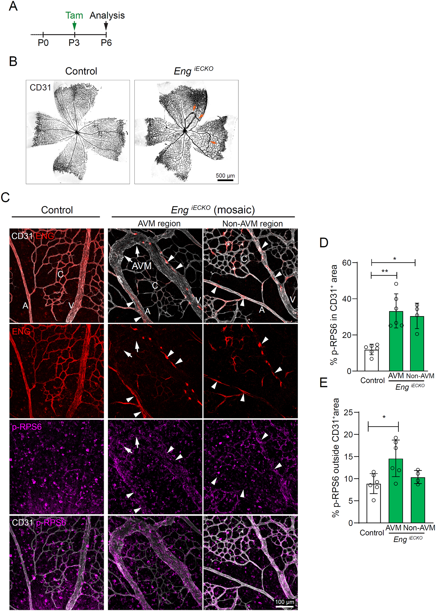
Method: We compared the effects of Perifosine, Uprosertib and VAD044 on angiogenesis focusing on the neonatal retina of Cdh5-CreERT2; Engflox/flox mice at P7. Mice received a single dose of 50 μg Tamoxifen at P2 to induce the formation of retinal AVMs and then were treated with AKT inhibitors at P3 and P5. The half maximal concentrations (EC50) were identified in mice and in human in vivo and in vitro by measuring AKT activity in primary endothelial cells and in platelets isolated from blood samples.
Result: VAD044 showed the best efficacy and safety profile. VAD044 at 2.5 mg.kg−1 of body weight strongly inhibited the formation of AVMs in Eng-iKOe mice. The blood exposure of VAD044 free base in the plasma of mouse neonates corresponded to a concentration of 55.1 nM over 48 h dosing interval. VAD044 IC50 on AKT phosphorylation was measured at 55 nM in control mouse endothelial cells and increased to 93 nM in mouse endothelial cells depleted for Eng. In human primary endothelial cells, VAD044 IC50 was comparable and measured at 87 nM. The values of those IC50 were similar to the average plasmatic concentration of VAD044 in mice and in human.
Conclusion: VAD044 showed efficacy in in preclinical models of HHT and was well tolerated. A phase II trial is currently ongoing to evaluate its efficacy to prevent bleeding in patients with HHT.
O8 Evaluating AAV vectors for HHT gene therapyLiang R, BS1; Press K, BS1; Gonzalez T, PhD2; Asokan A, PhD2; Su H, MD.1
1Center for Cerebrovascular Research, Department of Anesthesia and Perioperative Care, University of California, San Francisco, California, USA; 2Biomedical Engineering, Molecular Genetics and Microbiology, Department of Surgery, Duke University School of Medicine, Durham, North Carolina, USA
Background: Epistaxis from nasal telangiectasias and intracranial hemorrhage from brain arteriovenous malformations (bAVMs) are among the most devastating symptoms of HHT. All available managements for HHT have limitations. Adeno-associated viral vector (AAV) mediates long- term transgene expression with few adverse effects. We showed that intravenous delivery of soluble FMS-related tyrosine kinase 1 using an AAV9 vector (AAV9-sFLT1) reduced bAVM severity of endoglin deficient mice. However, minor liver inflammation and growth arrest in young mice were observed.
Objective: Identify AAV vectors and delivery methods that can best transduce brain and nasal tissue with minimum off-target transduction.
Methods: Three engineered AAV capsids (AAV.cc47, AAV.cc84 and AAV1RX) were compared with AAV9. A single-stranded CBA promoter driven tdTomato transgene were packaged in these capsids and delivered intravenously or intranasally to mice. Tissues were collected 4 weeks post-dosing.
Results: After intravenous injection, AAV9 and AAV.cc47 mediated transgene expression in different brain cells and hepatocytes; AAV1RX infected some brain endothelial cells (ECs) but no hepatocytes; and AAV.cc84 transduced a high percentage of brain ECs and a few hepatocytes. After intranasal delivery, AAV9 non-specifically transduced few brain cells and hepatocytes, 1RX transduced a few brain ECs but no hepatocytes, AAV.cc47 dosed animals showed robust transduction in the brain and the liver, while AAV.cc84 transduced brain perivascular cells and nasal epithelial cells, but no hepatocytes.
Conclusion: AAV.cc84 transduces brain perivascular cells and nasal epithelial cells after intranasal delivery without transducing hepatocyte and ECs predominantly after intravenous injection.
Therefore, AAV.cc84 is a promising candidate for HHT gene therapy.
Bleeding, Thrombosis, Anemia, and Iron.
O9 Antithrombotic therapy in hereditary hemorrhagic telangiectasia: a scoping reviewZhang E, BA1; Virk Z, MD2; Rodriguez-Lopez J, MD1,3; Al-Samkari H, MD1,2
1Harvard Medical School, Boston, MA, USA; 2Division of Hematology Oncology, Massachusetts General Hospital, Boston, MA, USA; 3Division of Pulmonary and Critical Care Medicine, Massachusetts General Hospital, Boston, MA, USA
Objective: Data describing safety and tolerability of anticoagulation and antiplatelet therapy in HHT is limited. We sought to better define the state of knowledge in this topic through literature review.
Methods: We performed a scoping review, searching MEDLINE and EMBASE from inception to November 2021 for eligible studies reporting detailed clinical data describing antithrombotic use in HHT. Data extracted included study design, patient population, and characteristics and outcomes of antithrombotic therapy.
Results: Of 575 unique manuscripts identified through database search, 72 manuscripts were included: 60 manuscripts reporting patient-level data on 61 patients and 11 reporting population-level data (Table). Inclusive of both patient-level and population-level manuscripts, data were extracted on a total of 401 patients. The most common reasons for antithrombotic therapy were VTE (56.2%), atrial arrythmias (14.4%) and stroke (10.1%). Anticoagulation was used in 287 episodes (76.1%), antiplatelet therapy in 70 episodes (18.6%), and both together in 11 episodes (2.9%). Complications of therapy included worsened HHT-associated bleeding (primarily epistaxis and gastrointestinal bleeding) in 154 antithrombotic treatment episodes (41.1%) and antithrombotic therapy discontinuation in 61 episodes (23.1%). Bleeding- directed therapy (local ablative therapy and systemic therapies) were employed to address worsening bleeding in 8.6% of episodes. No specific complications of therapy were reported in 198 total antithrombotic events (52.5%). Rates of bleeding, therapy discontinuation, and other complications ranged considerably from study to study.
Conclusion: Current publications vary widely on the outcomes and tolerability of antithrombotics in HHT. More formal studies are needed to better guide optimal antithrombotic use in these patients.
O10 Functional alterations involved in increased bleeding in hereditary hemorrhagic telangiectasia mouse modelsEgido-Turrión C, PhD1; Rossi E, PhD2; Ollauri-Ibáñez C, PhD1; Pérez-García M, PhD1; Silva-Sousa L, MS1; Bernabeu C, PhD3; Smadja DM, PhD2; López-Novoa J M, PhD1; Rodríguez-Barbero A, PhD1; Pericacho M, PhD.1
1Dpto. de Fisiología y Farmacología, Universidad de Salamanca. CARD-05. Grupo de Fisiopatología del Endotelio Vascular (ENDOVAS). Instituto de Investigación Biomédica de Salamanca (IBSAL), Salamanca, Spain; 2Inserm UMR-S1140, Faculté de Pharmacie. Paris Descartes University, Paris, France; 3Centro de Investigaciones Biológicas, Consejo Superior de Investigaciones Científicas (CSIC), and Centro de Investigación Biomédica en Red de Enfermedades Raras (CIBERER), Madrid, Spain.
Objectives: HHT patients present recurrent and difficult to stop bleeds that compromise patients’ life. The aim of this study is to assess possible alterations in hemostasis mechanisms in animal models of HHT.
Methods: Heterozygous murine models of HHT-1 (Eng±) and HHT-2 (ALK1±) were used to study different phases of hemostasis in vivo and ex vivo. Moreover, iKO-Eng and ENG+ mice were used to confirm the role of endoglin. Primary culture lung endothelial cells were obtained from these mice and in vitro platelet adhesion was assayed under static or shear stress conditions.
Results: Our results show that bleeding time is increased in both animal models of HHT, whereas endothelial-independent hemostasis show normal activity. Endoglin deficiency impairs platelet-endothelial adhesion, consequently, it is observed a reduction in the thrombus stabilization in Eng± animals, while it is increased in human endoglin transgenic mice (hEng+). On the other hand, the HHT-2 model presents alterations in fibrinolysis, as PAI-1 plasma level is decreased while t-PA is increased.
Conclusion: Both HHT murine models have defects in hemostasis, but the pathophysiologic mechanism underlying this effect seems to be different in HHT-1 and HHT-2. Endoglin deficiency leads to an impaired interaction between platelets and endothelium in HHT-1, resulting in a defective thrombus stabilization that associated with more severe hemorrhages. However, HHT-2 increased susceptibility to bleeding seems to be due to the acceleration of thrombus lysis due to an increased fibrinolysis. Both mechanisms would explain the common bleeding phenotype and should be considered as potential therapeutic targets in future investigations.
O11 Hereditary hemorrhagic telangiectasia is associated with a higher prevalence of heavy menstrual bleeding1Division of Hematology, University of North Carolina School of Medicine, Chapel Hill, NC, USA; 2Department of Biostatistics, University of North Carolina School of Medicine, Chapel Hill, NC, USA; 3Division of Pediatric Hematology/Oncology, Duke University, Durham, NC, USA; 4Department of Obstetrics and Gynecology, Duke University, Durham, NC, USA; 5Cure HHT Foundation, Monkton, MD, USA
Objective: (1) Determine the prevalence of heavy menstrual bleeding (HMB) and its impact on quality of life (QoL) (2) Compare HMB prevalence in women with HHT to the general population.
Method: A survey study was conducted and members of Cure HHT responded anonymously over 4 weeks. HMB present if: bleeding > 7 days, ≥ 1 product every hour for several consecutive hours, > 1 product-type to control bleeding, or required adult diapers. QoL negatively impacted if missed: work, school, family, or social activities.
Results: There were 633 respondents (Table): 352 (55.6%) of child-bearing age (Group A). HMB prevalence: entire cohort- 74% and Group A- 72%. In the entire cohort: 9.7% -hysterectomy, 8.7%-uterine AVMs and 23% -post-partum bleeding [4% required blood transfusions, 0.4% required hysterectomy and 5.1% required medications to control bleeding].
Group A: 49%- sought care for HMB, 56%- negative impact on QoL.
Prevalence of anemia in the last year was 67% and 79%- used oral iron, 26.5%- IV iron, and 9.5%- RBC transfusion. Interventions to manage HMB: 9.7%-IUD, 6.8%- progestin- only pills, 5.7%- antifibrinolytics, 3%- uterine ablation or equivalent, and 0.3%- hysterectomy.
Significant correlation noted between HMB and QoL (p < 0.001), anemia (p- 0.005), OCP use (p = 0.006), progestin-only pills (p = 0.015), IUD (p = 0.001), fibroids (p = 0.019), and endometriosis (p = 0.025).
Conclusions: We found a prevalence of HMB of 72% among women with HHT (56% reporting an adverse QoL), significantly higher than the reported 53% in the general population. This suggests HMB may be an HHT-related manifestation that is under-recognized and warrants further evaluation.
O12 Incidence of spontaneous pulmonary AVM rupture in HHT patientsFish A, MD.1; Henderson K, MS.1; Chan SM, BS1; Pollak, J, MD1; Schlachter T, MD.1
1Department of Interventional Radiology, Yale School of Medicine, New Haven, CT, USA
Objective: To determine the incidence and prevalence of spontaneous rupture of pulmonary AVMs in HHT patients.
Methods: This study retrospectively reviewed records of 2310 patients with known (1971) or possible (339) HHT according to the Curacao criteria or by genetic testing, referred to a single-center HHT clinic. Patients diagnosed with pulmonary AVMs were evaluated for a single lifetime episode of hemothorax or pulmonary hemorrhage secondary to spontaneous pulmonary AVM rupture. Medical records of the patients with spontaneous rupture were then further evaluated.
Results: Between July 2, 1996 and July 22, 2021, a total of 801 patients with HHT (759 known, 42 possible) were found to have pulmonary AVMs. Spontaneous rupture of the AVM occurred in 22 patients, identified over an average 16.3-year follow-up period (Range 0–25 years). The lifetime prevalence and incidence of spontaneous rupture in HHT patients with pulmonary AVMs was therefore estimated to be 2.7% and 0.16% respectively. Considering all HHT patients, the life-time prevalence was 1.1%. Spontaneous rupture of the AVM represented the initial presentation of 9 cases (40.9%), was life-threatening in 9 cases (40.9%), and occurred during pregnancy in five patients (22.7%). All cases of pulmonary hemorrhage were a result of lobar AVMs and all cases of hemothorax were a result of subpleural AVMs. All cases occurred in virgin lesions, and subsequent embolization was curative.
Conclusion: While a feared complication, pulmonary AVM rupture is rare and is likely effectively revented by existing embolization techniques and indications.
O13 Safety and efficacy of left atrial appendage closure for stroke protection from atrial fibrillation in hereditary hemorrhagic telangiectasiaAlghammass M, MD1; Dranow E, Ph D1; Whitehead K, MD.1
1Division of Cardiovascular Medicine, Department of Medicine, University of Utah, USA
Background: Atrial fibrillation (AF) is a common cause of stroke and occurs with increased incidence in patients with HHT. Anticoagulation can prevent stroke but is poorly tolerated in patients with HHT. Left atrial appendage occlusion (LAAO) is an alternative strategy for stroke prevention.
Objective: Evaluate the safety and efficacy of LAAO for stroke prevention in HHT patient with AF.
Method: Retrospective cohort study.
Results: We observed AF in 31 of 329 consecutive adult patients with definite diagnosis of HHT. A total of 11 patients underwent LAAO (9 AtriClip, 1 Watchman, 1 Lariat). The mean duration of AF in this group was 7.7 ± 4.4 years, with median follow-up post LAAO of 3.3 years (IQR 1.5—5.1 years). Median CHADS-VASc score was 4 (IQR 4—5). Anticoagulation was used by 3 patients (27.3%). The 20 patients without LAAO had a mean duration of AF of 4.4 ± 2.1 years and median CHA2DS2-VASc score of 4 (IQR 2.5—5); 3 patients were treated with anticoagulation (15.0%). We observed 7 ischemic strokes felt to be due to AF in this population, 3 in the control group, and 4 in the LAAO group prior to their procedure. No strokes were observed following LAAO. We expected to observe 7.48 strokes from all patients without or prior to LAAO (observed 7). We expected to observe 2.96 strokes following LAAO (observed 0). This difference approaches statistical significance (P = 0.12).
Conclusion: LAAO is a promising approach to stroke prevention in AF for patients with HHT.
 O14 Activation of coagulation and the impact of iron deficiency anemia in hereditary hemorrhagic telangiectasia (HHT)
Ghosh S, PhD*; Youkhana K, MD*; Smith K, RN*; Reeves BN, MD*; Pawlinski R, PhD*; Kasthuri RS, MD*
*Division of Hematology, University of North Carolina at Chapel Hill, Chapel Hill, NC, USA
Objective: (1) Determine whether there is evidence of coagulation activation in the plasma of patients with HHT; (2) Evaluate the impact of iron deficiency anemia on coagulation activation in the plasma of HHT patients.
Methods: Four groups of subjects were enrolled: HHT patients with and without iron deficiency anemia (IDA), patients with IDA but not HHT, and healthy controls. Pertinent clinical and demographic data were recorded and blood samples collected. The following lab tests were measured: complete blood count, ferritin, D-dimer, E-selectin and vascular endothelial growth factor (VEGF). In a cohort of 8 subjects representing the 4 study groups, differential expression of 55 angiogenesis-related proteins were explored using a protein array assay.
Results: Number of subjects in the 4 groups: HHT with IDA- 24, HHT without IDA- 11, IDA without HHT- 4, controls- 7. D-dimer levels were significantly higher in the HHT with IDA group compared to control (516.52 vs 210.19, p 0.049). VEGF levels were higher in both HHT groups. E-selectin levels were higher in the HHT groups compared to control but did not meet statistical significance. The angiogenesis protein array assay identified 9 upregulated and 11 downregulated proteins that were ≥ 50% control (Table), most notably fivefold increase in tissue factor.
Conclusion: We identified a significant increase in D-dimer in iron deficient HHT subjects that was independent of endothelial activation. Angiogenesis protein array reveled upregulation of tissue factor in the HHT cohort supporting a procoagulant state in addition to identifying additional differentially expressed proteins that warrant further study.
Table: Differentially expressed proteins (≥ 50%) in HHT subjects on angiogenesis protein array assay:
Down-regulated
Up-regulated
• Activin A
• Angiostatin
• ADAMTS-1
• FGF-4
• GM-CSF
• HB-EGF
• PDGF-AA
• TGF-beta 1
• Thrombospondin-2
• uPA
• Vasohibin
• Coagulation factor III (tissue factor)
• GDNF
• IL-1 beta
• Leptin
• MCP-1
• MIP-1a
• PIGF
• Serpin-B5
• VEGF
O15 Safety, tolerability, and effectiveness of anticoagulation and antiplatelet therapy in hereditary hemorrhagic telangiectasia: a multicenter studyVirk Z., MD1; Zhang E, BS2; Rodriguez-Lopez J, MD2,3; Witkin A, MD2,3; Wong A, MD2,3; Luther J, MD2,4; Lin A, MD2,5; Ning M, MD2,6; Grabowski E, MD, ScD2,7; Holbrook, MD2,8; Al-Samkari H, MD1,2
1Division of Hematology Oncology, Massachusetts General Hospital, Boston, MA; 2Harvard Medical School, Boston, MA; 3Division of Pulmonary and Critical Care Medicine, Massachusetts General Hospital, Boston, MA; 4Division of Gastroenterology and Hematology, Massachusetts General Hospital, Boston, MA; 5Department of Pediatrics, Massachusetts General Hospital, Boston, MA; 6Department of Neurology, Massachusetts General Hospital, Boston, MA; 7Division of Pediatric Hematology Oncology, Massachusetts General Hospital, Boston, MA; 8Division of Rhinology, Massachusetts Eye and Ear Institute, Boston, MA.
Objective: Antithrombotic therapy (anticoagulation and antiplatelet therapy) is frequently needed in patients with HHT, but data guiding its use is limited.
Methods: We evaluated outcomes in a 5-hospital observational cohort study of adults with HHT receiving antithrombotic therapy.
Results: 119 patients with 187 discrete antithrombotic therapy episodes were included. Treatment characteristics by patient and episode are given in Table 1 and Table 2. 59 patients (50%) prematurely discontinued and/or dose-reduced therapy (including 52 patients [44%] who discontinued) due to worsening bleeding. Initiation at reduced dose-intensity had a similar premature discontinuation rate (49%) as initiation at standard dose-intensity (43%). Difficulty receiving indicated therapy may have resulted in increased thromboembolic recurrence (20 patients, 17%). In a multivariable logistic model, a history of gastrointestinal bleeding was associated with 3.25-fold odds of discontinuation (P = 0.001, Fig. 1). Hemoglobin was significantly lower, and need for intravenous iron and RBC transfusion significantly higher, in the 3 months after antithrombotic therapy initiation versus the 3 months before (Table 3); ED visits and hospital admissions due to bleeding also increased. Rates of dose-reduction and/or premature discontinuation were similar regardless of anticoagulant class (warfarin, 46%; heparin- based, 48%; DOAC, 44%) or with multiple simultaneous agents (44%) but slightly lower with single-agent antiplatelet therapy (37%), Table 4.
Conclusions: Antithrombotic therapy remains challenging in HHT, resulting in objectively higher morbidity and healthcare utilization from worsened bleeding. Discontinuation rates approached 50% regardless of dose-intensity at initiation or type of antithrombotic agent used and were higher in patients with a history of gastrointestinal bleeding.
O16 Molecular Mechanisms in HHTVascular defects associated with hereditary haemorrhagic telangiectasia revealed in patient-derived isogenic iPSCs in 3D vessels-on-chip
Orlova, V.V.1*, Vila Cuenca M 1,2, van den Hil, F1, Nahon D.M.1, Freund C3, Lebrin F4, Mager H.J.5, Mummery C.L.1
Objective: Common anti-angiogenic and anti-inflammatory strategies (anti-VEGF, thalidomide) are used to treat patients with Hereditary Haemorrhagic Telangiectasia (HHT). However, some HHT patients do not respond at all to the treatment, others only transiently, as also observed in patients with other vascular conditions who do not always respond to anti-angiogenic therapy. Therefore, a preclinical human model that utilizes cells from HHT patients is needed.
Methods: Previously, we developed a multi-cell type 3D vessel-on-chip (VoC) model based entirely on human induced pluripotent stem cells (hiPSCs) (Vila Cuenca et al., SCR 2021). We have demonstrated that this 3D VoC model can capture vessel wall complexity and model endothelial-pericyte cell interactions. Here, we generated induced pluripotent stem cells (hiPSC) from a rare mosaic HHT1 patient with tissues containing both mutant (ENGc.1678C>T) and normal cells, enabling derivation of isogenic diseased and healthy hiPSCs respectively. We showed reduced ENG expression in HHT1-endothelial cells (HHT1-hiPSC-ECs), reflecting haploinsufficiency. HHT1c.1678C>T-hiPSC-ECs and the healthy isogenic control behaved similarly in 2D culture, forming functionally indistinguishable vascular networks.
Results: However, when grown in 3D organ-on-chip devices under microfluidic flow, lumenized vessels formed in which defective vascular organization was evident: interaction between inner endothelial cells (ECs) and surrounding pericytes was decreased and there was evidence for vascular leakage.
Conclusion: We are now at the stage when a cohort of HHT1 and HHT2 isogenic iPSC lines generated previously in our group can be tested in the 3D VoC model, and we are also working on the optimization of the throughput of the system for the drug screening.
O17 A cell resolution atlas of the human cerebrovasculature reveals angiogenic and inflammatory cell programs with arteriovenous malformationsEthan A. Winkler, MD, PhD1,2,3*, Chang N. Kim, BS2,4,5,*, Jayden M. Ross, BS1,2,4,5, Joseph H. Garcia, BS1, Eugene Gil, BS1,2, Irene Oh, PhD6, Lindsay Q. Chen, BS6, David Wu, PhD1,2, Joshua S. Catapano, MD3, Kunal Raygor, MD1, Kazim Narsinh, MD7, Helen Kim, PhD8, Shantel Weinsheimer, PhD8, Daniel L. Cooke, MD7, Brain P. Walcott, MD9, Michael T. Lawton, MD3, Nalin Gupta, MD, PhD1, Berislav V. Zlokovic, MD, PhD10,11, Edward F. Chang, MD1, Adib A. Abla, MD1, Daniel A. Lim, MD, PhD1,2,12, Tomasz J. Nowakowski, PhD.1,2,4,5,13
1Department of Neurological Surgery, University of California, San Francisco, CA, USA; 2Eli and Edythe Broad Center for Regeneration Medicine and Stem Cell Research, University of California, San Francisco, CA, USA; 3Department of Neurosurgery, Barrow Neurological Institute, Phoenix, AZ, USA; 4Department of Anatomy, University of California, San Francisco, CA, USA; 5Department of Psychiatry and Behavioral Sciences, University of California, San Francisco, CA, USA; 6Rebus Biosystems, Santa Clara, CA, USA; 7Department of Radiology and Biomedical Imaging, University of California, San Francisco, CA, USA; 8Center for Cerebrovascular Research, Department of Anesthesia and Perioperative Care, University of California, San Francisco, CA, USA; 9Department of Neurosurgery, North Shore University Health System, Evanston, Illinois, USA; 10Department of Physiology and Neuroscience, Keck School of Medicine, University of Southern California, Los Angeles, CA, USA; 11Zilkha Neurogenetic Institute, Keck School of Medicine, University of Southern California, Los Angeles, CA, USA; 12San Francisco Veterans Affairs Medical Center, San Francisco, CA, USA; 13Chan Zuckerberg Biohub, San Francisco, CA, USA.
Objective: Cellular dysfunction results in cerebrovascular diseases, a leading cause of death and disability. However, we lack a comprehensive atlas of cerebrovascular cells in the human brain to better understand disease mechanisms and therapeutic strategies.
Methods: To provide a human cerebrovascular cell atlas, we used single-cell mRNA sequencing (scRNAseq) using dissociated vascular cells isolated from the adult human brain and arteriovenous malformations (AVMs). Joint comparative analyses between scRNAseq datasets profiled patterns of aberrant gene expression in AVMs and to resolve cell states enriched in AVMs that bled.
Results: By performing scRNA-seq on 181,388 individual cells, we identified > 40 vascular or neighboring cell states from the human cerebrovasculature and AVMs. We identified an expanded diversity of endothelial and perivascular cells in humans. In AVMs, there was a loss of normal arteriovenous molecular zonation among endothelial cells characterized by the emergence of a distinct cell state with heightened angiogenic potential and immune cell cross-talk spatially confined to the AVM nidus. We characterized the cellular ontology of the cerebrovascularly derived immune cell response and identified infiltration of distinct immune cell states, such as GPNMB + monocytes, which deplete stabilizing smooth muscle cells in AVMs that bled.
Conclusion: Our single-cell atlas highlights the heterogeneity underlying cell function and interaction in the human cerebrovasculature and defines molecular and cellular perturbations in arteriovenous malformations, a leading cause of stroke in young people. The identified interplay between vascular and immune cells may aid the development of therapeutics targeting angiogenic and inflammatory programs in vascular malformations.
O18 Mutation of platelet-derived growth factor receptor β causes cerebrovascular malformation and enhances brain arteriovenous malformation severity in micePrado D, PhD1, Tang C, MD, PhD1, Shaligram S, PhD1, Liang R1, Winkler E, MD,PhD2, Su H, MD1
1Center for Cerebrovascular Research, Department of Anesthesia and Perioperative Care, University of California, San Francisco, California, USA; 2Department of Neurosurgery, University of California, San Francisco, California, USA
Objective: Reduction of pericytes is correlate with brain arteriovenous malformation (bAVM) hemorrhage. The platelet-derived growth factor B and its receptor β (PDGF- B/PDGFRβ) play important roles in regulating pericyte recruitment during angiogenesis. We hypothesize that mutation of PDGFRβ causes cerebrovascular malformation and enhances bAVM severity in endoglin (Eng) deficient mice.
Methods: Three mouse models were used: (1) Pdgfrβ F7 (F7) mice that have mutations disrupting Pdgfrβ signaling, (2) Pdgfb-icreER;En f/f mice that have Pdgfb promoter driving, tamoxifen inducible cre expression in endothelial cells and an floxed Eng gene; and (3) Pdgfb- icreER;Engf/f ;F7+/- mice. Brain angiogenesis was induced by intra-brain injection of an adeno-associated viral vector expressing vascular endothelial growth factor (AAV- VEGF). Brain AVMs were induced in Pdgfb-icreER;Engf/f and Pdgfb-icreER;Engf/f ;F7 mice by tamoxifen induced endothelial Eng deletion and intra-brain AAV-VEGF injection. Brain AVM phenotypes were analyzed 8-weeks after model induction by latex vascular cast to detect arteriovenous shunts, immunostaining and Prussian blue staining to quantify dysplasia vessels and hemorrhage.
Results: Compared to WT mice, F7+/- and F7+/+ mice have more dysplastic vessels and fewer vascular pericyte before and after AAV-VEGF injection. F7+/- and F7+/+ mice showed minor hemorrhage on the AAV-VEGF injection sites and arteriovenous shunts in 40% F7+/- and 86% of F7+/+ mice. Pdgfb-icreER;Engf/f mice showed dysplastic vessels and hemorrhage at AAV-VEGF injection sites and arteriovenous shunts in 100% of mice. Compared to Pdgfb-icreER;Engf/f mice, Pdgfb-icreER;Engf/f ;F7+/- had more dysplastic vessels.
Conclusion: F7 mutation cause AVM like structure in mouse brains and exacerbates bAVM phenotype in Eng mutant mice.
O19 Structural basis for biosynthesis of BMP9 and BMP10 in a monomeric formSchwartze T, BS1; Hinck C, PhD1; Morosky S, BS2; Rosato T, PhD2; Lin G, PhD1; Calero G, PhD1; Roman B, PhD2; Hinck, A, PhD.1
1University of Pittsburgh, School of Medicine, Pittsburgh, PA 15208 USA; 2University of Pittsburgh, School of Public Health, Pittsburgh, PA 15208 USA
Objective: BMP9 and BMP10 activate the ALK1 pathway in endothelial cells (ECs), yet in some cell types they are secreted as monomers. In this study we investigated the structural basis for monomer formation and the signaling activity of monomer relative to dimer in ECs.
Method: proBMP9 and proBMP10 were expressed in expi293 cells and purified from the medium. Monomeric and dimeric BMP9 and BMP10 growth factor were isolated and were characterized using mass spectroscopy (MS), X-ray crystallography, and induction of pSmad1 in human umbilical vein ECs (HUVECS).
Results: proBMP9and proBMP10 were secreted as a mixture of monomers and dimers, but the dimer: monomer ratio was lower for proBMP9 compared to proBMP10. MS analysis showed that BMP9 and BMP10 monomers are cysteinylated and crystallographic analysis of BMP9 showed that attachment is through Cys73. Crystallographic analysis of the BMP9 dimer showed that the inter-chain disulfide is radiation-sensitive. BMP10 monomers retained significant pSmad signaling activity, but their EC50 was reduced about 14-fold relative to BMP10 dimers.
Conclusion: X-ray structural studies showed that the interchain disulfide bond is stressed in BMP9, presumably due to a shift in the registration of monomers, and that formation of cysteinylated monomers is a consequence of this. The propensity of BMP9 and BMP10 to form monomers, and the reduced activity of the BMP10 monomer, may be relevant to the formation of the BMP9/10 heterodimer, which has been reported to be the dominant form of BMP9 and BMP10 in the blood.
Support: This research was supported by the U.S. Department of Defense (W81XWH-17–1-0429 awarded to A.P.H.).
Disclosure: The authors have no competing financial interests.
O20 ALK1 heterozygosity is not sufficient for driving hht pathogenesisAl Tabosh T1; Liu H, PhD1; Koca D1; Dupuis-Girod S, MD1,2,3; Tu L, PhD4,5; Giraud S, MD2; Beaudoin M2; Rivière S, MD6; Grobost V, MD7; Rondeau-Lutz M, MD8; Dupuis O, MD9,10; Ricard N, PhD1; Salomon A1; Guignabert C, PhD4,5; Battail C, PhD1; Desroches-Castan A, PhD1; and Bailly S, PhD1
1Biosanté unit U1292, Grenoble Alpes University, INSERM, CEA, F-38000 Grenoble, France; 2Genetics Department, Femme-Mère-Enfants Hospital, Hospices Civils de Lyon, F-69677 Bron, France; 3National Reference Center for HHT, F-69677 Bron, France; 4School of Medicine, Paris-Saclay University, Le Kremlin-Bicêtre, France; 5INSERM UMR_S 999, Marie Lannelongue Hospital, Le Plessis-Robinson, France; 6Internal Medicine Department, CHU of Montpellier, St Eloi Hospital and Center of Clinical Investigation, INSERM, CIC 1411, F-34295 Montpellier CEDEX 7, France; 7Internal Medicine Department, CHU Estaing, 63,100 Clermont-Ferrand, France; 8Internal Medicine Department, University Hospital of Strasbourg, 67,091 Strasbourg CEDEX, France; 9Obstetrics & Gynecology Department, Hospices Civils de Lyon, Lyon-Sud Hospital, Pierre-Bénite, France; 10Faculty of Medicine, Lyon University, Lyon, France.
Objective: Heterozygous ALK1 mutations are responsible for nearly half HHT cases and few PAH cases. Here, we aimed to understand the impact of such mutations on the downstream BMP9/10-ALK1-Smad signaling pathway by studying the transcriptome of patient-derived endothelial cells.
Methods: Endothelial colony-forming cells (ECFC) and microvascular endothelial cells (HMVEC) carrying heterozygous loss-of-function ALK1 mutations were isolated from the cord blood of newborn HHT patients and the explanted lungs of PAH patients, respectively. RNA-sequencing was performed on each type of cells compared to control counterparts following an overnight stimulation with BMP9 or BMP10.
Results: In control ECFC, BMP9 and BMP10 stimulations induced around 1200 differentially expressed genes (DEGs). Interestingly, none were differentially regulated between BMP9 to BMP10 stimulation. Comparison of the transcriptome between control and patient ECFC showed very similar profiles at the basal level, and stimulation with BMP9/10 in patients induced a transcriptomic response highly similar to controls. Consistently, patient ECFC displayed a normal Smad response, which could not be explained by a compensation in cell-surface ALK1 level. Conversely, patient HMVEC revealed strong transcriptional deregulations compared with controls with >1600 DEGs at the basal level. Because our study involved two variables (genotype and BMP stimulation), we performed two-factor differential expression analysis and identified 49 genes with impaired BMP9 regulation in patient HMVEC but none in patient ECFC.
Conclusion: ALK1 heterozygosity does not seem sufficient for driving HHT pathogenesis, and the difference observed in patient pulmonary HMVEC could probably be attributed to second hits (mutation/inflammation) found in the sick lung microenvironment.
O21 Hemodynamics, Cell Biology, and Animal ModelsAn HHT-on-a-chip microphysiological model that recapitulates vascular lesions of patients
Fang, J.S.,1,3 Ph.D., Hatch,2 C.J., B.S., van Trigt, W., B.S.,1 Andrejecsk, J., Ph.D.,1 and Hughes, C.C.W., Ph.D.1,2
1Department of Molecular Biology and Biochemistry, School of Biological Sciences, University of California-Irvine, Irvine, CA, USA; 2Department of Biomedical Engineering, Henry Samueli School of Engineering, University of California-Irvine, Irvine, CA, USA; 3Department of Cell and Molecular Biology, School of Science and Engineering, Tulane University, New Orleans, LA, USA
Objective: In Hereditary Hemorrhagic Telangiectasia (HHT), mutations in endothelial-expressed genes such as ACVRL1 (Alk1) drive vascular malformations (VM) including telangiectasias and arteriovenous shunts in tissues including liver, brain, and skin, and rupture of these aberrant vessels significantly compromises patient quality-of-life. There is currently no cure for HHT and efforts to develop such treatments are challenged by lack of available in vivo and in vitro models that completely mimic the microenvironment in which healthy and diseased blood vessels form in human HHT patients.
Methods: Here, we present a novel HHT-on-a-chip microfluidic model wherein primary human EC spontaneously self-organize into a perfused blood vessel network. Inclusion of stromal cells from brain (astrocytes, neurons) or liver (hepatocytes) produces microvasculature that exhibits specific characteristics of these respective tissues.
Results: Using this platform, we show that primary human EC engineered (via RNA silencing) to lack ACVRL1 (Alk1) expression form aberrant blood vessel networks, including structures reminiscent of both telangiectasias and arteriovenous shunts typical of VM in human HHT. We also show compatibility of the HHT-on-a-chip platform with HHT patient-derived cells. Next, we determined that arteriovenous shunts are mosaic structures comprised of both Alk1-intact and Alk1-deficient EC, and that Alk1 is protective against malformations in growth- activated vessels. We also assessed whether the HHT-on-a-chip could detect potentially efficacious HHT drugs and found that VM are prevented following exposure to pazopanib.
Conclusions: Taken together, we describe a robust, scaleable, tissue-specific microphysiological disease model of HHT that will enable further studies into the pathophysiology of HHT as well as drug discovery and testing.
Disclosures: None.
O22 Altered cerebrovascular dynamics in endoglin deficient mice measured by functional ultrasound localization microscopyThalgott JH, MS1; Deffieux T, PhD2; Fangchen L, MS1; Rabelink TJ, MD, PhD1; Tanter M, PhD2 and Lebrin F, PhD1, 2
1Einthoven Laboratory for Experimental Vascular Medicine, Department of Internal Medicine (Nephrology), Leiden University Medical Center, 2333 ZA Leiden, The Netherlands; 2Physics for Medicine, INSERM U1273, ESPCI Paris, CNRS, PSL Research University, 75012 Paris, France
Objective: We have investigated how acute genetic deletion of Endoglin in endothelial cells impairs the regulation of cerebral blood flow (CBF) in adult mice.
Method: functional Ultrasound Localization Microscopy (fULM) was introduced to analyze the functional blood capillary response during task-evoked brain activity through the neurovascular coupling. In parallel, electrophysiology and confocal microscopy combined with chemical inhibitors/activators were used to decipher the mechanisms by which endoglin modulates mural cell functions and vasoreactivity along the arteriovenous axis.
Results: Depletion of endoglin had no effect on canonical vascular smooth muscle cells that cover arterioles or on pericytes that extend thin processes longitudinally along capillaries but strongly affected the attachment of Ensheathing (EP) and mesh (MP) pericytes to the endothelium. These cells are located at the pre-capillary arteriolar zone and regulate blood flow. EPs and MPs exhibited a reduced capacity to constrict blood vessels when electrically stimulated ex vivo in whole retina, defect that was rescued by exogenous treatment with active TGF-β1. Alk5 and Rho kinase inhibitors blocked the effects of TGF-β1. Finally, genetic deletion of Endoglin impaired neurovascular coupling in the barrel cortex of mice under whisker stimulation. Pharmacological treatment with SRI-011381, an orally active TGF-β1 signaling agonist rescued the vasoreactivity of the endothelium to changes in neural activity.
Conclusion: Defective TGF-β1 signaling in mural cells is responsible for the structural defects of the vessel walls in HHT1. Impaired CBF could be detected by fULM opening new avenue for the identification of ultrasound markers to detect microvascular dysfunctions in patients with HHT.
O23 BMP9 deletion induces vessel enlargement and arteriovenous malformations in multiple organsDesroches-Castan A PhD1; Lenoir O PhD2; Liu H PhD1; Roelants C1; Resmini L2; Ricard N PhD1; Tillet E PhD1.; Koça D1; Tharaux PL PhD2; Battail, C PhD1; Bailly S PhD.1, 1Biosanté lab, Unit U1292, Health Department of IRIG, F-38000 Grenoble, France; 2 PARCC, INSERM, Université de Paris, Paris, France
Objective: BMP9 and BMP10 are the two high affinity ligands of the ALK1 and endoglin receptors mutated in HHT. We have previously shown that loss of Bmp9 leads to spontaneous liver fibrosis via capillarization of liver sinusoidal endothelial cells (LSEC) and kidney lesions. Here, we aimed to further characterize the vascular defects in the kidney, to extend the analysis to other organs and to define the molecular mechanisms by which BMP9 controls endothelial quiescence.
Method: For this, we have studied the vascularization of different organs in adult Bmp9-KO mice in the 129/Ola strain by different microscopic approaches. In parallel, we have performed an RNAseq analysis of LSEC from WT and Bmp9-KO animals.
Results: We found that loss of BMP9 leads to vessel enlargement in the lungs, brain and kidney glomeruli. In the latter, vascular defects are associated with alteration of the podocytes. Interestingly, the loss of Bmp9 led to spontaneous arteriovenous malformations (AVM) in the uterus, intestine and liver. RNAseq analysis of LSEC in adult WT versus Bmp9-KO mice identified over 2000 differentially expressed genes. Gene ontology analysis showed that Bmp9 deletion led to a decrease in LSEC differentiation markers, in BMP and Notch signaling as well as an activation of the cell cycle.
Conclusion: Altogether, these results demonstrate that BMP9 plays an important role in vascular quiescence of many organs by regulating endothelial differentiation markers. It also demonstrates that loss of Bmp9 is sufficient to induce spontaneous AVM, supporting a
Comments (0)