Animals
All experimental procedures were conducted in accordance with the National Institutes of Health Guide for the Care and Use of Laboratory Animals and approved by the Purdue University Animal Care and Use Committee. Male and female mice were used for all experiments. Mice were group-housed in a temperature- and humidity-controlled vivarium on a 12-h reverse light/dark cycle, with ad libitum access to food and water. All behavioral procedures were performed during the dark phase.
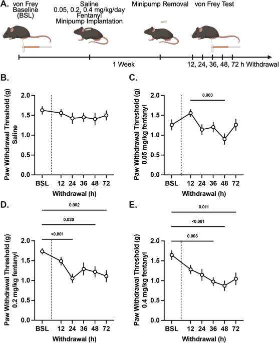
C57BL/6J mice (Jackson Laboratory, stock #000664) were used for validation of the minipump implantation fentanyl dependence model (saline, n = 14 [6 males, 8 females]; fentanyl 0.05 mg/kg, n = 13 [7 males, 6 females]; fentanyl 0.2 mg/kg, n = 14 [7 males, 7 females); fentanyl 0.4 mg/kg, n = 15 [7 males, 8 females]) and for whole-brain network analysis during naloxone-precipitated fentanyl withdrawal (n = 11 [5 males, 6 females]).
Vgat-IRES-Cre mice (Jackson Laboratory, stock #028862) were bred in-house and used for Fos/GAD67 immunohistochemistry (SAL, n = 13 [7 males, 6 females]; WD, n = 13 [7 males, 6 females]), chemogenetic manipulation (Sham::VEH, n = 10 [5 males, 5 females]; Sham::CNO, n = 9 [5 males, 4 females]; Gi::VEH, n = 10 [6 males, 4 females]; Gi::CNO, n = 11 [6 males, 5 females]), and intravenous self-administration (Sham, n = 13 [9 males, 4 females]; Gi, n = 15 [9 males, 6 females]) experiments.
Vgat-ChR2-EYFP mice (Jackson Laboratory, stock #014548) were bred in-house and used for CRACM electrophysiology experiments (SAL, n = 6 (3 males, 3 females); WD, n = 6 (3 males, 3 females)). Mice were between 8 and 10 weeks of age at the start of each experiment.
Drugs
Fentanyl citrate (Cayman Chemical, #22659) was dissolved in sterile 0.9% saline, and all doses are reported as the free base equivalent. Naloxone hydrochloride (Millipore Sigma, #BP548) was dissolved in sterile 0.9% saline and administered intraperitoneally (10 mg/kg). Clozapine‑N‑oxide (CNO; TOCRIS, #6329) was dissolved in sterile 0.9% saline and administered intraperitoneally (3 mg/kg) 30 min prior to behavioral testing. Vehicle injections consisted of an equivalent volume of sterile saline.
von Frey Test for mechanical pain sensitivity
Mechanical pain sensitivity was assessed using the von Frey test. Mice were habituated for 30 min in individual Plexiglas enclosures on an elevated wire mesh platform. Nylon monofilaments (0.04–1.4 g; Stoelting Co.) were applied to the plantar surface of the hind paw using the simplified up-down method (SUDO) [19,20,21]. Detailed procedures are provided in the Supplementary Methods.
Osmotic minipump implantation and withdrawal paradigm
To induce fentanyl dependence, osmotic minipumps (Alzet LLC, model 1002) were filled with either fentanyl citrate or sterile 0.9% saline and implanted subcutaneously between the scapulae. Minipumps provide continuous, controlled drug delivery at a constant rate and are widely used to produce sustained systemic opioid exposure in rodents [22,23,24,25,26,27]. Prior studies have demonstrated that this method yields measurable and stable plasma opioid concentrations sufficient to induce canonical opioid effects, including tolerance and physical dependence [25, 28, 29]. For initial testing, to establish the ideal dose for fentanyl dependence, 0.05, 0.2, and 0.4 mg/kg were evaluated, and 0.4 mg/kg was selected for subsequent experiments. Mice were briefly anesthetized with isoflurane (3–4% induction, 1–2% maintenance) during minipump implantation. At 7 days, minipumps were surgically removed under isoflurane anesthesia to initiate spontaneous withdrawal. All mice were monitored daily throughout the infusion and withdrawal period for signs of distress or complications. For naloxone-precipitated withdrawal, at 7 days, mice were injected with naloxone to precipitate withdrawal, and brains were collected 90 min after injection.
Whole-brain clearing and network analysis
Brains were immunolabeled and cleared using the iDISCO+ protocol [22, 30,31,32,33], imaged with light-sheet microscopy, and processed with ClearMap to detect Fos+ cells and register them to the Allen Mouse Brain Atlas. Fos quantification, data normalization, and criteria for excluding low-signal regions are described in the Supplementary Methods. Due to the resolution and atlas registration constraints of iDISCO+ and ClearMap, Fos signals were quantified at the level of the entire PAG, and individual PAG subregions (e.g., vlPAG) could not be reliably distinguished in this analysis.
Group-level correlation matrices were constructed from log10-transformed Fos counts, organized by major brain divisions, and converted to distance matrices for hierarchical clustering. Functional modules were defined by trimming dendrograms at 50% tree height, consistent with prior studies. Graph-theoretical metrics, including within-module degree z-score (wMDz) and participation coefficient (PC), were then calculated to identify hub regions and intermodular connectivity. Detailed pipelines, thresholding criteria, and visualization procedures are provided in the Supplementary Methods. Region abbreviations, full names, and anatomical group assignments are provided in Supplementary Table 1, and all PC and wMDz values are reported in Supplementary Table 2.
Immunohistochemistry and imaging
To assess neuronal activity in the vlPAG, Fos/GAD67 immunohistochemistry was performed on brain sections from male and female Vgat-IRES-Cre mice treated with fentanyl or saline via osmotic minipump and collected 48 h after pump removal. Sections were immunostained using rabbit anti-c-Fos (1:500; Cell Signaling, #2250) and mouse anti-GAD67 (1:250; Millipore, #MAB5406), followed by goat anti-rabbit Alexa Fluor 488 (1:500; Thermo Fisher, A-11008) and goat anti-mouse Alexa Fluor 594 (1:500; Thermo Fisher, A32742). Images were acquired using a Zeiss LSM 880 inverted confocal microscope at 63×. Detailed immunohistochemistry procedures are provided in the Supplementary Methods.
Electrophysiology: channelrhodopsin-assisted circuit mapping (CRACM)
To assess GABAergic synaptic transmission in the vlPAG, whole-cell voltage-clamp recordings were performed in brain slices from male and female Vgat-ChR2-EYFP mice subjected to fentanyl dependence (0.4 mg/kg/day for 7 days) or saline controls, collected 48 h after pump removal. Recordings were obtained from 6 mice per group (3 males, 3 females), with multiple cells recorded per animal (SAL: 73 cells; WD: 91 cells). Cells were sampled across animals and slices to minimize clustering within subjects. Recordings were made from visually identified vlPAG neurons using IR-DIC optics, with ChR2-EYFP expression used to guide targeting. Cells were voltage-clamped at 10 mV to isolate IPSCs. ChR2-expressing GABAergic terminals were stimulated using 470 nm light pulses (1–2 ms). Optogenetic stimulation activated vGAT-positive axon terminals within the slice, capturing both local and long-range inhibitory inputs; thus, recordings reflect overall inhibitory synaptic input rather than a presynaptic source. IPSC amplitudes were quantified and used to generate synaptic input maps. Cells with direct photocurrents or unstable baselines were excluded. Additional experimental details are provided in the Supplementary Methods and are consistent with previously described CRACM approaches [34].
Stereotaxic surgery
Mice were anesthetized with isoflurane (3–4% induction, 1–2% maintenance) and placed in a stereotaxic frame (Kopf Instruments, Tujunga, CA, USA). For mice with active virus, AAV5-hSyn-DIO-hM4D(Gi)-mCherry (Addgene, #44362-AAV5, 5 ×10¹² GC/mL; Gi) was bilaterally injected into the vlPAG of Vgat-IRES-Cre mice. For control mice with inactive virus, AAV5-hSyn-DIO-mCherry (Addgene, #50459-AAV5, 5 × 10¹² GC/mL) was bilaterally injected instead. Injections were delivered using a Hamilton 7000 series 0.5 µL syringe (Hamilton, #65457-01) at a volume of 150 nL per side. Coordinates relative to bregma were AP − 4.85 mm, ML ± 0.4 mm, DV − 2.8 mm, based on prior studies [12, 35]. Virus was infused at a rate of 50 nL/min, and the syringe was left in place for an additional 5 min to allow for diffusion. Intraperitoneal injections of cefazolin (400 mg/kg) during the surgery and subcutaneous injections of carprofen (5 mg/kg) during and 3 days after the surgery were given. Mice were monitored daily and allowed to recover for a minimum of 3 weeks before behavioral testing.
Whole-cell patch clamp recordings
To assess functional inhibition of GABAergic vlPAG neurons, whole‑cell voltage‑clamp recordings were performed in brain slices from Vgat-IRES-Cre mice injected with Gi virus into the vlPAG. Brain slices were prepared as described in the Channelrhodopsin-Assisted Circuit Mapping section. After baseline acquisition in ACSF, CNO (10 µM) was added to the bath and responses were recorded for 10 min.
Intravenous catheterization and self-administration
Mice underwent jugular catheterization before fentanyl self-administration. Fentanyl IVSA was conducted in operant chambers under an FR1 schedule, in which each active lever press delivered a 2.5 µg/kg infusion followed by a 20-s timeout; inactive lever presses had no programmed consequence. Mice completed 14 consecutive 2-h sessions. On Days 11 and 14, mice received saline injections, whereas on Days 12 and 13, mice received CNO 30 min before testing. This IVSA procedure was adapted from our previous work [19]. Detailed surgical, catheter maintenance, and patency procedures are provided in the Supplementary Methods.
Image analysis and quantification
Image analysis was performed using FIJI/ImageJ. Fos+ cells and double-labeled cells were manually counted within the vlPAG ROIs. Counts were averaged across left and right hemispheres per section. All analyses were performed blind to treatment.
Statistical analysis
Total animal numbers and sex distribution for each experiment are reported in the Methods and corresponding figure legends. Statistical analyses were performed in GraphPad Prism (version 10.5.0). Data were tested for normality. Analyses included t-tests, one-way, two-way, three-way, or repeated-measures ANOVA, with Bonferroni post hoc tests as appropriate. For all experiments, sex differences were tested; unless otherwise noted, sexes were combined for analysis. Significance was defined as p < 0.05. Data are presented as mean ± SEM with individual data points shown where applicable.


Comments (0)