
Remember me
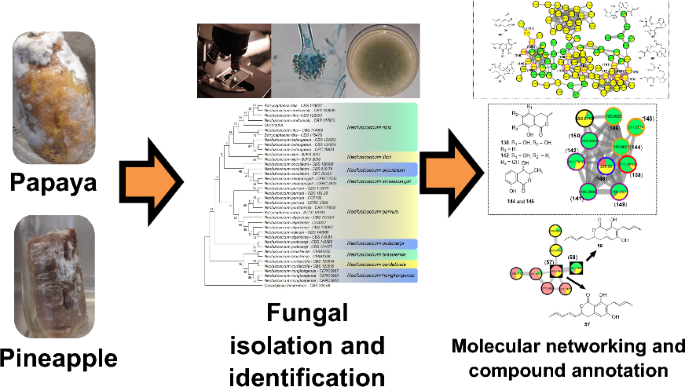
The methodology employed for isolating endophytic and phytopathogenic fungi proved effective for obtaining new environmental fungal strains for the laboratory collection. The isolates were initially evaluated based on macroscopic colony characteristics, allowing grouping according to macromorphological similarities. One representative strain from each group was selected for further analyses (Fig. S1). These strains were assigned the code LMC2300 and subjected to micromorphological examination (Fig. S2). Distinctive conidiogenous structures enabled preliminary characterization, confirming the presence of five Fusarium species and two Aspergillus species [27, 28]. For strain LMC23009, however, it was not possible to photograph the reproductive structures due to the specific developmental pattern of this species.
For precise identification, phylogenetic trees were constructed for each isolate using reference loci obtained from the literature [27,28,29,30,31,32,33,34,35] (Tables S1-8). The dataset included fungal species names, strain codes, and corresponding GenBank accession numbers for each locus. Distinct fungal strains, along with an outgroup, were incorporated in each phylogenetic reconstruction. Maximum Likelihood (ML) analysis enabled species-level resolution, with fungal strain codes, host fruit, and isolation conditions detailed in Table 1.
Table 1 Molecular identification of fungal isolates from papaya and pineapple, with corresponding GenBank accession numbersOne isolate, LMS23017 (Trichoderma sp.), could not be resolved to the species level, indicating the need for additional loci to achieve accurate classification. Notably, a higher proportion of phytopathogenic fungi was recovered compared to endophytic strains, suggesting the predominance of opportunistic environmental fungi that proliferate under favorable conditions.
The predominance of Fusarium species among the isolated fungi was particularly notable, with five out of the twelve obtained fungal specimens belonging to this genus. This finding underscores the ecological prevalence of Fusarium, a fungus renowned for its ability to persist in soil for extended periods while awaiting favorable conditions for development and proliferation. Such ecological resilience is attributed mainly to its capacity to produce resistant conidia that remain viable until environmental factors such as humidity, temperature, and the availability of suitable hosts trigger their germination and subsequent growth [4, 6, 36].
For precise taxonomic identification, two phylogenetic trees were constructed: one for the Fusarium solani species complex (Fig. 1), which enabled the identification of strains LMC23007.2 and LMC23008; and another for the Fusarium fujikuroi species complex (Fig. 2), which facilitated the classification of strains LMC23012, LMC23015, and LMC23018. This phylogenetic approach provided robust species-level resolution for the Fusarium isolates, confirming their placement within these well-characterized species complexes.
Fig. 1 The alternative text for this image may have been generated using AI.
Phylogenetic tree of the Fusarium solani species complex (FSSC). Maximum Likelihood phylogenetic tree based on RPB2 sequences showing the placement of isolates LMC23007.2 (Fusarium falciforme) and LMC23008 (Fusarium petroliphilum) within the Fusarium solani species complex. Bootstrap support values (1000 replicates) are indicated at the nodes. Reference sequences were retrieved from GenBank, with species names and corresponding strain codes provided
Fig. 2 The alternative text for this image may have been generated using AI.
Phylogenetic tree of the Fusarium fujikuroi species complex (FFSC). Maximum Likelihood phylogenetic tree based on RPB2 sequences showing the placement of isolates LMC23012 (Fusarium sacchari), LMC23015 (F. sacchari), and LMC23018 (Fusarium verticillioides) within the Fusarium fujikuroi species complex. Bootstrap support values (1000 replicates) are indicated at the nodes. Reference sequences were retrieved from GenBank, with species names and corresponding strain codes provided
In Fig. 2, the close phylogenetic relationship between the Fusarium sacchari strains LMC23012 and LMC23015 is evident, despite their isolation from different host fruits. Strain LMC23012 exhibited a macroscopically white mycelium with the production of purple pigments, whereas strain LMC23015 produced yellow-pigmented mycelia, as depicted in Fig. S1. In addition, the laboratory collection included F. guttiforme (F1-MMBF- 04/07), a member of the F. fujikuroi species complex, which was cultivated under the same experimental conditions as the five isolated Fusarium species.
The identity of isolate LMC23006 (N. ribis) was confirmed through phylogenetic analysis (Fig. 3). The phylogenetic tree includes representatives of different species complexes within the genus Neofusicoccum, highlighting synonymous designations (e.g., N. parvum) and taxonomic relationships relevant to the classification of N. ribis.
Fig. 3 The alternative text for this image may have been generated using AI.
Phylogenetic tree of the LMC23006 fungus. Maximum Likelihood phylogenetic tree based on ITS, TUB2, and RPB2 sequences confirming the identity of isolate LMC23006 as Neofusicoccum ribis. Bootstrap support values (1000 replicates) are indicated at the nodes. The tree encompasses representatives of several Neofusicoccum species complexes and reveals synonymous species within the genus, such as N. parvum
For the remaining isolates, phylogenetic trees were also constructed to confirm their identification, including Aspergillus flavus, Aspergillus terreus, Mucor circinelloides, Talaromyces funiculosus, and Trichoderma sp. These analyses are provided in Figs. S3–S7.
2.2 Molecular networking and isolated compoundsThe molecular networking for each Fusarium species was constructed using the GNPS2 platform to study the metabolites produced under different culture conditions, and the GNPS workflow link for each analysis is provided in Table S9 with its respective conditions. The Fusarium strains were co-cultivated with the endophytic fungus N. ribis to promote fungal interaction and competition, since all isolates originated from fruit samples. This strategy aimed to investigate their ecological relationships and potentially activate silent biosynthetic pathways. Annotation was performed based on spectral similarity to the GNPS2 fragmentation database, and all matches and predictions were verified using.
ChemWalker and/or SIRIUS v. 6.2.2. Additionally, compounds annotated by these tools were cross-referenced with literature to confirm their natural origin. For each fungus, a molecular networking Fig., annotated compounds, and a corresponding retention time-ordered compound table were generated. Isolation of selected compounds further enhanced the reliability of the results. The compounds were analyzed and annotated using molecular networking. Some extracts were fractionated, and for the discussion, the molecular networks were divided by fungal species to examine the influence of the culture medium on metabolic production and chemical diversity. Finally, the molecular network of the co-cultures of the six Fusarium strains with N. ribis was analyzed. All NMR data for the isolated compounds are presented in the experimental section, together with literature references. The data for common, well-documented metabolites are provided there as well. In contrast, spectra were only included in the Supplementary Material for the new compound and for those exhibiting structures that differed from the metabolites observed in the different culture media.
2.2.1 Fusarium falciformeFor the fungus LMC23007.2, 34 compounds were annotated (Fig. 4, Table S10), with significant clusters displayed in Fig. 5. A dominant cluster corresponded to the bis-alkenoic class, including fusaridioic acid A (16)[37] and its derivatives—compounds well-documented for the Solani complex [38] Side-chain esterification via water loss yielded Hymeglusin (20) [39] Fragmentation analysis enabled the annotation of additional group members (Fig. 5), revealing diverse derivatives with hydroxylations at varying positions, dehydration, double-bond modifications, and potential dimerization/trimerization, complicating annotation. Nevertheless, computational tools facilitated the identification of metabolites such as halymecin C (22) [40] Dimers like fusariumester A1 (24) and its isomer A2 (28) were distinguished by retention times [38], while a trimer (31) was also detected 2,4-Tetradecadienedioic acid, 12-[[13-carboxy-3-[(13-carboxy-12,14-dihydroxy-3,5,7-trimethyl-1-oxo-2,4-tetradecadien-1-yl)oxy]-2-(hydroxymethyl)-8,10,12-trimethyl-1-oxo-10,12-tridecadien-1-yl]oxy]-13-(hydroxymethyl)-3,5,7-trimethyl.
Fig. 4 The alternative text for this image may have been generated using AI.
Compounds annotated by molecular networking from the LMC23007.2 fungus. Highlighted compounds (1, 2, 3, 4) were isolated and structurally characterized
Fig. 5 The alternative text for this image may have been generated using AI.
Molecular Network generated by the GNPS2 platform of metabolites produced by the LMC23007.2 fungus in rice and corn culture media
Fusaric acid derivatives—previously isolated by our group (2–4) [41]—were observed alongside esterified forms: Methyl 5-[(3S)-3-hydroxybutyl]-2-pyridinecarboxylate (5) and methyl dehydrofusarate (8) [42]. From the GNPS2 spectral match and ChemWalker, two diastereomers were annotated: isomarticin (11, RT 15.23 min) and marticin (15, RT 16.09 min), and also of an ester compound formed by acid methylation, annotated as isomarticin methyl ester (17) [43] and annotated by SIRIUS. Cyclic depsipeptide, assembled from diverse amino acids like sansalvamide (29) [44] and N-methylsansalvamide (30) [45], and larger cyclosporin derivatives (A (23), C (33), and B (34)), frequently reported in this fungus [46].
The GNPS2 network revealed metabolite variation across conditions. Rice medium (Fig. 5, green nodes) exhibited higher metabolic diversity and abundance, whereas corn medium induced exclusive compounds, such as the fusaric acid derivative cluster (4).
2.2.2 Fusarium petroliphilumThe fungus LMC23008, another member of the Solani complex, yielded the annotation of 21 metabolites, as shown in Fig. 6 and Table S11. Its molecular network revealed a predominant cluster composed mainly of polyketide skeleton, within which GNPS2 enabled the identification of NG-391 (41) [47] (Fig. 7). Further ChemWalker analyses annotated six derivatives of this compound, including fusarin A (47) [48] and fusarin F (44) [48] both were previously reported from Fusarium species.
Fig. 6 The alternative text for this image may have been generated using AI.
Compounds annotated by molecular networking from the LMC23008 fungus. Highlighted compounds (35, 37, 38, 40, 43, 49, 50) were isolated and structurally characterized
Fig. 7 The alternative text for this image may have been generated using AI.
Molecular Network generated by the GNPS2 platform of metabolites produced by the LMC23008 fungus in rice and corn culture media
Using SIRIUS, three additional metabolites belonging to distinct clusters were annotated: chaetiacandin (45), 8-acetylneosolaniol (51) [49] and unguisin B (53). No further annotations were obtained for the remaining members of these clusters, suggesting the occurrence of new derivatives or compounds not yet represented in current databases. Despite belonging to the same species complex, LMC23008 and LMC23007.2 shared only two metabolites, isomarticin (11) and marticin (15) [50].
For LMC23008, a high production of aromatic metabolites was detected. The isolation of some compounds from the fungal extract allowed the identification of anthraquinones and aza-anthraquinones. The first compound (37) [51] displayed characteristic 1H NMR signals, including resonances typical of an aromatic ring at δH 6.67 (1H, d, J = 2.1 Hz) and δH 6.69 (1H, d, J = 2.1 Hz), a vinylic hydrogen at δH 5.97 (1H, s), and a hydroxylated proton at δH 4.21 (1H, m). All NMR data for the isolated compounds, which are commonly found in the literature, are presented in the experimental section, together with literature references used for comparison, supporting its identification as 7-hydroxy-3-(2-hydroxypropyl)-5-methyl-epiisocromen-1-one (37) [51]. This metabolite was also annotated in the GNPS2 molecular network. Another compound, citreoisocoumarin (36) [52] was annotated through SIRIUS and ChemWalker.
Compound (38) exhibited a 1H NMR spectrum with a vinylic proton conjugated to a carboxyl group at δH 6.46 (1H, s), as well as chelated hydroxyl protons at δH 12.51 (1H, s) and δH 13.00 (1H, s), consistent with an aromatic ring with two hydroxyl groups. Additional non-equivalent methylene protons with geminal coupling constants were observed at δH 2.59 (1H, m) and δH 2.77 (1H, brd, J = 17.9 Hz), along with another deshielded CH₂ group adjacent to oxygen at δH 4.66 (1H, dt, J = 17.6; 2.3 Hz) and δH 4.72 (1H, d, J = 17.6 Hz). Based on these data and comparison with the literature, this metabolite was identified as fusarubin (38) [53]. Compound (49) displayed similar spectral features to (38), with the additional presence of a methoxyl group at δH 3.20 (3H, s). The absence of the δH 6.16 (1H, d, J = 1.5 Hz) signal indicated substitution of a hydroxyl group by a methoxyl group. Comparison with the NMR data from literature supported its identification as 3-O-methylfusarubin (49) [53]. Compound (35) exhibited chelated hydroxyl signals at δH 12.26 (1H, s) and δH 12.00 (1H, s), similar to other members of the series. However, the presence of a distinctive aromatic proton at δH 6.82 (1H, s) suggested a different structural feature. The oxygenated CH₂ group in the cyclic system appeared as a doublet of doublets at δH 4.11 (1H, dd, J = 11.4; 4.7 Hz) and δH 3.75 (1H, dd, J = 10.9; 11.1 Hz), indicative of vicinal coupling. Additional CH₂ protons were observed at δH 2.30 (1H, dd, J = 13.6; 3.6 Hz) and δH 1.70 (1H, dd, J = 13.6; 11.4 Hz) with another vicinal coupling. Together with literature comparisons, this compound was assigned as dihydrofusarubin (35) [54].
Compound (40) presented characteristic signals of a pyridine nucleus conjugated with a carbonyl group at δH 9.29 (1H, s) and δH 7.89 (1H, s), in addition to a vinylic proton at δH 5.57 (1H, s). A singlet at δH 2.66 (3H, s) corresponded to a methyl group bound to an unsaturated system. Based on 1H NMR from the literature, the metabolite was identified as 5,7,10-trihydroxy-3-methylbenzo[g]isoquinoline-6,9-dione (40). Compound (43) also exhibited diagnostic protons of a pyridine nucleus at δH 9.38 (1H, s) and δH 8.00 (1H, s), as well as an aromatic hydrogen at δH 6.97 (1H, s), consistent with an aza-anthraquinone skeleton. Chelated hydroxyl groups were confirmed by the signals at δH 13.51 (1H, s) and δH 13.10 (1H, s). These features, together with comparative NMR data from literature, enabled its identification as bostrycoidin (43) [53].
A structurally related metabolite displayed the same aza-anthraquinone skeleton, but with an additional aromatic hydrogen, evidenced by coupling at δH 7.34 (1H, d, J = 2.5 Hz) and δH 6.86 (1H, d, J = 2.5 Hz), indicating the absence of one hydroxyl group when compared with compound (43). Comparison with reported data supported its assignment as 5-deoxybostrycoidin (50) [55]. This metabolite formed a cluster with its positional isomer (52), which exhibited the same molecular mass but a slightly different retention time, and was annotated through SIRIUS as 7-O-desmethylescorpinone.
The molecular networking analysis demonstrated that the main secondary metabolites were produced in rice medium (highlighted in green, Fig. 7), while only a few were detected in corn medium (highlighted in yellow). Bostrycoidin (43) was the dominant metabolite in corn medium, accompanied by several derivatives that formed a molecular cluster, although these compounds could not be annotated yet.
2.2.3 Fusarium sacchari isolated from papayaThe species LMC23012, belonging to the Fusarium fujikuroi species complex, exhibited a diversified metabolic profile, with 20 compounds annotated (Fig. 8, Table S12). Molecular networking analysis (Fig. 9) revealed that the predominant cluster consists of beauvericin derivatives.
Fig. 8 The alternative text for this image may have been generated using AI.
Compounds annotated by molecular networking from the LMC23012 fungus. Highlighted compounds (2, 43, 57, 58, 59, 70) were isolated and structurally characterized
Fig. 9 The alternative text for this image may have been generated using AI.
Molecular Network generated by the GNPS2 platform of metabolites produced by the LMC23012 fungus in rice and corn culture media
The fungal extract separation primarily yielded beauvericin (70), a well-characterized metabolite previously reported by our group and commonly described in Fusarium species [20]. GNPS2 analysis facilitated the annotation of beauvericin analogs, including beauvericin D (68)[52] and beauvericin A (71) [56].
Further structural elucidation via ChemWalker and SIRIUS led to the identification of six additional derivatives. However, several cyclic depsipeptides remained uncharacterized, due to closely similar molecular masses, suggesting subtle variations in amino-acid positioning or substitution within the core structure. Notably, production profiles varied significantly among different culture media. In both rice-based (green, Fig. 9) and corn-based (yellow) media, the production of beauvericin derivatives was balanced and occurred in similar proportions. In contrast, cultivation on SCG (pink) led to a dramatic reduction in these metabolites, with only two compounds detected within the cluster. These two metabolites appeared to be unique to the SCB medium, indicating a distinct metabolic response to the nutritional environment provided by this substrate.
Among the identified metabolites, fusarinolic acid (2) and bostrycoidin (43)—both previously reported in other fungal species—showed higher production in corn-based medium for this isolate, whereas lower quantities were observed in SCB medium.
Structural analysis of a separate cluster uncovered a metabolite containing the distinctive N-methyl-4-hydroxy-2-pyridone core, annotated as oxosporidinone (64) [57] through GNPS. A structurally related compound, featuring two additional hydrogen atoms, was characterized as an oxosporidinone derivative (60) using SIRIUS and ChemWalker.
Within the class of diketomorpholines, bassiatin (54) [58] was annotated via SIRIUS, although several other cluster constituents remain unresolved. A distinct cluster, predominantly composed of compounds produced in corn medium (with minor presence in rice medium), enabled the annotation of the diterpenoid lactone 7,19-dihydroxy-6,18-epoxicaur-16-en-18-one (55) [59] via GNPS, while the identities of other cluster members remain to be determined. Fusapyrone (61) [60] was predominantly detected in the SCB medium and annotated through the SIRIUS platform.
Compound (57) exhibited distinct spectroscopic features in 1H NMR (Fig. S8) an aromatic proton at δH 6.37 (s, 1H) and a conjugated trans-double bond system evidenced by signals at δH 5.47 (dd, J = 6.3, 15.7 Hz), δH 6.12 (dd, J = 10.4, 15.7 Hz), δH 6.40 (dd, J = 10.4, 15.3 Hz), and δH 5.83 (dq, J = 6.8, 15.3 Hz). Additional olefinic protons were observed at δH 5.55 (dq, J = 1.4, 6.3, 15.3 Hz) and δH 5.73 (dd, J = 6.5, 15.3 Hz). Multiplets at δH 2.97 and 2.88 (1H each) suggested a cyclic CH₂ group, while a signal at δH 5.10 (brdd, J = 6.3, 12.2 Hz, 1H) is consistent with an ester moiety within a six-membered lactone ring. Comparison with published data allowed assignment of this compound (57) as 7-but-15-enyl-6,8-dihydroxy-3(R)-penta-9,11-dienylisocoumarin [61].
A structurally related compound, designated as (58), exhibited a similar 1H NMR (Fig. S9) profile but lacked the conjugated double bond system. High-resolution mass spectrometry confirmed the presence of two additional hydrogen atoms relative to compound (57), permitting assignment of (58) as 7-but-2-enyl-6,8-dihydroxy-3-pent-3-enyl-3,4-dihydroisocoumarin [62]. Both metabolites clustered together in the molecular network, sharing analogous fragmentation patterns, although other related metabolites in the group remain uncharacterized.
A third derivative, compound (59), showed structural variation via hydrogenation of the side chain, yielding an n-butyl group—supported by proton signals at δH 2.62 (t, J = 7.5 Hz, 2H), 1.51 (quint, J = 7.5 Hz, 2H), 1.36 (sext, J = 7.4 Hz, 2H), and 0.91 (t, J = 7.4 Hz, 3H). Comparison across all NMR data (Fig. S10) with literature enabled its identification as 7-butyl-6,8-dihydroxy-3(R)-pent-11-enylisochroman-1-one [62]. This structural shift yielded a divergent mass fragmentation pattern that placed compound (59) in a separate cluster. Through complementary analyses using ChemWalker, a fourth, non-isolated derivative was annotated as versicoumarin C (56), further enriching the structural scope of this metabolite class.
Comparative profiling across cultivation media revealed marked differences in specialized metabolite production. While rice and corn media favored the predominant biosynthesis of beauvericin derivatives (Fig. 9), cultivation in SCB medium led to production of isocoumarin derivatives (compounds 56–59), coincident with a substantial decline in beauvericin synthesis (highlighted in pink in Fig. 9). This metabolic shift underscores the plasticity of secondary metabolism in Fusarium sp, demonstrating its ability to activate distinct biosynthetic pathways in response to nutritional ingredients. These results highlight the strategic importance of medium selection in accessing the chemical diversity of filamentous fungi and advancing their biotechnological potential for the discovery of new compounds.
2.2.4 Fusarium sacchari isolated from pineappleThe fungus LMC23015 exhibited a markedly distinct chemical profile, characterized by 16 annotated metabolites (Fig. 10 and Table S13). Its major metabolite featured a pyridine core (Fig. 11), structurally related to the mycotoxin fusaric acid (4), which was not detected in this cluster, although its derivatives were isolated.
Fig. 10 The alternative text f
Comments (0)