
Remember me


Human papillomavirus (HPV) is a double-stranded DNA virus that infects both the skin and mucous membranes, with over 400 identified genotypes. It is one of the most common sexually transmitted infections globally, transmitted through direct skin-to-skin contact, including during sexual activity [28]. HPV causes a wide range of cutaneous and mucocutaneous lesions, ranging from benign warts to cancers of the anogenital and oropharyngeal regions.
Cutaneous manifestations of HPV include several distinct types of warts, which vary in appearance and viral genotype. Common warts (verruca vulgaris), caused by HPV types 2 and 4, present as rough, hyperkeratotic papules typically found on the hands, knees and elbows. Plantar warts (verruca plantaris), often due to HPV types 1, 2 and 4, are flat lesions on the soles that may be painful and contagious [29]. Flat warts (verruca plana), caused by types 3, 10 and 28, are small, smooth, brown papules usually seen on the face and extremities. Filiform warts, linked to types 1, 2, 4 and 7, appear as long, narrow projections on the face or neck. Genital warts (condyloma acuminata), primarily caused by HPV types 6 and 11, are soft, flesh-colored growths in the anogenital area, spread through sexual contact [30]. Patients who are immunocompromised, including people living with advanced HIV, are at increased risk of developing extensive or treatment-refractory warts [31].
Beyond benign lesions, high-risk HPV types, especially HPV-16 and HPV-18, are oncogenic and associated with squamous cell carcinoma (SCC) in multiple mucosal sites. These cancers arise through a dysplasia-carcinoma sequence driven by persistent HPV infection and integration of viral DNA into host cells, disrupting tumor suppressor pathways such as p53 and Rb. Cervical cancer remains the most well-established and globally prevalent HPV-associated malignancy, typically preceded by cervical intraepithelial neoplasia (CIN), with CIN III carrying the highest risk of progression to invasive cancer [32, 33]. Oropharyngeal SCCs commonly arise in the crypt epithelium of the palatine and lingual tonsils and may present with cystic cervical lymphadenopathy. In the anal region, the precursor lesion is anal intraepithelial neoplasia (AIN), also graded I to III, with AIN III most likely to progress to invasive SCC [34]. HPV-16 and HPV-18 are the most frequent causative types in these malignancies. Risk factors for anal SCC include HIV infection, immunosuppression, tobacco use and receptive anal intercourse, particularly among men who have sex with men.
Early recognition of HPV-associated skin findings is important for the diagnosis and prevention of STI-related malignancies. Vaccination remains a critical tool in reducing the burden of both benign and oncogenic HPV-related disease [28] (Fig. 6).
Fig. 6 The alternative text for this image may have been generated using AI.
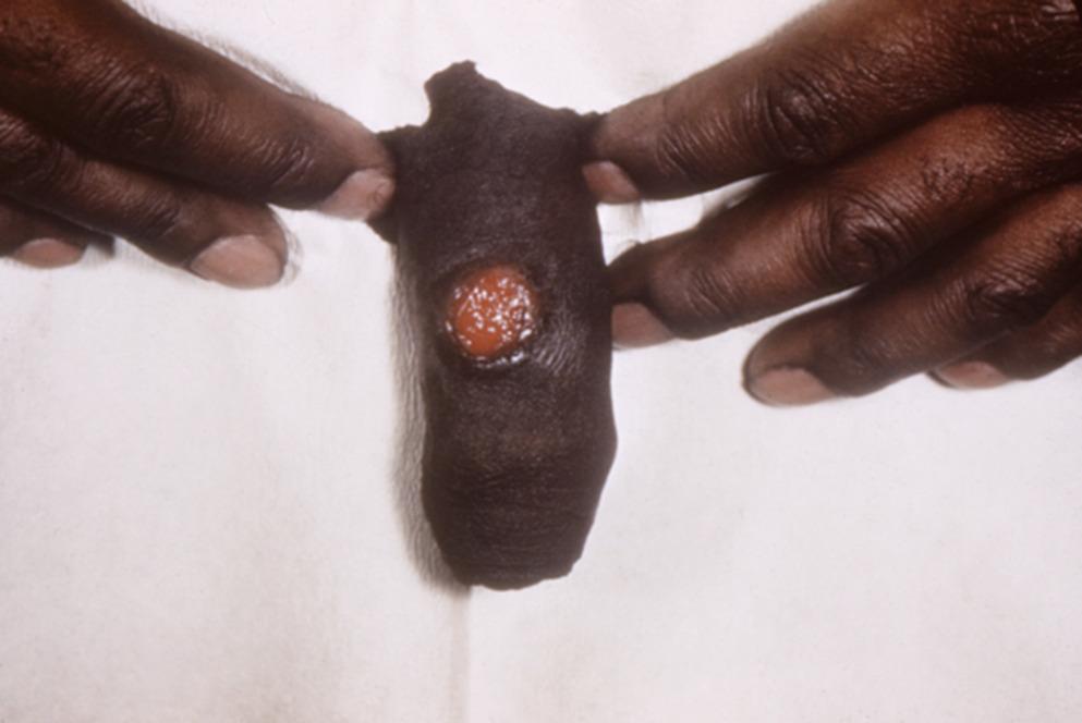
Female patient’s perineal area with many cutaneous outgrows of the perianal and labial area, due to condylomata acuminata which is caused by HPV infection [35]
Herpes Simplex VirusHerpes simplex virus refers specifically to Herpes Simplex Virus 1 (HSV-1) and 2 (HSV-2)[36]. They are members of the Herpes DNA virus family (Herpesviridae) and occur both in immunocompetent and immunocompromised individuals and may present with heterogeneous mucocutaneous symptoms [37]. After primary infection through direct contact with skin or bodily fluids with the virus present, the virus establishes latency in neurons, with the potential for reactivation, usually near the site of initial acquisition [36, 37]. However, most infections are asymptomatic (~ 2/3 of individuals) [36, 37]. Primary infection may present at primary gingivostomatitis (fever, sore throat, cervical lymphadenopathy, oral cavitary vesicular enanthem), a mononucleosis type syndrome (pharyngitis, fever, cervical lymphadenopathy), or with traditional ulceration in the oral cavity or genital lesions. In rare cases it may lead to viral encephalitis, often with classic temporal lobe localization and associated seizures [36,37,38]. Aseptic meningitis, caused predominantly by HSV-2, is a less severe neurologic syndrome, characterized by self-limited fever and meningismus, and can recur [39].
Following primary infection, it may reactivate with a small number of painful, clustered vesicles with an erythematous base, that often heal spontaneously without treatment over 4–10 days. The lesions are usually localized to the oropharynx, preferentially affecting the gingiva or oral labia, but may be found throughout the oral cavity. In females they may be found in the vaginal region, including the vulva and cervix, perianal region, buttocks or thighs. In males the ulcers or vesicles may be found on the penis, perianal or on buttocks and thighs.
Another cutaneous form, Herpes dermatitis, may be seen in athletes (herpes gladiatorum), health care workers and children (herpetic whitlow) and patients with eczema who became superinfected with HSV (Kaposi’s varicelliform eruption) [40, 41]. Erythema multiforme often occurs ~ 5% of individuals with recurrent or symptomatic HSV [40, 41].
Treatment of mucocutaneous Herpes involves systemic therapy with acyclovir or valacyclovir for 7–10 days for the first clinical episode, and then 2–5 days for recurrences [12, 42]. People living with HIV with recurrent disease or use in pregnancy at 36-week gestation or later, warrant suppressive daily therapy with acyclovir or valacyclovir [43,44,45]. Prophylaxis is beneficial for acutely immunosuppressed patients including organ or bone marrow transplant patients [12]. If recurrences occur despite adequate therapy or on suppressive therapy, resistance mutations should be investigated and alternative agents such as intravenous foscarnet and/or topical imiquimod may be required [12, 36].
Ocular herpes is a medical emergency and can involve both HSV 1 and 2[46]. Treatment often requires a combination of topical and systemic acyclovir or valacyclovir, along with topical steroids with prednisolone acetate [12, 46].
In neonates, Herpes can present in a variety of ways with cutaneous disease, central nervous system (CNS) disease, or disseminated disease. Untreated, neonates have >80% mortality with CNS or disseminated disease, with high risk of debilitating morbidity including cognitive deficits, neuromuscular deformities and end stage organ failure [5] (Fig. 7).
Fig. 7 The alternative text for this image may have been generated using AI.
Infant at 12-hours old with maculopapular skin rash due to herpes simplex neonatorum [47]
Molluscum ContagiosumMolluscum contagiosum virus (MCV) is a poxvirus with four different subtypes (I, II, III, IV) which results in collections (1–20) of 2–5 mm dome-shaped papules with a depressed center called mollusca [2, 5, 48]. These lesions differ from other pox virus lesions as they are epidermal tumors rather than necrotic pox lesions [2]. They are typically painless but can become inflamed and even rupture. These are commonly seen in children but are also transmitted sexually via skin-to-skin contact. They can appear on the trunk, face or genitals. In people living with HIV, hundreds of mollusca can occur on the face or other areas [2]. An eczematous reaction called molluscum dermatitis can erupt and encircle the lesions [5]. In patients with darker complexions, post-inflammatory hyperpigmentation can occur. The lesions are self-limited in patients without compromising conditions and resolve in 6–12 months but curettage, cryosurgery, electrodesiccation or topical chemical agents can be used to treat bothersome lesions or lesions in areas likely to be transmitted to others [2, 5]. In people living with HIV, treatment includes immune reconstitution. Cidofovir has in vitro activity against MCV and intravenous treatment has been reported [5]. Differential diagnosis includes flat warts, condylomata acuminata, sebaceous hyperplasia or SCC. Lesions in people with advanced HIV may resemble disseminated fungal infections caused by cryptococcus or histoplasma [2].
MpoxMpox infection is caused by the monkeypox virus (MPXV) which is a double-stranded DNA zoonotic virus within the Orthopoxvirus genus with two different clades (clade I is endemic to central Africa; clade II is endemic to west Africa and historically less severe) [49, 50]. The first human case reported in 1970 in a child from the Democratic Republic of the Congo (DRC) with most of the cases up until 2022 occurring in endemic areas via zoonotic transmission [50,51,52,53]. In 2022, a multi-national outbreak of clade IIb MPXV spread rapidly across 118 countries, with a global health emergency declared by the World Health Organization [49,50,51]. Most cases of the 2022 outbreak involve men (98%) who have had sex with other men (95%) and are aged 30–39[49, 50]. Human-to-human transmission occurs via contact with skin lesions, large respiratory droplets and exposure to fomites [49, 50]. The incubation period is wide-ranging, from 2 to 21 days with a median of 7 to 10 days [49, 50]. The classic infectious syndrome includes a prodrome with fever, fatigue, headache, myalgias and lymphadenopathy which may last for 1 to 5 days. This is followed by development of skin lesions, which has historically occurred in a more centrifugal distribution, with involvement of the face and trunk [51]. The recent global outbreak and associated with sexual transmission had led to common involvement of the genito-inguinal area, peri-anal region, and perioral area. Other commonly involved sites include the face, lips, or hands. In 54% of cases, skin lesions are the first sign of infection [49]. Less than 5% of patients present with a single skin lesion without other systemic symptoms [49]. Patients typically notice papules, vesicles or pustules on days 1–5 of symptoms, followed by pustules, erosions or ulcers, and crusts or scabs from days 6–10. After day 11, crust or scabs are most common, although the various stages of progression can last up to 4 weeks [49, 51]. Patients may also uncommonly develop abscesses or a morbilliform skin rash. Many patients (13%) develop scarring, typically one to four months after infection. Some patients can develop severe disease with liver function test abnormalities, pneumonia, encephalitis or bacterial superinfection [51]. Differential diagnosis includes HSV, VZV, molluscum contagiosum, LGV, and secondary syphilis. Diagnosis is made most commonly with real-time PCR of skin lesions. For most patients, the symptoms are self-limited and treatment is with supportive care. There are no current FDA approved antiviral treatments. For immunocompromised patients including those with advanced HIV or patients with severe disease, therapies which inhibit viral DNA synthesis including tecovirimat, cidofovir or brincidofovir may be available via expanded access but efficacy data is limited [51] (Fig. 8).
Fig. 8 The alternative text for this image may have been generated using AI.
Palms with crusted vesicular lesions caused by the MPXV [54]
Acute Retroviral SyndromeAcute retroviral syndrome develops in an estimated 40–90% of individuals within the first few weeks following initial HIV infection. The syndrome is associated with a morbilliform eruption on the trunk or limbs and typically consists of pink papules and macules up to 1 cm in diameter [2, 55]. Patients may also develop oropharyngeal lesions consisting of red macules or aphthous-like ulcers of the pharynx or genitals. Differential diagnosis includes drug rash or other viral exanthem. There are many dermatologic diseases that are associated with immune deficiency of HIV infection (i.e. opportunistic infections, neoplasm, ART-associated skin eruptions) which are not sexually transmitted and will not be covered in this text.
Comments (0)