Aortopulmonary window
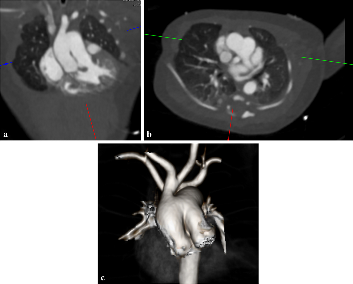
A 2-day-old boy with a prenatal diagnosis of a large-caliber aortopulmonary window (APW) presents for cardiac CT angiography (CTA) with prospective ECG gating. An oblique coronal reformat (a) demonstrates an abnormal communication between the ascending aorta and main pulmonary artery, consistent with APW. Similar findings are present on the oblique axial reformat (b). Three-dimensional reconstruction (c) further delineates the spatial relationship of the ascending aorta and pulmonary artery, aiding anatomic assessment. APW is a rare congenital anomaly, occurring in approximately 0.1% of congenital heart defects, and is frequently associated with additional anomalies such as tetralogy of Fallot, aortic coarctation, and transposition of the great arteries. Although suspected prenatally, the diagnosis is typically confirmed postnatally with echocardiography. Cardiac CTA provides high-resolution anatomic detail and 3D visualization useful for surgical planning. Treatment consists of surgical repair via sternotomy or, in select simple cases without associated defects, transcatheter closure.


Comments (0)