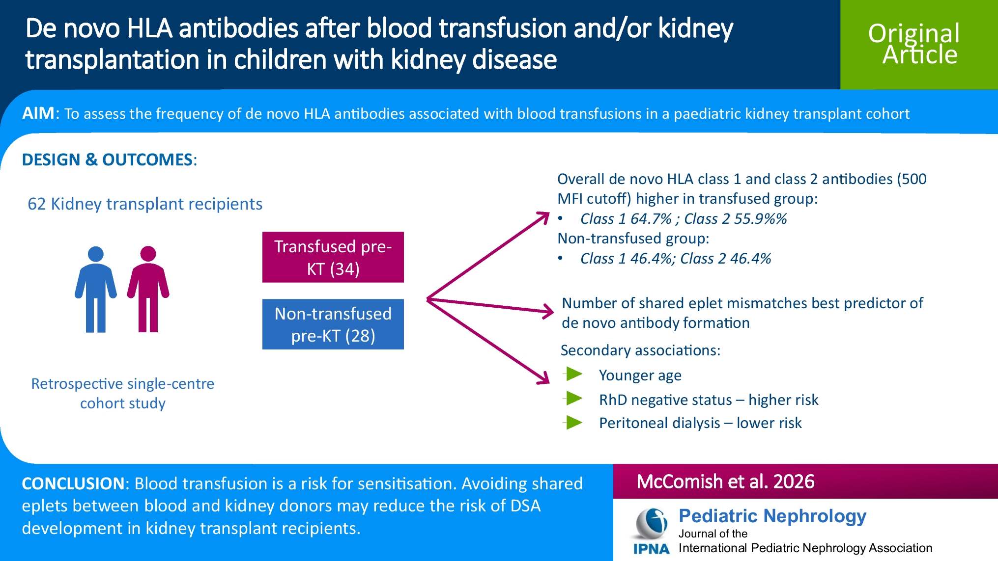
In this multicenter study, we retrospectively enrolled all children attending our pediatric rheumatology units between January 1998 and January 2025 with a diagnosis of JIA. Written informed consent was obtained from all data subjects prior to any clinical procedure, and the Research Ethics Committee of the University of Campania “Luigi Vanvitelli” approved this retrospective study.
Inclusion criteria were (i) diagnosis of JIA established according to the International League Against Rheumatism (ILAR) criteria [23]; (ii) a minimum of two follow-up visits within a 1-year interval; and (iii) age < 18 years at diagnosis of JIA. Exclusion criteria were (i) refusal to undergo the follow-up and investigations we proposed, in line with our clinical practice; (ii) missing data; (iii) loss to follow-up; (iv) known primary kidney disease; (v) other underlying diseases; (vi) used ACE inhibitors, angiotensin receptor blocker, and aminoglycosides before JIA diagnosis; and (vii) features of “COPA” syndrome.
Patients diagnosed with JIA before the introduction of the ILAR criteria were included only if their clinical charts provided evidence that they met the ILAR diagnostic criteria at the time of diagnosis [23].
None of the patients showed features of or were diagnosed with COPA syndrome (autosomal dominant disease characterized by lung disease, arthritis, and glomerulonephritis that can mimic JIA) during follow-up.
Follow-up schedule
Patient follow-up was conducted in accordance with standard clinical practice at the participating centers. Individuals with a newly established diagnosis of JIA were evaluated monthly until clinical remission was achieved; subsequent visits were scheduled every 3 or 6 months according to disease activity and ongoing treatment.
At each visit, clinical data—including height, weight, body mass index, blood pressure, and presence and duration of morning stiffness—were recorded, together with laboratory parameters such as complete blood count, hemoglobin, platelet count, serum creatinine, blood urea nitrogen, glucose, electrolytes (sodium, potassium, chloride), liver enzymes (aspartate and alanine aminotransferase), C-reactive protein (CRP), procalcitonin, erythrocyte sedimentation rate (ESR), urine dipstick analysis, and the albumin-to-creatinine ratio (ACR). Antinuclear antibodies (ANA) were assessed at the time of JIA diagnosis and subsequently on a yearly basis.
Serum creatinine was measured using an IDMS-traceable assay, beginning in 2009 at one center and in 2019 at the other. Before these time points, the Jaffe method was used. Blood samples were obtained after overnight fasting, and first-morning urine samples were routinely collected for analysis.
Overall disease activity was quantified using the Juvenile Arthritis Disease Activity Score based on 10 joints (JADAS-10). This composite index incorporates the count of active joints (range 0–10), the physician’s global assessment of disease activity measured on a 10-cm visual analog scale (where 0 indicates no activity and 10 indicates maximum activity), and the parent’s or patient’s global assessment of well-being (10-cm VAS, where 0 indicates “very well” and 10 indicates “very poor”) [24]. The ESR was included after normalizing to a 0–10 scale using a standardized formula: (ESR[mm/hour] − 20)/10 [24]. The estimated glomerular filtration rate (eGFR) was calculated using the Hoste (age) equation [25] for creatinine measured with the IDMS-traceable method and the Schwartz equation for creatinine measured using the Jaffe method [25, 26]. A summary of all variables collected for the study is provided in Table 1.
Table 1 Clinical and laboratory characteristics of all enrolled patients, and of the patients with and without AKITreatment of JIA
In our cohort, JIA treatment was assigned according to recommendations of the American College of Rheumatology [27]. NSAIDs and intra-articular corticosteroids were prescribed as first-line treatments for oligoarticular JIA. Disease-modifying antirheumatic drugs (DMARDs), such as methotrexate, were prescribed for patients with polyarticular JIA or for those with oligoarticular disease unresponsive to NSAIDs or intra-articular corticosteroids [27].
Biological drugs were prescribed for patients with systemic JIA and for those with polyarticular or oligoarticular JIA unresponsive to methotrexate or in cases of methotrexate intolerance. In our centers, biologics have been used to treat JIA since 2001. Systemic corticosteroids were administered for short courses in patients with systemic JIA or with high disease activity to achieve remission [27]. The available treatments for JIA are summarized in Table 2.
Table 2 Main drugs used in JIA treatmentIn patients who developed AKI after NSAID exposure, NSAID therapy was discontinued and corticosteroids were initiated, either as intra-articular injections or oral prednisone at a dose of 1 mg/kg for a short course, depending on the JIA subtype. Subsequently, DMARDs were started.
Primary outcome definition
The primary endpoint of the study was AKI, which was identified in accordance with the Kidney Disease: Improving Global Outcomes (KDIGO) guidelines based on serum creatinine criteria [28]. Baseline creatinine values were derived using previously validated back-estimation approaches [29]. As previously described, height-dependent and height-independent basal serum creatinine estimation methods were comparable [29]. Therefore, we applied the Hoste (age) equation [25] to back-calculate baseline serum creatinine, assuming median age-based estimated glomerular filtration rate (eGFR) values for children ≤ 2 years [30] and eGFR = 120 mL/min/1.73 m2 for those >2 years [30]. AKI staging was defined as follows: (i) no AKI: all serum creatinine values < 1.5 × baseline; stage 1 AKI: 1.5 to <2 × baseline; stage 2 AKI: 2 to <3 × baseline; stage 3 AKI: ≥3 × baseline.
Secondary outcome definition
The secondary outcome was the occurrence of CKD and/or hypertension (combined under the term “Kidney damage, KD” in this manuscript) at follow-up. CKD was defined as either: (i) ACR ≥ 30 mg/g persisting for >3 months, confirmed in three separate samples; or (ii) eGFR < 90 mL/min/1.73 m2 for >3 months [31].
To simplify the reading of the manuscript, although an ACR between 30 and 300 mg/g indicates microalbuminuria, in this manuscript we defined an ACR ≥ 30 mg/g as proteinuria.
Blood pressure was recorded after 15 min of seated rest. Hypertension in this study was defined retrospectively following the methods described by Flynn et al. [32, 33]. According to these guidelines, we used the mean of three auscultatory readings only when initial oscillometric measurements were elevated. Hypertension was diagnosed if elevated auscultatory readings were recorded on three different occasions. All patients identified as hypertensive received standard follow-up assessments for JIA, including evaluation for lipid profile, hemoglobin A1c, plasma renin activity, and kidney ultrasound [34].
Statistical analysis
Statistical significance was defined as a p value below 0.05. Continuous variables were compared using an independent-samples t-test when normally distributed, or the Mann–Whitney test for non-normal distributions. Categorical variables were compared using the chi-squared test.
Primary outcome
Logistic regression models were applied to explore associations with AKI. Parameters showing a significant association with AKI (p < 0.05) in the initial comparison between patients with and without AKI (Table 1) were included in the univariate logistic regression analysis. Proteinuria at JIA diagnosis was excluded from the logistic regression models because it was observed in only four patients.
Variables with p < 0.05 in the univariate analysis were entered into the multiple logistic regression model. Procalcitonin was excluded from the multiple analysis due to availability in only 46 patients, while ferritin was excluded because of collinearity with CRP and a wider confidence interval compared to CRP. For the multiple logistic regression, the significance threshold was adjusted using Bonferroni correction and set at p < 0.017.
Secondary outcome
The secondary outcome was analyzed using Kaplan–Meier survival methods. Time-to-event was calculated from the date of hospital admission for JIA diagnosis. Differences between survival curves were assessed with the log-rank test. Cox regression analysis was used to calculate the hazard ratio (HR) for the secondary outcome according to AKI status at JIA diagnosis, adjusting for sex, cumulative dose and duration of methotrexate and NSAIDs treatment, low birth weight, and preterm birth. We also calculated the odds ratio (OR) for developing the secondary outcome in patients with AKI at JIA diagnosis, adjusted for sex, age at last follow-up, disease duration, cumulative dose and duration of methotrexate and NSAIDs treatment, low birth weight, and preterm birth. NSAID and methotrexate exposure were not analyzed as primary determinants of KD, since their association with KD in children with JIA has already been established in a previous study [7]. Accordingly, in the present analysis, NSAIDs and methotrexate were included as covariates to adjust the association between AKI and long-term KD in both Cox and logistic regression models in children with JIA. We did not adjust these models for the use or cumulative dose of biological agents because these factors have not previously been associated with the development of KD [7].
All statistical analyses were performed using Stat-Graph XVII software for Windows, except for the Kaplan–Meier analysis, which was conducted with GraphPad Prism 7 software for Windows.


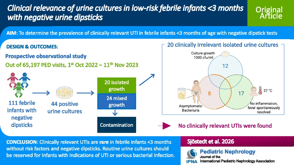
Comments (0)