
Remember me
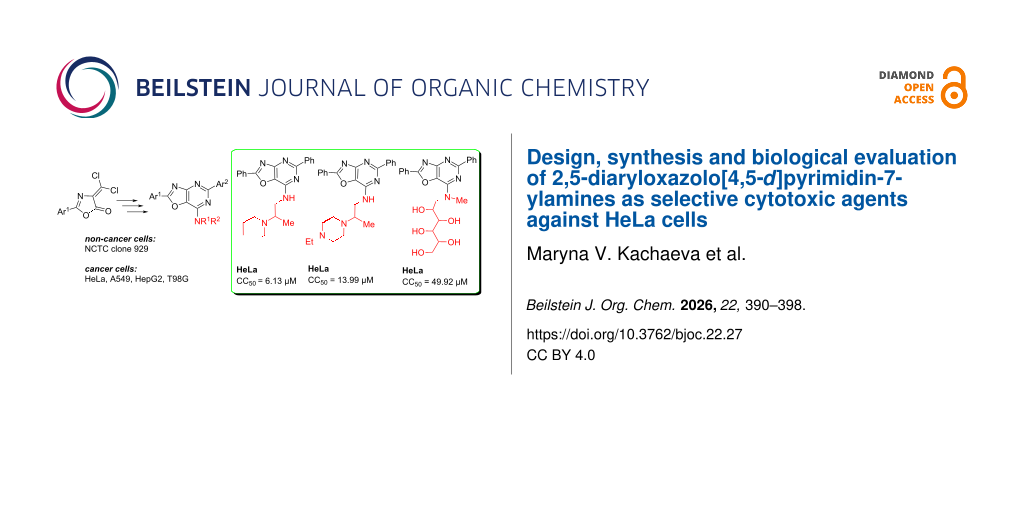
Purine-mimicking scaffolds are a proven strategy in the design of anticancer drugs. Many cancer-related proteins (e.g., kinases, ATPases, DNA/RNA polymerases) have binding pockets designed for purine nucleotides (ATP, GTP). Oxazolopyrimidines can compete with purines or their analogues, inhibiting enzymatic activity. They combine purine-like recognition features with the synthetic flexibility of heterocycles, offering a platform for selective targeting of tumor-related enzymes and receptors. Tumor cells overexpress kinases, DNA/RNA polymerases, and metabolic enzymes that bind purine nucleotides. Cancer cell lines such as HepG2 (liver), HeLa (cervix), A549 (lung), and glioblastoma models rely on enhanced nucleotide metabolism to sustain rapid DNA/RNA synthesis. Oxazolopyrimidines can inhibit these overactive enzymes in cancer cells, while normal cells (with lower demand) are less affected. This explains why oxazolopyrimidine derivatives may demonstrate selective cytotoxicity against cancer cell lines like HeLa, HepG2, A549, and central nervous system (CNS) tumor models .
The introduction of heterocyclic molecules with different amino groups as hydrogen-bond donors (and sometimes acceptors via lone electron pairs) can increase water solubility and facilitate salt formation. Introducing amino groups into heterocycles can have a significant impact on the anticancer activity of a compound .
Many studies have investigated the anticancer potential of heterocycle derivatives containing cytisine, glucamine, and the Strecker amine (aminoethylamine) moieties. Cytisine has been shown to inhibit the proliferation of A549, HepG2, EC109, K562, HL-60, and U937 cells. Following a 48 h treatment, IC50 values were approximately 2 mM in HL-60 and U937 cells, whereas higher concentrations were required to inhibit K562 and EC109 cells (IC50 > 12 mM). In A549 and HepG2 cells, cytisine exhibited activity, with IC50 values of 4.40 ± 1.70 mM and 5.92 ± 2.77 mM, respectively . Heterocycle hybrids with sugar moieties have shown selective cytotoxicity against liver cancer (HepG2 – IC50 values of 1.32–9.43 µM) and breast cancer cells (MCF-7 – IC50 1.18–11.81 µM) by increasing intracellular accumulation .
A significant number of studies has been published on the anticancer activity of oxazolo[5,4-d]pyrimidines, showing their significant activity as agonists and antagonists of signaling pathways involved in the regulation of the cell life cycle . On the contrary, oxazolo[4,5-d]pyrimidines represent a poorly studied class of compounds because of limited access to this scaffold . An in silico study showed that both isomeric forms of oxazolopyrimidines can form stable complexes due to hydrogen bonds between a lone electron pair at the nitrogen atoms and protonated amino acids of proteins. However, oxazolo[5,4-d]pyrimidine forms more stable complexes, which is in good agreement with in vitro studies .
We have already shown the anticancer potential of 7-N-derivatives of 2,5-diaryl[1,3]oxazolo[4,5-d]pyrimidines and a series of 7-N-(1,4-diazepane)- and 7-N-piperazine-substituted 2,5-diaryl[1,3]oxazolo[4,5-d]pyrimidines were highly effective anticancer agents, as proven in a five-dose assay in NCI60 cancer cell line screen . The most active derivatives of both series displayed high total antiproliferative and cytotoxic activity against the tested NCI60 cancer cell lines. Also, several other 7-(1,4-diazepan)- and 7-piperazine-substituted [1,3]oxazolo[4,5-d]pyrimidines (structures A and B in Scheme 1) showed cytotoxic activity in the micromolar concentration range against most breast cancer cell lines represented in the corresponding NCI60 subpanel derived from leukemia, melanoma, non-small-cell lung carcinoma, and cancers of the brain, ovary, breast, colon, kidney, and prostate . These results provided evidence that the compound could be helpful in developing new anticancer drugs. The combination of the [1,3]oxazolo[4,5-d]pyrimidin-7-amine fragment (structures A and B) with cytisine, glucamine, and aminoethylamine represents a promising strategy for the rational design of multifunctional hybrids with improved biological activity, water solubility, lower toxicity and target engagement potential.
![[1860-5397-22-27-i1]](https://www.beilstein-journals.org/bjoc/content/inline/1860-5397-22-27-i1.svg?scale=2.0&max-width=1024&background=FFFFFF)
Scheme 1: Rational design of [1,3]oxazolo[4,5-d]pyrimidin-7-amine derivatives 1–9.
Therefore, further functionalization of 2,4-diaryl[1,3]oxazolo[4,5-d]pyrimidines at position 7 of the structure-forming core was carried out in this work.
Results and Discussion ChemistryThe synthesis of 1,3-oxazolo[4,5-d]pyrimidine derivatives 1–9 was accomplished according to the previously described methods as depicted in Scheme 2.
![[1860-5397-22-27-i2]](https://www.beilstein-journals.org/bjoc/content/inline/1860-5397-22-27-i2.svg?scale=2.0&max-width=1024&background=FFFFFF)
Scheme 2: Synthesis of new 2,5-diaryloxazolo[4,5-d]pyrimidin-7-ylamines 1–9.
The 7-Amino-substituted 1,3-oxazolo[4,5-d]pyrimidines 1–9 were obtained with high yields (65–80%) by the reaction of 2,5-diaryl-7-chloro-1,3-oxazolo[4,5-d]pyrimidines II with the corresponding amine (aminoethylamines , cytisine and N-methyl-ᴅ-glucamine). Compounds II were obtained by the sequence of reactions starting from available 2-aryl-4-dichloromethylene-1,3-oxazol-5(4H)-ones I . Thus, treatment of compounds I with arylamidine hydrochlorides in the presence of triethylamine, followed by heating with pyridine, afforded the cyclocondensation products – [1,3]oxazolo[4,5-d]pyrimidines. Subsequent reaction with phosphoryl chloride in the presence of N,N-dimethylaniline converted these intermediates into 2,5-diaryl-7-chloro[1,3]oxazolo[4,5-d]pyrimidines II.
The structures of compounds 1–9 were proven and confirmed by 1H and 13C NMR, IR spectroscopy, LC–MS and elemental analysis (Supporting Information File 1, Figures S1–S36). The IR spectra of compounds 1–5 showed the presence of NH absorption bands in the range 3381–2936 cm−1, OH at 3396 cm−1 (compound 9), and C=O at 1601–1610 cm−1 (compounds 6-8).
Cytotoxic potency of the compounds 1–9Cytotoxic properties of the studied compounds were assessed on the non-cancer cell line NCTC clone 929 (Mus musculus normal subcutaneous connective tissue cells), as well as four human cancer cell lines: HeLa (Human cervix adenocarcinoma cells), A549 (Human lung carcinoma cells), HepG2 (Human hepatocellular carcinoma cells), and T98G (Human glioblastoma multiforme cells). Cytotoxicity was determined by measurement of 50% inhibition of cell growth by the MTT (3-(4,5-dimethylthiazol-2-yl)-2,5-diphenyltetrazolium bromide) assay. The selectivity index (SI) was calculated for the investigated compounds. The cytotoxicity results (CC50, µM) for the investigated compounds and the calculated selectivity index values are presented in Table 1.
Table 1: Results of in vitro anticancer screening of the compounds 1 9.
Compd. Non-cancer cells Cancer cells NCTC clone 929 HeLa A549 HepG2 T98G CC50a IC50 SIb IC50 SI IC50 SI IC50 SI 1 51.33 ± 7.91 6.13 ± 1.95 8.37 32.76 ± 2.63 1.57 85.43 ± 4.37 0.60 245.26 ± 19.17 0.21 2 >1000 >1000 ndc >1000 nd >1000 nd >1000 nd 3 21.60 ± 4.61 13.99 ± 1.80 1.54 56.23 ± 11.10 0.38 39.02 ± 3.74 0.55 161.93 ± 7.36 0.13 4 81.19 ± 9.52 98.05 ± 4.11 0.82 251.86 ± 39.85 0.32 223.16 ± 31.97 0.36 >1000 <0.10 5 72.63 ± 15.73 58.26 ± 7.57 1.25 465.88 ± 26.13 0.16 >1000 <0.10 >1000 <0.10 6 >1000 >1000 nd >1000 nd >1000 nd >1000 nd 7 >1000 62.59 ± 8.73 >15.98 922.3 ± 12.94 >1.08 >1000 nd >1000 nd 8 >1000 >1000 nd >1000 nd >1000 nd >1000 nd 9 706.50 ± 48.51 49.92 ± 3.98 14.15 920.23 ± 79.92 0.77 >1000 <0.71 >1000 <0.71 PTX 54.67 ± 5.77 0.02 ± 0.00 2733.50 36.00 ± 6.54 1.52 56.67 ± 8.08 0.96 0.03 ± 0.00 1822.33aCC50 and IC50 are expressed in µM; bSI, the selectivity index is calculated as SI = CC50/IC50; cnd, not defined; data are presented as mean ± S.D. of three replicates. Paclitaxel (PTX) was used as a positive control.
The compounds 2, 6, and 8 were found to be non-toxic towards all the cancer and non-cancer cell lines investigated (CC50 > 1000 µM). In general, cancer cells form the following rows in the order of decreasing sensitivity to the active compounds:
HeLa: 1 > 3 > 9 > 5 > 7 > 4
A549: 1 > 3 > 4 > 5 > 7 = 9
HepG2: 3 > 1 > 4
T98G: 3 > 1
It was observed that compounds 1 and 3 showed the highest cytotoxicity against all cancer cells. Compound 1 exhibited the highest selectivity towards HeLa cells (SI = 8.37), suggesting potential selective toxicity. Its activity against other cancer lines was less pronounced (SI < 2). Moreover, compounds 7 and 9 showed high selectivity (SI > 10), and 1 demonstrated only moderate one (SI < 10) to the HeLa cell line. Compound 9 also showed noteworthy selectivity for HeLa cells (SI = 14.15), with a high CC50 value for non-cancer NCTC clone 929 cells (706.50 µM), suggesting low general toxicity. Compound 3 displayed low selectivity towards HeLa cells (SI = 1.54). All compounds showed very low SI values for other cancer lines (SI ≤ 0.6), indicating limited therapeutic potential.
Analyzing the structure–activity relationship with respect to the HeLa cancer cell line, it can be seen that among the diphenyloxazolopyridine derivatives, the substitution of the piperidine functional motif at position 7 in compound 1 by 4-ethylpiperazine leads to a decrease in the cytotoxicity by two times (compound 3). Whereas its substitution by morpholine lead to inactive compound 2. The same result was obtained by substituting 2-piperidin-1-ylpropylamine in 1 with hexahydro-1,5-methanopyrido[1,2-a][1,5]diazocin-8-one (6). However, methylation of the phenyl group at the 5-position of compound 6 resulted in restoration of activity (7). In contrast, simultaneous introduction of a methyl group in both phenyl residues of the oxazolopyridine ring of compound 6 did not give a positive result (8). The combined replacement of the 5-phenyl substituent in compound 1 by a 4-methylphenyl group and addition of a 4-chlorophenyl group instead of the methyl group as a functional motif at position 7 (4) sharply reduced the cytotoxicity of compound 1. The substitution of piperidine by morpholine in this motif in compound 4 resulted in a 2-fold increase in the activity of the obtained derivative 5. However, compound 5 was one order less potent than compound 1 in terms of its cytotoxic potency.
It is important to note that among the compounds that have demonstrated high activity against cancer cells, a significant indicator of their potential as drug candidates is cytotoxic selectivity relative to non-cancer cells. Therefore, regardless of the concentration in effect, the compound with greater selectivity should be favored. According to this criterion, compounds 1, 7, and 9 show the most significant appeal for further testing as potential agents against cervical cancer. Unfortunately, none of these compounds was anywhere near the activity of paclitaxel against HeLa cells (SI = 2733.50).
ADMET analysisADMET (absorption, distribution, metabolism, excretion, toxicity) analysis performed among active in vitro anticancer compounds allows to identify the most promising agents to be sent to further stages of drug candidate development, taking into account the expected pharmacokinetic and toxicological, as well as undesirable properties of the molecules. At the same time, drug similarity filters developed by pharmaceutical companies based on the physicochemical properties of approved drugs with known mechanisms of action have utilitarian value, as they do not account for the probability of including targets not inherent to existing drugs in their molecular mechanisms of anticancer action. The physicochemical properties of such compounds may not fit the parameters developed from databases of approved drugs, thereby hindering the search for drug candidates with unique anticancer properties . Practically, this means that the failure of any new compounds with in vitro anticancer activity and acceptable pharmacokinetic and toxicological properties to pass these filters requires a tailored approach. It should be noted, however, that different ADMET platforms may yield conflicting results, especially regarding the metabolism and cytotoxic molecular targets of the analyzed compounds, due to different approaches used to virtually evaluate these parameters. Therefore, indeed, the equivalence of results for a particular parameter obtained from different platforms increases the reliability of the prediction . In addition, during the development of chemotherapeutics, oral bioavailability is desirable but not critical, given the preference for the parenteral route of administration.
Due to the probabilistic nature of the evaluation, parameters for which the probability of a compound's relevant property is predicted to occur in the range 0.3 < p < 0.7 does not allow a confident conclusion to be drawn and require further evaluation . The same applies to the conflicting results obtained by different platforms.
Pharmacokinetic propertiesADMET analysis enables the prediction of a compound's pharmacokinetic profile, which is crucial for assessing its pharmacodynamic activity. The predicted pharmacokinetic properties of compounds 1, 7 and 9 are given in Table 2.
Table 2: Complex pharmacokinetic profile of compounds 1, 7, and 9 predicted by ADMETlab3.0.
Parameter ADMETlab3.0 1 7 9 HIAa 0.000 0.000 0.000 BBB permeantb 0.999 0.998 0.754 Pgp substratec 0.004 0.000 0.001 Pgp inhibitor 0.950 0.949 0.013 CYP1A2 substrated 0.623 0.053 0.000 CYP2C19 substrate 0.119 0.636 0.000 CYP2C9 substrate 0.000 0.142 0.000 CYP2D6 substrate 0.401 0.088 0.000 CYP3A4 substrate 0.242 0.208 0.000 CYP2B6 substrate 0.021 0.019 0.020aHIA, human intestinal absorption; bBBB, blood–brain barrier penetration; cPgp, glycoprotein P; dCYP, cytochrome P450.
For human intestinal absorption, the virtual platforms gave mutually exclusive results. However, as mentioned above, this parameter is not critical for anticancer compounds.
It should be noted that the training samples of medicines used by all ADMET platforms are generic, i.e., medicines used to treat a wide range of diseases whose medical requirements may be mutually exclusive. Consequently, the underlying training samples used to calibrate the data may also differ depending on the application route of the medicine. Therefore, due to the conflicting results of BBB permeability predicted for compound 9 by common ADMET platforms, a specific drug identity filter for CNS-penetrating compounds was utilized (Table 3) .
Table 3: The parameters of physicochemical properties of compounds 1, 7, and 9 determine the threshold values of BBB permeability according to the CNS rule descriptor.
Descriptor Compound CNS rule 1 7 9 MWa 413.2 475.2 466 135–582 logPb 5.6 4.7 5.4 −0.2 to 6.1 nHAc 6.0 7.0 10.0 ≤5 nHDd 1.0 0.0 5.0 ≤3 tPSAe 67 77 156 3–118aMW, molecular weight; bLogP, octanol/water distribution coefficient; cnHA, number of H–bond acceptors; dnHD, number of H–bond donors; etPSA, topological polar surface area.
It follows from the presented data that, according to the value of physicochemical parameters used to predict the BBB permeability of anticancer CNS targeted compounds, compound 9 (two deviations: descriptors nHA and nHD), unlike 1 and 7 (one deviation: nHA, that is acceptable for decision-making), does not pass the CNS filter. In other words, it belongs to the group of compounds that do not pass the blood–brain barrier. This finding is also entirely consistent with the predicted ADMETlab3.0 neurotoxicity of these compounds (Table 3). With regard to the substrate specificity of Pgp, it is highly probable that these compounds are not substrates of this protein, although compounds 1 and 7, unlike 9, appear to be capable of inhibiting it (Table 3). A low probability of oxidative metabolism of these compounds by the studied cytochromes is also predicted.
Human toxicity of compounds 1, 7 and 9The results of predicting the human toxicity of tested compounds 1, 7, and 9 are presented in Table 4 and Table 5.
Table 4: Predicted parameters of human toxicity of compounds 1, 7, and 9.
Parameter ADMETlab3.0 1 7 9 cardiotoxicity (hERG blocker) 0.802 0.469 0.172 DILI (drug-induced liver injury) 0.832 0.967 0.651 carcinogenicity 0.480 0.644 0.072 respiratory toxicity 0.755 0.587 0.003 nephrotoxicity 0.441 0.566 0.310 neurotoxicity 0.888 0.953 0.280 hematotoxicity 0.202 0.661 0.014 genotoxicity 0.517 0.989 0.082 RPMI-8226 immunitoxicity 0.043 0.085 0.021Table 5: Predicted probability of interaction of compound 1, 7, and 9 with nuclear receptor (NR) signaling, and stress response (SR) pathways.
Target ADMETlab3.0a Deep-PK 1 7 9 1 9 NR-AhRb ++ - --- 0.069 0.079 NR-ARc -- --- --- 0.170 0.203 NR-AR-LBDd ++ --- --- 0.041 0.013 NR-Aromatase + -- --- 0.010 0.020 NR-ERe ++ -- --- 0.235
Comments (0)