Horseshoe kidney (HSK) is a congenital renal malformation—originally described by Berengario da Carpi in 1522 [1]—characterized by the fusion of two functional kidneys across the midline, with resultant abnormalities in renal position, rotation and vascular supply [2,3,4,5]. The isthmus connecting the two renal masses can be either functional renal parenchyma, in about 80% of cases, or a fibrous band, in the remaining 20%. In over 90% of cases the fusion is between the lower poles, and it can be symmetrical or, more commonly (70%), asymmetrical with left-sided dominance [3, 6,7,8,9]. The isthmus usually lies anterior to the great vessels at the level of the third to fifth lumbar vertebrae and is found immediately below the inferior mesenteric artery in 40% of cases [3].
HSK forms during embryogenesis between the 4th and 6th weeks of gestation [3, 6, 9] It is the most common renal malformation associated with chromosomal anomalies [10,11,12], occurring in 17–24% of patients with trisomy 18 and in as many as 45% of females with Turner syndrome. HSK may also occur within the spectrum of developmental anomalies seen in Fanconi pancytopenia, a monogenic autosomal recessive disorder [13], or in approximately 2% of patients with hepatocyte nuclear factor 1β (HNF1β)-associated disease [14], an autosomal dominant condition that represents the most frequent monogenic cause of congenital anomalies of the kidney and urinary tract (CAKUT) [15].
Despite being the most prevalent renal fusion anomaly, HSK is a relatively rare condition with prevalence estimates ranging from 1 in 300 to 1 in 1000 individuals [8, 16,17,18,19]. In a large autopsy series, Campbell reported HSK in 61 of 19,046 (0.32%) paediatric and 61 of 32,834 (0.18%) adult autopsies [20]. HSK is about twice as common in males than females [8, 16, 17, 19], and typically occurs sporadically with rare occurrence of familial cases [21].
Nearly one third of individuals with HSK remain clinically asymptomatic, with the condition often discovered incidentally during radiologic examinations [4, 9, 22]. HSK can occur as an isolated condition, but in approximately one-third of cases, it is associated with a range of anomalies [10]. These may include genitourinary anomalies, such as ureteropelvic junction obstruction, hydronephrosis, nephrolithiasis, and recurrent urinary tract infections [2, 4, 9, 10, 12, 16, 22,23,24,25]. In addition, HSK can coexist with abnormalities affecting other systems, including the central nervous, gastrointestinal, cardiopulmonary, and musculoskeletal systems [2, 4, 10, 12]. When HKS is diagnosed in adults, it is typically an isolated condition [23, 26]. A variety of benign and malignant tumours are associated with HSK with renal cell carcinoma being the most commonly reported malignancy in patients with HSK [4, 26, 27].
Polycystic horseshoe kidney (P-HSK) is an extremely rare condition [10, 28,29,30]—with fewer than 20 cases reported in the literature before 2013 [31]—resulting from the coexistence of a HSK and autosomal dominant polycystic kidney disease (ADPKD), a genetically heterogeneous renal tubular disorder characterized by progressive formation of fluid-filled cysts arising from any segment of the renal epithelium [32]. HSK and ADPKD are considered pathogenetically unrelated, and their coexistence is rare, with estimated frequencies ranging from 1 in 134,000 to 1 in 8 million, based on the assumption of their independent occurrence [29]. In Campbell’s series of 51,880 autopsies, only two cases of P-HSK were identified [20], while Trapp et al. reported one case in 8026 autopsies [33].
ADPKD is the most common inherited kidney disorder, accounting for approximately 5–10% of end-stage kidney disease (ESKD) cases requiring renal replacement therapy (RRT) [32, 34]. By the age of 60, nearly half of affected individuals have initiated RRT. The progressive increase in kidney volume due to cyst expansion correlates closely with the gradual loss of renal function. The clinical prevalence of ADPKD in the European Union ranges from 0.25 to 0.45 per 1000 population [32, 35], while autopsy-based estimates suggest a higher prevalence (1.5 per 1000) [36,37,38], likely reflecting undiagnosed asymptomatic cases. In southern Portugal, the clinical prevalence has been estimated at 0.33 per 1000 [39], but it can be significantly higher in Northern Portugal [40].
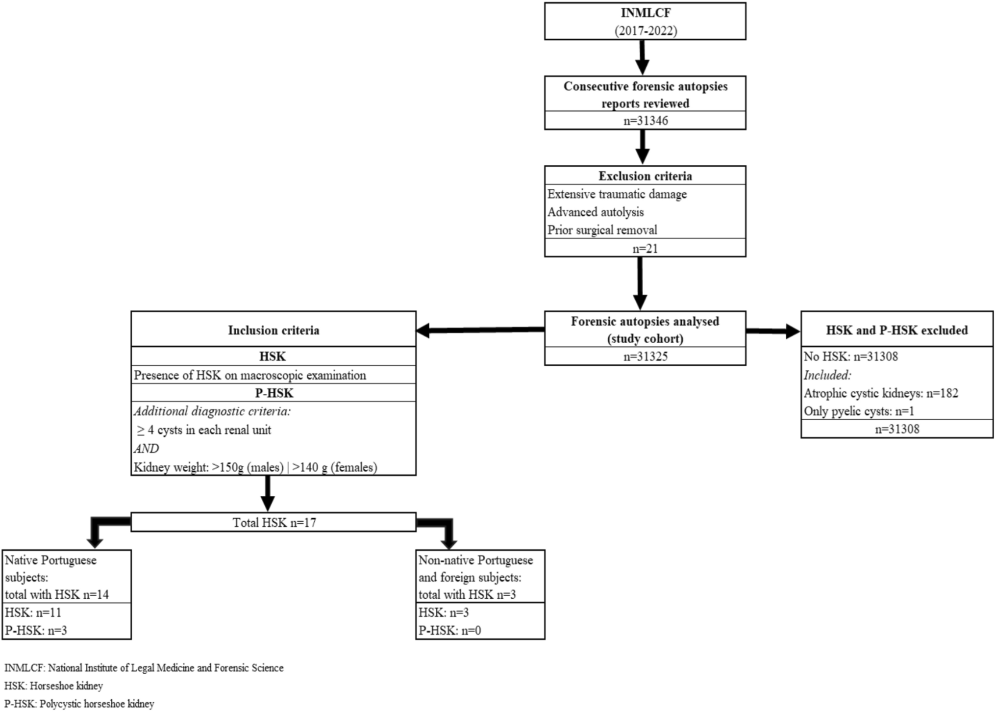
Forensic autopsies offer a unique opportunity to uncover epidemiological data on congenital anomalies such as HSK, which often go undiagnosed during life. Similarly, despite its progressive nature, ADPKD may remain unrecognized until late adulthood, particularly in families with genetically milder forms. In this study, we analysed an extensive, systematically collected forensic autopsy dataset from mainland Portugal and the Islands to estimate the prevalences of HSK and P-HSK, and to explore their associations with other systemic pathologies. Our findings underscore the importance of interdisciplinary collaboration among forensic pathologists, clinical geneticists, and other medical specialists in improving the diagnosis and management of rare renal anomalies.

Comments (0)