
Remember me
Inflammatory breast cancer (IBC) is a rare and aggressive form of invasive breast cancer characterized by the rapid onset of skin discoloration and edema involving more than one-third of the breast within six months [1, 2]. The clinical aggressiveness of the disease is compounded by frequent delays in diagnosis and a marked propensity for rapid progression, contributing to Stage IV presentation in approximately 30% of patients [3, 4]. Diagnosis is disproportionately enriched among tumors with biologically aggressive subtypes, particularly Her2-overexpressed and triple negative breast cancers [3, 5].
The pathophysiology of IBC is thought to involve excessive obstruction by tumor emboli of the vasculature of the breast and dermal lymphatics, leading to the hallmark clinical features of skin thickening and edema. The characteristic diffuse infiltration of disease throughout the lymphovascular channels of the breast and skin rather than a discrete tumor mass, as more commonly appreciated in non-inflammatory breast cancer, dictates the importance of a comprehensive multi-modal treatment approach. Lymphovascular obstruction may not account for all aspects of the disease’s phenotype, however; preclinical models have demonstrated skin changes in the absence of lymphatic emboli, suggesting alternative or complementary pathways of disease manifestation [6].
Compared to individuals with non-inflammatory breast cancer (non-IBC), patients diagnosed with IBC tend to be younger, have a higher likelihood of prior pregnancies, and are disproportionately African American [7]. Incidence varies widely internationally, ranging from close to 10% of breast cancer cases in North Africa [8] to as low as 0.5% in The Netherlands [9, 10]. Despite substantial research efforts, molecular drivers specific to IBC, remain elusive. Extensive genomic and transcriptomic comparative analyses consistently demonstrate an overlap with non-inflammatory aggressive breast cancer subtypes, with minimal consensus across studies supporting one or more unique molecular signatures [11,12,13,14,15].
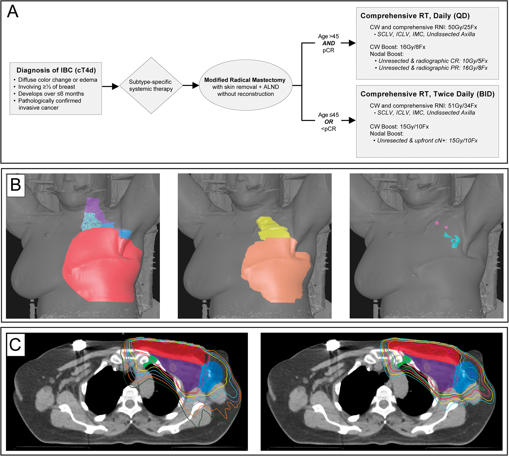
The 5-year overall survival (OS) for patients with stage III IBC has remained around 50% for many years [16]. Despite its distinct clinical presentation, biological profile, and guarded prognosis, guideline-concordant care (GCC) for IBC largely mirrors stage-matched non-IBC [2, 17]. The standard treatment recommendation includes neoadjuvant chemotherapy tailored to receptor subtype, modified radical mastectomy (MRM) with removal of all clinically involved skin and axillary lymph node dissection (ALND), followed by post mastectomy radiotherapy (PMRT) to the chest wall and regional draining lymph node basins (Fig. 1). Adjuvant systemic therapy is recommended based on residual disease and subtype-specific standard-of-care.
Fig. 1
Schematic and Dosimetric Illustration of Guideline-Concordant Locoregional Therapy for Inflammatory Breast Cancer (IBC). (A) Clinical pathway for standard-of-care, guideline-concordant trimodality therapy in non-metastatic IBC. All patients receive subtype-specific neoadjuvant systemic therapy followed by modified radical mastectomy (MRM) with excision of clinically involved skin and axillary lymph node dissection (ALND), without immediate reconstruction. Postmastectomy radiotherapy (PMRT) is prescribed using once-daily or twice-daily fractionation to deliver a minimum equivalent dose in 2 Gy (Gy) fractions (EQD2) of ≥ 60 Gy. Comprehensive regional nodal irradiation (RNI) includes the supraclavicular (SCLV), infraclavicular (ICLV), internal mammary chain (IMC), and undissected axilla, with individualized boosts to the chest wall flaps and any clinically or radiographically involved unresected nodal disease. (B) Skin renderings from radiation treatment planning scans demonstrating comprehensive target volume delineation. Left: All primary target volumes, including the chest wall and comprehensive regional nodal basins. Center: Boost volumes encompassing the chest wall flaps, including all clinically involved skin, as well as any unresected involved nodal stations. Right: Isolated clinically involved, unresected lymph nodes identified for dose escalation with a sequential boost. C) Representative axial slices of a volumetric modulated arc therapy (VMAT) radiation plan. Left: Primary treatment plan demonstrating conformal coverage of the chest wall and regional nodal basins. Right: Boost plan illustrating dose escalation to the chest wall flaps and involved unresected lymph nodes, consistent with an EQD2 ≥ 60 Gy to all high-risk target volumes
Systemic therapy advances, particularly for Her2-overexpressing tumors, have improved response rates and clinical outcomes in IBC, resulting in more long-term survivors. Concurrently, prospective randomized trials of patients with non-IBC have demonstrated the safety and efficacy of de-escalating surgery [18,19,20] and radiotherapy [21] for certain patients. These findings raise the question of whether aggressive locoregional therapy (LRT) as traditionally recommended by clinical guidelines remains essential for all patients with IBC.
Indeed, recent data indicate a decline in adherence to GCC for patients with IBC. Two 2025 analyses of the National Cancer Database (NCDB) report compliance rates as low as 25%, with up to 37% of patients not receiving PMRT [22, 23]. A national survey performed in 2023 indicated that while 99% of United States breast surgeons acknowledge the importance of GCC for patients with IBC, de-escalation of axillary surgery was frequently practiced even in the setting of advanced nodal disease [24]. Adherence to GCC was associated with significantly improved 3- and 5-year OS, while patients who did not receive guideline-concordant PMRT were 3 times more likely to die within 12 months [23].
In this review, we critically examine the current evidence and notable gaps regarding the safety of de-escalation of LRT for patients with IBC. We argue that improved systemic control, by increasing the likelihood of freedom from distant metastatic disease, provides a critical opportunity for guideline-concordant LRT to eradicate locoregional minimal residual disease and achieve cure. Furthermore, we emphasize that the morbidity associated with local progression for patients with IBC underscores the need for guideline-concordant LRT, even for many stage IV patients.
Improved Diagnosis Will Potentiate the Impact of Locoregional TherapyHistorical data demonstrate that surgery was inconsistently utilized for patients with IBC as recently as the 1980s [25]. As the efficacy of systemic therapies improved, mastectomy became more standard, and was strongly linked to improved locoregional control (LRC). Two large retrospective series from France and Canada demonstrated that rising implementation of mastectomy in the 1980s was correlated with improved LRC but not OS, likely due to the competing risk of uncontrolled subclinical systemic disease [26, 27].
Improved time to diagnosis and the increased ability to control distant metastatic disease with modern systemic therapies amplifies the potential OS benefit of optimal LRC. Importantly, this is because systemic control renders LRC more curative. This phenomenon is particularly relevant for biologically aggressive tumors as diagnostic delays can allow microscopic disease to spread before systemic therapy starts. Furthermore, it is important to highlight that durable LRC is not just an “added benefit” for patients with IBC but should be considered a primary goal in management. Local progression of IBC can be particularly devastating with the development of fungating disease or extensive cutaneous metastatic spread (carcinoma en cuirasse) that can be challenging to palliate and cause significant patient distress even in the metastatic setting (Fig. 2).
Fig. 2
Morbidity of Locoregional Failure in Inflammatory Breast Cancer. (A) Patient with de novo metastatic inflammatory breast cancer (IBC) with locoregional progression and extension of disease to the shoulder, neck, and back. (B) Patient diagnosed with non-metastatic IBC treated with late locoregional recurrence. These clinical cases highlight the morbidity of locoregional failure and the importance of durable locoregional control
Early diagnosis of IBC has been historically challenging due to the subjectivity of the relatively ill-defined criteria comprising this clinical diagnosis. To address this key issue, a collaborative initiative involving the Komen Foundation, the Milburn Foundation, and the IBC Breast Cancer Research Foundation (BCRF) convened a multidisciplinary panel of IBC experts and clinical stakeholders to develop a quantitative scoring system to aid physicians in discriminating IBC from other forms of locally advanced breast cancer, particularly non-inflammatory clinical T4 (cT4a-T4c) disease [28]. The IBC Scoring System incorporates 7 key clinical, radiographic, and pathologic features characteristic of IBC to achieve a final score that classifies patients based on the “likelihood of IBC.” In a multi-institutional validation study including over 1,300 patients treated at the high-volume, dedicated IBC programs at MD Anderson Cancer Center (MDACC) and Dana Farber Cancer Institute (DFCI), the scoring system demonstrated high diagnostic sensitivity to distinguish IBC from other forms of breast cancer [29].
Importantly, clinical presentation remains the most heavily weighted component of the score, reinforcing the foundational understanding of IBC as a clinical diagnosis. Critically, neither dermal lymphovascular space invasion nor skin erythema is required; many patients will exhibit edema without discoloration or discoloration that is not “red”, and while the presence of dermal lymphatic emboli is appreciated in up to 75% of cases, its absence does not preclude the diagnosis [1, 30]. The IBC Scoring System is publicly available online (https:/www.komen.org/ibc-calc) and has already been accessed from over 80 countries (personal communication, Susan G. Komen). It is anticipated that a validated quantitative scoring system will facilitate earlier recognition of IBC, thereby reducing the time from symptom onset to diagnosis. The ultimate goal of this scoring system is that earlier diagnosis will lower the percentage of patients presenting with de novo metastatic disease, thereby improving survival through the timely delivery of effective systemic therapy and guideline-concordant LRT to achieve cure.
Surgical Considerations for IBCThe primary role of surgery in breast cancer is to achieve complete resection of malignant tissue with clear margins. In IBC, the disease typically involves a majority of the breast and often the overlying skin. As a result, the recommended surgical approach is a modified radical mastectomy (MRM), which includes removal of the entire breast and nipple-areolar complex, the clinically involved skin, and the axillary lymph nodes. To accomplish this, pretreatment medical photography is essential to document the extent of skin involvement at presentation. Coordination with plastic surgery may be needed for wound coverage, particularly in cases for which there is insufficient skin laxity to allow for primary closure following excision of the involved skin. Breast- or skin-preserving surgical approaches are ill-advised in this setting due to the increased risk of positive margins and local recurrence [31]. Due to the imperative to excise the breast and all involved skin, immediate reconstruction, placement of tissue expanders, “Goldilocks mastectomies,” and so-called “cleavage sparing” mastectomies are contraindicated for IBC [31, 32].
Despite these principles, a meta-analysis of 19 retrospective studies evaluating mastectomy (17 studies) versus breast conserving surgery (BCS, 2 studies) in IBC reported no survival benefit for MRM over BCS [33]. We suggest caution, however, with regards to extrapolation of these data to clinical practice. In this analysis there was significant imbalance in patient characteristics between groups without an attempt for propensity-score matching, and the sample size of neither group was provided. A prospective trial is currently underway in Italy to evaluate the safety of BCS in this population [34]. Until new data from that trial become available, GCC for IBC remains MRM.
Several prospective efforts have attempted to determine if axillary surgery can be de-escalated for patients with IBC. In a prospective trial by DeSnyder et al. to evaluate the safety and efficacy of sentinel lymph node biopsy (SLNB) in patients with IBC, lymphatic mapping was found to be unsuccessful in 75% of patients [35]. While SLNB was technically feasible for patients who achieved a pathologic complete response (pCR), the inability to accurately predict pCR preoperatively is not yet adequate to safely support the use of SLNB. A subsequent study performed by the Translational Breast Cancer Research Consortium (TBCRC) reached similar conclusions with a false negative rate of 20% in this patient population [36]. As a result, despite the clinical appeal for de-escalation of nodal surgery, the standard of care remains complete ALND until preoperative prediction of nodal pCR can be reliably demonstrated.
Unfortunately, ALND is associated with a substantially higher rate of lymphedema for patients with IBC than what is observed for patients with non-IBC, with rates reported to be as high as 50% [37]. Early data from Memorial Sloan Kettering Cancer Center (MSKCC) suggest that prophylactic lymphovenous bypass may reduce the incidence of lymphedema in a high-risk patient cohort [38]. While this technique may be an important solution to reduce the risk of lymphedema, longer-term follow-up is needed to determine the durability, safety, and broader applicability of this approach.
Given the high incidence of de novo metastatic presentation, it is important to remember that definitive LRT remains an important component of care even for patients with Stage IV disease and is associated with favorable outcomes [39,40,41,42,43,44,45]. Notably, for patients with Her2-overexpressing IBC, 5-year OS rates now approach 50% when definitive LRT is a component of a comprehensive treatment approach [46]. Although existing data generally do not support routine LRT to metastatic sites for patients with non-inflammatory disease, IBC frequently presents with oligometastatic disease involving regional sites such as the contralateral axilla. In these instances, definitive LRT to the contralateral axilla yields outcomes comparable to those of patients with Stage III disease, suggesting that aggressive locoregional management may be justified in carefully selected stage IV patients [47].
Radiotherapy Considerations for IBCA clear historical trend towards more aggressive radiotherapy regimens for IBC is apparent as early as the mid-1970s [25]. Barker et al. provided initial indications of a dose-response relationship with LRC, suggesting that while prescription doses less than 6000 rads were insufficient for local control for patients with larger tumors, those who received twice-daily fractionation experienced longer time to locoregional or distant recurrence.
Prior to the routine adoption of trimodality therapy (TMT), definitive radiotherapy with concurrent chemotherapy was commonly recommended for patients with IBC who remained inoperable following upfront systemic therapy or experienced a complete clinical response [26]. An analysis of patients with IBC who were treated during the era of transition towards increased adoption of MRM demonstrated that patients who received TMT experienced significantly longer 5-year LRC compared to patients who did not complete surgery (79% vs. 66%) [26]. The benefit of TMT was appreciated across subgroups, including patients who achieved either a complete clinical or pathologic response (84% vs. 68%). Interestingly, a 10-year OS of approximately 40% in the non-surgical cohort suggests that definitive radiotherapy may indeed be adequate for a select cohort of patients. This success, however, was counterbalanced by the meaningful toxicity experienced with the significant dose escalation of 70–75 Gy (Gy) prescribed to gross disease.
Prior to the routine availability of mastectomy, the historical MD Anderson Cancer Center (MDACC) approach to definitive radiotherapy for inoperable IBC utilized a twice-daily (BID) schedule. This regimen delivered 1.5 Gy per fraction to the chest wall and regional lymph nodes to a total of 51 Gy, with an additional BID boost to gross disease, culminating in a total dose of 66 Gy. As improvements in systemic therapy increased the proportion of patients eligible for surgical resection, a transition to conventional once-daily fractionation was trialed. This latter approach, however, was associated with significantly higher local failure rates than those seen in patients with non-IBC, leading to the readoption of the BID regimen for high-risk subgroups following mastectomy [48].
A subsequent MDACC analysis evaluating the twice-daily regimen to 66 Gy versus conventional fractionation demonstrated that dose-escalation primarily benefited high-risk patients, defined as age ≤ 45 years, poor response to systemic therapy, or close/positive surgical margins (Fig. 1) [49]. Accordingly, twice-daily treatment is now reserved for these high-risk subgroups. Standard risk patients receive conventional fractionation (50 Gy/25Fx) with a generous boost (10–16 Gy/5-8Fx) directed to the chest wall flaps and skin suspicious for involvement at diagnosis (as determined by 18 F-Fluorodeoxyglucose positron emission tomography (PET)/computed tomography (CT) and medical photography at time of diagnosis) as well as all advanced nodal basins with clinical or pathological involvement that were not surgically dissected. This approach, employed alongside modern systemic therapy and MRM without immediate reconstruction, has yielded a 5-year locoregional recurrence (LRR) rate of just 6.9% and negative surgical margins in 100% of patients. This patient cohort represents the lowest reported LRR rate for IBC in the modern era and is comparable to LRR rates following PMRT for patients with non-IBC [31].
Table 1 summarizes 5-year rates of LRC in published series of IBC patient cohorts treated with trimodality therapy, supporting a dose response for improved LRC with receipt of an equivalent dose in 2 Gy fractions (EQD2) ≥ 60 Gy to the chest wall and involved nodal basins. This compilation supports a standard-of-care recommendation of, at minimum, a prescription dose of 50 Gy in 25 daily fractions (Fx) followed by a 10 Gy boost to the chest wall and unresected clinically involved nodal basins, with consideration for further dose escalation for patients with higher risk factors, including younger age (≤ 45yo), absence of pCR, or persistence of disease on diagnostic imaging studies in regions that are not amenable to resection.
Table 1 Locoregional control in inflammatory breast cancer by PMRT dose in retrospective seriesAlthough randomized trials comparing hypofractionation versus conventional PMRT have yet to report cancer control endpoints, interest in hypofractionated regimens is growing, mirroring the trend observed for treatment of patients with non-IBC. An early Canadian series that incorporated hypofractionated regimens reported a LRR-free survival (LRFS) of only 63%; however, this outcome is likely reflective of the suboptimal systemic therapies of the era and may not represent anticipated outcomes today [27]. More recently, a retrospective study from Calgary compared hypofractionation (EQD2 43.5–54.3 Gy) to conventional fractionation (EQD2 50 Gy), both without a boost [50]. This study found no significant difference by fractionation scheme, yet reported unacceptably high rates of LRR (30–50%) across all molecular subtypes.
A large French series comparing standard radiotherapy to “intensified” regimens, defined as inclusion of boost (6% of patients), bolus (26% of patients), or hyperfractionation (0% of patients), found no difference in either local or distant recurrence [51]. This study, however, possess limitations beyond the significant potential group imbalances inherent in its retrospective design. Notably, most patients in the “intensified radiotherapy” group received only a bolus (without a boost), which facilitates therapeutic dose to the skin but is not a method for dose escalation. The authors have subsequently collaborated with the MDACC group on a propensity score-matched analysis utilizing their “standard” cohort (EQD2 45–50 Gy) and a cohort of patients from the MDACC registry (EQD2 60–66 Gy) in which all patients receive a boost to the chest wall boost and indicated nodal regions. After balancing for pCR, triple-negative subtype, and cN3 nodal stage, dose-intensification was associated with significantly improved 5-year LRC. Furthermore, multivariable analysis revealed that only increased dose and treatment response remained independently associated with improved LRC (unpublished data).
Based on current evidence, providing a minimum EQD2 of 60 Gy for patients with IBC undergoing PMRT remains important, especially for patients who fail to achieve a complete pathologic response. Should hypofractionation be the only feasible option, regimens achieving an EQD2 of at least 50 Gy (e.g. 43.5–44 Gy/15Fx; EQD2 = 51 Gy) would be advisable, in contrast to regimens common for non-inflammatory disease, such as 40 Gy in 15 fractions, which only yield an EQD2 of 45 Gy [52]. Furthermore, while large retrospective studies have questioned the utility of bolus in unselected breast cancer populations, its use in IBC patients is critical. Bolus ensures the skin dose approaches 100% of the intended prescription, addressing the characteristic extensive dermal lymphatic involvement. Given the high risk of residual disease following surgical resection, prompt initiation of PMRT is essential. If significant delays in commencing PMRT are anticipated, consideration may be given to initiating systemic adjuvant therapy.
Radiotherapy Technique and Treatment PlanningAlthough prospective data regarding optimal radiotherapy technique for IBC are limited, evolving technologies and institutional experience have guided best practices. Factors for consideration aside from dose prescription and fractionation include treatment modality, target volume delineation, boost strategies, and usage of bolus.
Treatment ModalityOver the past decade, there has been an expansion of available radiotherapy modalities in addition to 3-dimensional conformal radiotherapy (3D-CRT), with more widespread adoption of intensity-modulated radiation therapy (IMRT)/volumetric modulated arc therapy (VMAT), and proton beam therapy (PBT) using both passive scatter and pencil beam/intensity modulated photon therapy (IMPT) technique. Optimal selection of treatment modality is an opportunity for greater personalization based on a patient’s anatomy and disease features to achieve comprehensive coverage of clinically implicated target volumes with minimization of dose to adjacent organs at risk (OARs).
Common techniques for comprehensive PMRT using 3D-CRT include electron/photon fields matched to shallow photon tangents and partially wide photon tangents. In contrast, IMRT, and its rotational variant, VMAT, offer superior dose conformality and homogeneity compared to 3D-CRT. Increased adoption of VMAT has enabled more intentional target volume delineation, facilitating anatomical volumes clinically determined to be at risk to receive high EQD2 (≥ 60 Gy) regimens for better LRC, with reduced compromise regarding dose heterogeneity, hot spots, and off-target toxicity. The result is more personalized and precise targeting of the large, complex volumes typically requiring treatment for patients with IBC with potentially improved sparing of OARs. It is important to acknowledge, however, that while VMAT may reduce the volume of OARs receiving high doses, it may also expose a larger volume of normal tissue to low-dose radiation [53]. The long-term consequences of this low-dose exposure, including the risk of secondary malignancies, remain under active investigation.
Proton therapy has emerged as a promising approach for IBC due to its dosimetric advantage in delivering approximately 100% skin dose while minimizing the integral dose to uninvolved tissues. A recent small prospective study of proton therapy in IBC (n = 19) reported no locoregional failures at 2 years despite high-risk features in this cohort [54]. While the local control in this study is encouraging, 4 patients experienced rib fractures, of which 3 received a simultaneous integrated boost (SIB) to the chest wall, and an additional patient developed a fistula. Given these findings, dose intensification with SIB should be limited to prospective protocols until further evidence supports its safety and efficacy in this population.
Target Volume DelineationRegardless of treatment modality, meticulous and comprehensive target volume delineation is paramount in IBC due to the diffuse nature of the disease and its propensity for widespread lymphatic involvement. Cross-sectional pre-treatment imaging, ideally including 18 F-Fluorodeoxyglucose positron emission tomography (PET)/computed tomography (CT), should be fused with the CT simulation planning scan to accurately define the extent of disease prior to any treatment within the skin and nodal stations.
As tumor infiltration within dermal and breast lymphatics is a primary mechanism of metastatic spread in IBC, the full thickness of the skin should be considered a therapeutic target. Medical photographs taken at diagnosis and prior to systemic therapy are critical for identifying the gross involvement of the skin at diagnosis. The chest wall clinical target volume (CTV) will encompass the entire pre-chemotherapy extent of skin involvement, guided by imaging and clinical documentation, and will include the entirety of the chest wall flaps and adjacent chest wall tissue defined by surgical clips and tissue changes (Fig. 1). Clinical practice at MDACC incorporates generous margins on the surgical drain sites (1–2 cm) and the mastectomy scar (typically 3 cm) to mitigate the risk of chest wall recurrence, even when this necessitates partial inclusion of the contralateral breast tissue [55].
To achieve adequate superficial coverage, bolus application should be considered to ensure a therapeutic dose is delivered to the skin surface. Type and thickness of bolus, extent of coverage, and frequency can be individualized based on the individual patient’s disease presentation, anticipated dose to the skin based on therapeutic modality, as well as the development of acute toxicity during treatment. Patients should be closely monitored at weekly visits throughout treatment for an evolving skin reaction. A sequential boost of 10–16 Gy to the chest wall flaps and all areas of appreciated skin involvement at diagnosis is biologically supported to achieve adequate dose to gross disease.
Comprehensive nodal irradiation incorporating the internal mammary lymph node chain, axillary levels I-III and the supraclavicular fossa is a standard component of treatment for IBC. The Radiotherapy Comparative Effectiveness (RADCOMP) contouring atlas, developed for the Radiation Therapy Oncology Group (RTOG) 3509/3510 studies, is currently the most comprehensive guideline for advanced nodal targeting appropriate for patients with IBC [56]. This atlas ensures contiguous coverage between adjacent nodal echelons (e.g. from the SCV region to the superior internal mammary chain [IMC]), which can be involved in patients with IBC and are associated with marginal failures when omitted [57].
A “one-size-fits-all” CTV approach is insufficient for IBC; target volume delineation must be personalized, integrating pre-treatment imaging, surgical details, and consideration of risk-adapted inclusion of less common but at-risk nodal basins. All available diagnostic studies should be utilized for accurate gross tumor volume (GTV) definition of initially involved nodal disease, with particular attention to biopsy-proven or suspicious nodes in the undissected internal mammary chain, level III/infraclavicular (ICV) fossa, and supraclavicular (SCV) fossa (cN3). Utilization of PET/CT in radiation therapy (RT) planning is supported by the 2024 Joint European Association of Nuclear Medicine and Society of Nuclear Medicine and Molecular Imaging (EANM-SNMMI) guidelines, noting its potential to change RT plans in 18–20% of IBC patients by identifying disease outside of conventional RT fields not visualized with conventional imaging [58]. Any clinically involved disease that remains surgically unresected should be delineated and expanded to a CTV for dose escalation with a boost of 10–16 Gy based on clinical (radiologic) and pathologic response (Fig. 1). Given the importance of LRC even in the setting of metastatic disease, comprehensive LRT may be offered to patients with de novo stage IV disease when their expected survival exceeds six months and systemic therapy can be safely interrupted for the time required for MRM and delivery of radiation.
Advances To Improve Patient SelectionWhile the efficacy of modern systemic therapies prompts interest in treatment de-escalation, such advances arguably heighten the importance of aggressive LRT, particularly for patients with a high probability of cure achievable with systemic control. From a biological perspective, patients who demonstrate a meaningful response to systemic therapy are precisely those most likely to benefit from definitive locoregional management. Supporting this, emerging studies on circulating tumor material have shown high rates of detectable circulating tumor cells (CTCs) and circulating tumor DNA (c
Comments (0)