2.1 Materials
Chitosan oligosaccharides (MW, 3500 Da) and 5,10,15,20-tetrakis(4-carboxyphenyl)-porphine-Mn(III) (Mn-TCPP; > 98%) were obtained from Kittolife (Pyeongtaek, Korea) and Por-Lab (Scharbeutz, Germany), respectively. NH2-PEG-maleimide (MW: 2000 g/mol, > 99%) and 4arm-PEG-maleimide (4arm-PEG-MAL; MW: 10,000 g/mol, > 95%) were purchased from JenKem Technology (TX, USA). N-Hydroxy succinimide (NHS; > 98%), 3,3-dithiodipropionic acid (99%), dialysis tubing (benzylated, 2 kDa molecular weight cutoff (MWCO)), O-(benzotriazol-1-yl)-N,N,N′,N′-tetramethyluronium hexafluorophosphate (HBTU; > 98%), dimethyl sulfoxide (DMSO; anhydrous; 99.9%), hydrogen peroxide (H2O2; 30% w/w in H2O) were purchased from Sigma-Aldrich (St. Louis, MO, USA). 1-(3-Dimethylaminopropyl)-3-ethylcarbodiimide (EDAC; > 98%) and N, N-diisopropylethylamine (DIPEA; > 99%) were purchased from TCI (Chuo-ku, Japan). Dithiothreitol (DTT; > 99%), snakeskin™ dialysis tubing (7 kDa MWCO), fetal bovine serum (FBS), and penicillin–streptomycin (10,000 U/ml) were obtained from Thermo Fisher Scientific (MA, USA). Disodium salt dihydrate (EDTA) was purchased from Santa Cruz Biotechnology (TX, USA). Mytomycin C was purchased from Thermo Fisher Scientific (MA, USA). A hematoxylin & eosin (H&E) staining kit was purchased from Abcam (Cambridge, UK). Zoletil50 was purchased from Virbac Korea (Seoul, South Korea).
2.2 Synthesis and characterization of functional polymers.
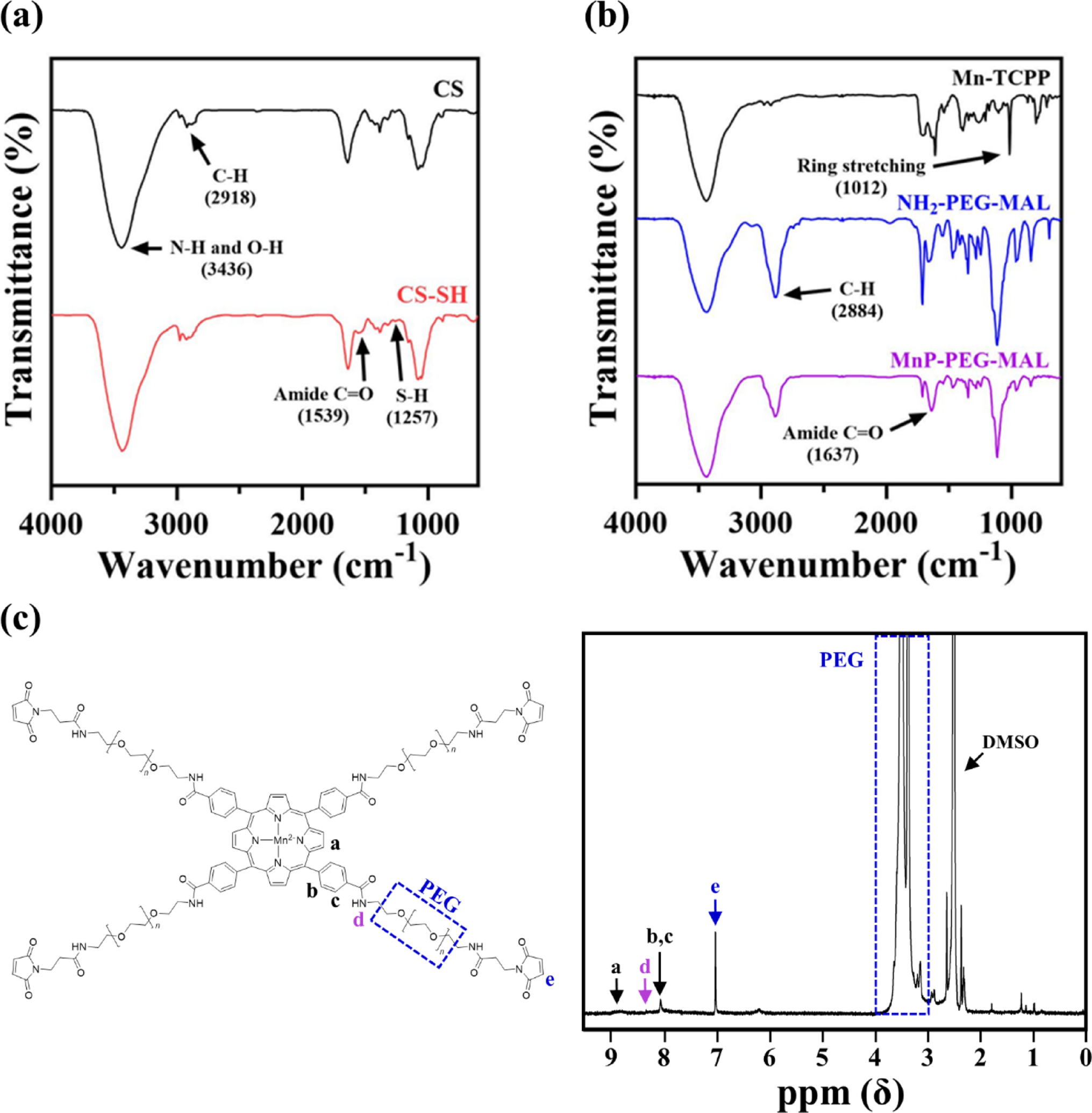
The CS-SH and MnP-PEG-MAL were synthesized as described in our previous study, with modifications [7]. Briefly, to synthesize thiolated chitosan, 100 mg of chitosan oligosaccharide (CS), 60 mg of 3,3-dithiodipropionic acid, 64.4 mg of NHS and 107.3 mg of EDAC·HCl were dissolved in 10 ml of distilled water. The reaction was conducted at 60 °C and pH 5 for 12 h. Then, 400 mg of DTT was added, and the mixture was stirred for 3 h under dark conditions. The resulting solution was purified by dialysis in 5 mM HCl using a 2 kDa MWCO membrane for 5 days, and subsequently freeze-dried to yield the final CS-SH. To synthesize MnP-PEG-MAL, 27 mg of Mn-TCPP, 16.2 mg of NH2-PEG-MAL, 324 mg of HBTU, and 125 μl of DIPEA were dissolved in 9.6 ml of anhydrous DMSO and stirred at room temperature for 1 h. The resulting solution was dialyzed using 7 kDa MWCO tubing, sequentially in DI water for 24 h, 90% methanol for 3 h, and again in DI water for an additional 4 days. The purified solution was subsequently freeze-dried to obtain MnP-PEG-MAL in solid form.
The thiol content of the lyophilized CS-SH was quantified using Ellman’s assay according to standard protocols [12]. The Fourier transform infrared (FT-IR) spectra for both CS and CS-SH were obtained at room temperature over the range of 4000 − 600 cm−1 with a spectral resolution of 8 cm−1 (Tensor27, Bruker, Billerica, MA, USA). The FT-IR spectra of Mn-TCPP, NH2-PEG-MAL, and MnP-PEG-MAL were also obtained under the same conditions. Nuclear magnetic resonance (1H NMR) spectroscopy was performed at room temperature using DMSO-d6 as the solvent to characterize the chemical structure of MnP-PEG-MAL (Avance III HD 400, Bruker, Germany).
2.3
Hydrogel preparation
The Ch_MnP hydrogel was prepared via in situ gelation by mixing the CS-SH solution with the solution containing maleimide-functionalized compounds [7]. Each solution was separately prepared in phosphate buffered saline (PBS; pH 7.4) at equimolar concentrations of thiol and maleimide groups (16.8 mM). The pH of the CS-SH solution was adjusted to 6.5. In the maleimide solution, the molar ratio of MnP-PEG-MAL and 4-arm PEG-MAL was set to 75:25 and 0:100 to produce the hydrogel with and without MnP (Ch_MnP(+) and Ch_MnP(-), respectively). Considering injection into the bulla inside the ear, each solution was loaded into a dual syringe and then injected through a 5 cm mini-volume line (1 mm inner diameter), where it formed a gel upon exiting a 23-gauge needle. To maintain sufficient fluidity under these conditions, a 12 wt% CS-SH solution combined with 3 v/v% 1 N NaOH was employed.
2.4 Hydrogel characterization
The cross-sectional morphology of the hydrogels was analyzed by field-emission scanning electron microscopy (FE-SEM; SGMA, Carl Zeiss, Cambridge, UK). The hydrogel was lyophilized and sliced to expose its cross-section [13]. The swelling ratio of the hydrogel was assessed using a standard method [14]. A 60 μl of the hydrogel was prepared as described above, and then fully submerged in PBS (pH 7.4) at 37 °C for 48 h. The swelling ratio was calculated using the following equation:
$$ }\,}\,\left( \% \right) \, = \, \left( }_}} - }_}} } \right)/}_}} \times 00 $$
where Ws represents the weight of the swollen hydrogel, and Wi represents its initial weight immediately after in situ gelation. The experiment was conducted in triplicate for each hydrogel type.
To evaluate the ROS scavenging efficacy, superoxide anion (O2·) scavenging capability of the hydrogels was assessed using a superoxide dismutase (SOD) assay kit (Dojindo Molecular Technologies, Tokyo, Japan), following the manufacturer's protocol [15]. Briefly, 20 µl of the hydrogel was added to a working solution containing xanthine, xanthine oxidase, and Water-Soluble Tetrazolium (WST)-1, and then incubated at 37 °C for 30 min. The absorbance of WST formazan was measured at 450 nm using a microplate reader, and the SOD activity was calculated as previously described [16]. The ability to generate oxygen was assessed using a dissolved oxygen meter (OxyLiteTM PRO; Oxford Optronix, Adderbury, UK). The prepared hydrogel was immersed in 4 ml of 0.3 and 1 mM H2O2 solution and incubated at 37 °C for 1 h, after which the dissolved oxygen concentration was measured. The experiment was conducted in triplicate for each hydrogel type.
2.5 Animals
All protocols of animal experiments were approved by the Institutional Animal Care and Use Committee of Seoul National University Hospital Biomedical Research Institute (IACUC no. 24–0050-S1SA1). Animals were maintained in the facility accredited AAALAC International (#001169) in accordance with Guide for the Care and Use of Laboratory Animals 8th edition, NRC (2010).
Twenty male Sprague Dawley Rats, 6 weeks old and weighing 130–190 g, were used for in vivo experiments (a total of 40 ears). The animal was housed in a semi-pathogen-free facility with controlled temperature, humidity, and a light/dark cycle (12 h/12 h). Prior to the experiments, chronic TM perforation was induced [17]. For this, the animal was anesthetized with an intramuscular injection of Zoletil (1.2 ml/kg; Zoletil 50, Virbac, Carros, France) and xylazine (5 mg/kg). After examining the ear under a microscope, a TM perforation was created using a pick involving the inferior quadrant of the TM. Then, mitomycin C-soaked gelatin foam was applied to the perforated wound via the external ear canal for 30 min, with additional applications on days 2 and 4 after TM perforation. Ears with TM perforations that persisted for 5 weeks were selected for further studies using the Ch_MnP hydrogel (Online Resource 1 and 2).
2.6 In vivo hydrogel application
Ears with chronic TM perforation were randomly assigned to three different treatment groups: (1) Control group: ears that received no treatment (n = 20); (2) Ch_MnP(−) group: ears treated with Ch_MnP(−) hydrogel (n = 13); and (3) Ch_MnP(+) group (n = 7): ears treated with Ch_MnP(+) hydrogel. For the groups treated with the hydrogel, we injected 0.03 mL of CS-SH solution and 0.03 mL of the maleimide solution using a dual syringe through a 5 cm mini volume line and a 23-gauge needle via the transtympanic route to reach the bulla, where the hydrogel formed in situ as the mixed solutions exited the needle. The hydrogel administration was performed three times on days 0, 3 and at week 1 for each ear in both the Ch_MnP(−) and Ch_MnP(+) groups.
2.7 Endoscopic TM evaluation
An endoscope (GD-060, Chammed, Gunpo, South Korea), 2.7 mm in diameter, was paired with a smartphone (iPhone 4, Apple Inc., Cupertino, CA, United States) to capture images of the rats’ external auditory canal and TM. Observations were made for signs of inflammation, swelling, congestion, perforation, or other adverse effects. The surface integrity, perforation healing, and transparency of the TM were also evaluated. Perforation size was assessed using Image J (National Institutes of Health, Bethesda, MD, USA). TM endoscopic images were obtained immediately before the hydrogel application (Before hydrogel) and at week 3 after the first hydrogel application.
2.8 Computed tomography (CT) analysis
A micro-CT system (NFR PolarisG90; Nanofocusray, Jeonju, South Korea) was used to assess the remaining hydrogel in the bulla. Images were assessed at the base of the bulla and between the ossicles, where the hydrogel was typically detected. Micro-CT images were processed and evaluated using a DICOM Viewer (Radiant DICOM Viewer Version 2024.1, Poznan, Poland). 2D images were stacked to form a 3D volume. The volume of the remaining hydrogel was calculated by integrating the planimetric measurements of 2D images (mm2) and reformatting them into a 3D volume (mm3). The micro-CT images were acquired immediately before the hydrogel application (Before hydrogel), immediately after the first hydrogel application (week 0) and week 1 and 3 after the first hydrogel application.
2.9 Auditory brainstem response (ABR) evaluation
The Smart EP system (Intelligent Hearing Systems, Miami, FL, United States) was utilized to measure the ABR threshold at click and 16 kHz frequencies within a soundproof chamber [18, 19]. Before measurements, rats were anesthetized, and subdermal needle electrodes were placed at the vertex (active electrode) and behind both the ipsilateral (reference electrode) and contralateral ears (ground electrode). The speaker was positioned in line with the external auditory canal, and the earphone tube was gently inserted into the ear canal. Hearing thresholds were assessed using click stimuli and 16 kHz pure tone stimuli. The hearing threshold was measured immediately before the hydrogel application (Before hydrogel) and at week 3 after the first hydrogel application, which was compared with the baseline threshold measured from the intact TM before perforation.
2.10 Histology and measurement of the bulla mucosa
All animals were sacrificed at the end point of experiments (week 3) for histological analysis of the bulla mucosa, and external auditory canal (EAC) skin. Bilateral temporal bones were harvested, embedded in paraffin wax, and cut into 5-μm-thick sections. Sections were stained with hematoxylin and eosin. The bulla mucosa and EAC skin were observed with an optical microscope (CX31, Olympus, Tokyo, Japan), and the images were obtained to measure the thickness of bulla mucosa and EAC skin with the DP2-BSW software (Olympus).
2.11 Statistics
Continuous variables were expressed as means ± standard deviations (SD) in the tables and figures. Statistical analyses were performed using SPSS software (version 26.0; SPSS Inc., IBM Corp., Armonk, NY, USA). The Mann–Whitney U test, crosstab and t test were used to compare the outcomes in each group. p values < 0.05 were considered to indicate statistical significance.

Comments (0)