McCutcheon RA, Reis Marques T, Howes OD. Schizophrenia—an overview. JAMA Psychiatry. 2020;77:201–10.
Article
PubMed
Google Scholar
Howes OD, Kambeitz J, Kim E, Stahl D, Slifstein M, Abi-Dargham A, et al. The nature of dopamine dysfunction in schizophrenia and what this means for treatment. Arch Gen Psychiatry. 2012;69:776–86.
Article
CAS
PubMed
PubMed Central
Google Scholar
Valdés-Tovar M, Rodríguez-Ramírez AM, Rodríguez-Cárdenas L, Sotelo-Ramírez CE, Camarena B, Sanabrais-Jiménez MA, et al. Insights into myelin dysfunction in schizophrenia and bipolar disorder. World J Psychiatry. 2022;12:264–85.
Article
PubMed
PubMed Central
Google Scholar
Zucca FA, Segura-Aguilar J, Ferrari E, Muñoz P, Paris I, Sulzer D, et al. Interactions of iron, dopamine and neuromelanin pathways in brain aging and Parkinson’s disease. Progress in Neurobiology. 2017;155:96–119.
Article
CAS
PubMed
Google Scholar
Saghazadeh A, Mahmoudi M, Shahrokhi S, Mojarrad M, Dastmardi M, Mirbeyk M, et al. Trace elements in schizophrenia: a systematic review and meta-analysis of 39 studies (N = 5151 participants). Nutrition Reviews. 2020;78:278–303.
Article
PubMed
Google Scholar
Georgieff MK. Iron deficiency in pregnancy. Am J Obstet Gynecol. 2020;223:516–24.
Article
CAS
PubMed
PubMed Central
Google Scholar
Stevens JR. Neuropathology of schizophrenia. Arch Gen Psychiatry. 1982;39:1131–9.
Article
CAS
PubMed
Google Scholar
Casanova MF, Waldman IN, Kleinman JE. A postmortem quantitative study of iron in the globus pallidus of schizophrenic patients. Biol Psychiatry. 1990;27:143–9.
Article
CAS
PubMed
Google Scholar
Kornhuber J, Lange KW, Kruzik P, Rausch WD, Gabriel E, Jellinger K, et al. Iron, copper, zinc, magnesium, and calcium in postmortem brain tissue from schizophrenic patients. Biol Psychiatry. 1994;36:31–34.
Article
CAS
PubMed
Google Scholar
Kral VA, Lehmann HE. Further studies on the iron content of the cerebrospinal fluid in psychoses. AMA Arch Neurol Psychiatry. 1952;68:321–8.
Article
CAS
PubMed
Google Scholar
Lehmann HE, Kral VA. Studies on the iron content of cerebrospinal fluid in different psychotic conditions. AMA Arch Neurol Psychiatry. 1951;65:326–36.
Article
CAS
PubMed
Google Scholar
Lotan A, Luza S, Opazo CM, Ayton S, Lane DJR, Mancuso S, et al. Perturbed iron biology in the prefrontal cortex of people with schizophrenia. Mol Psychiatry. 2023;28:2058–70.
Article
CAS
PubMed
PubMed Central
Google Scholar
Xu M, Guo Y, Cheng J, Xue K, Yang M, Song X, et al. Brain iron assessment in patients with First-episode schizophrenia using quantitative susceptibility mapping. Neuroimage Clin. 2021;31:102736.
Article
PubMed
PubMed Central
Google Scholar
García Saborit M, Jara A, Muñoz N, Milovic C, Tepper A, Alliende LM, et al. Quantitative susceptibility mapping MRI in deep-brain nuclei in first-episode psychosis. Schizophr Bull. 2023;49:1355–63.
Article
PubMed
PubMed Central
Google Scholar
Ravanfar P, Syeda WT, Jayaram M, Rushmore RJ, Moffat B, Lin AP, et al. In vivo 7-Tesla MRI investigation of brain iron and its metabolic correlates in chronic schizophrenia. Schizophr. 2022;8:1–11.
Article
Google Scholar
Sonnenschein SF, Parr AC, Larsen B, Calabro FJ, Foran W, Eack SM, et al. Subcortical brain iron deposition in individuals with schizophrenia. J Psychiatr Res. 2022;151:272–8.
Article
PubMed
Google Scholar
Sui YV, McKenna F, Bertisch H, Storey P, Anthopolos R, Goff DC, et al. Decreased basal ganglia and thalamic iron in early psychotic spectrum disorders are associated with increased psychotic and schizotypal symptoms. Mol Psychiatry. 2022;27:5144–53.
Article
CAS
PubMed
Google Scholar
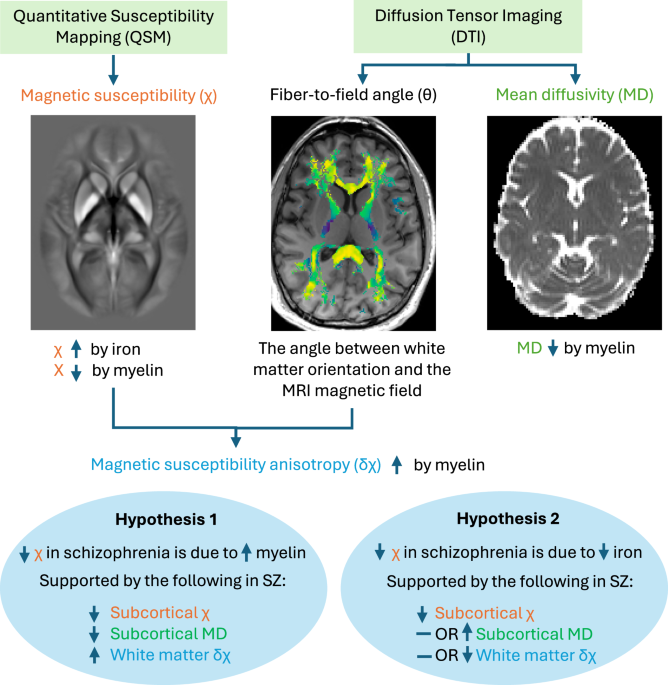
Duyn JH, Schenck J. Contributions to magnetic susceptibility of brain tissue: magnetic susceptibility of brain tissue. NMR Biomed. 2017;30:e3546.
Article
Google Scholar
Seehaus A, Roebroeck A, Bastiani M, Fonseca L, Bratzke H, Lori N, et al. Histological validation of high-resolution DTI in human post mortem tissue. Front Neuroanat. 2015;9:98.
Article
PubMed
PubMed Central
Google Scholar
Hallgren B, Sourander P. The effect of age on the non-haemin iron in the human brain. J Neurochem. 1958;3:41–51.
Article
CAS
PubMed
Google Scholar
Hametner S, Endmayr V, Deistung A, Palmrich P, Prihoda M, Haimburger E, et al. The influence of brain iron and myelin on magnetic susceptibility and effective transverse relaxation - a biochemical and histological validation study. NeuroImage. 2018;179:117–33.
Article
CAS
PubMed
Google Scholar
Lee J, Shmueli K, Fukunaga M, van Gelderen P, Merkle H, Silva AC, et al. Sensitivity of MRI resonance frequency to the orientation of brain tissue microstructure. Proc Natl Acad Sci USA. 2010;107:5130–5.
Article
CAS
PubMed
PubMed Central
Google Scholar
Wharton S, Bowtell R. Effects of white matter microstructure on phase and susceptibility maps. Magn Reson Med. 2015;73:1258–69.
Article
PubMed
Google Scholar
Li X, Vikram DS, Lim IAL, Jones CK, Farrell JAD, van Zijl PCM. Mapping magnetic susceptibility anisotropies of white matter in vivo in the human brain at 7 T. Neuroimage. 2012;62:314–30.
Article
PubMed
Google Scholar
Li W, Wu B, Avram AV, Liu C. Magnetic susceptibility anisotropy of human brain in vivo and its molecular underpinnings. NeuroImage. 2012;59:2088–97.
Article
PubMed
Google Scholar
Vano LJ, McCutcheon RA, Rutigliano G, Kaar SJ, Finelli V, Nordio G, et al. Mesostriatal dopaminergic circuit dysfunction in schizophrenia: a multimodal neuromelanin-sensitive magnetic resonance imaging and [18F]-DOPA positron emission tomography study. Biol Psychiatry. 2024;96:674–83.
Article
CAS
PubMed
Google Scholar
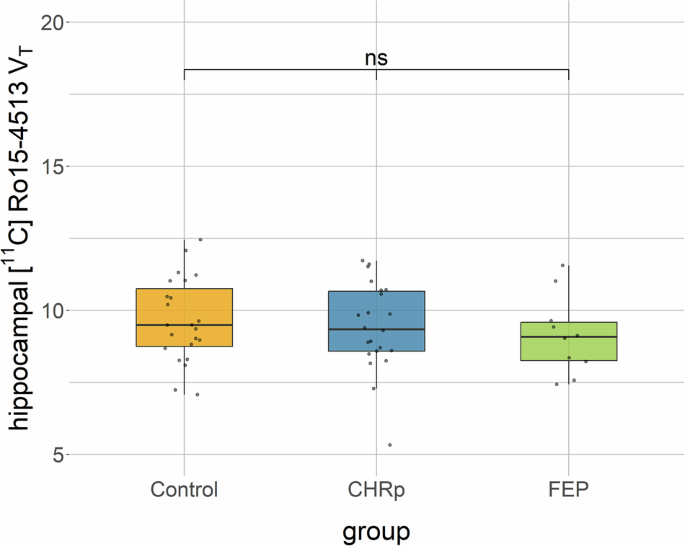
Vano LJ, McCutcheon RA, Sedlacik J, Kaar SJ, Rutigliano G, Nordio G, et al. Reduced brain iron and striatal hyperdopaminergia in schizophrenia: a quantitative susceptibility mapping MRI and PET study. Am J Psychiatry. 2025;182:830-9.
Hawrylycz MJ, Lein ES, Guillozet-Bongaarts AL, Shen EH, Ng L, Miller JA, et al. An anatomically comprehensive atlas of the adult human brain transcriptome. Nature. 2012;489:391–9.
Article
CAS
PubMed
PubMed Central
Google Scholar
Howes O, Marcinkowska J, Turkheimer FE, Carr R. Synaptic changes in psychiatric and neurological disorders: state-of-the art of in vivo imaging. Neuropsychopharmacol. 2024;50:164–83.
Article
Google Scholar
First MB. Structured clinical interview for DSM-5 disorders – clinician version (SCID-5-CV). Arlington, VA: American Psychiatric Association; 2016.
Google Scholar
Kim E, Howes OD, Veronese M, Beck K, Seo S, Park JW, et al. Presynaptic dopamine capacity in patients with treatment-resistant schizophrenia taking clozapine: an [18F]DOPA PET study. Neuropsychopharmacology. 2017;42:941–50.
Article
CAS
PubMed
Google Scholar
Smith-Kielland A, Skuterud B, Mørland J. Urinary excretion of 11-nor-9-carboxy-delta9-tetrahydrocannabinol and cannabinoids in frequent and infrequent drug users. J Anal Toxicol. 1999;23:323–32.
Article
CAS
PubMed
Google Scholar
Elbejjani M, Auer R, Jacobs DR, Haight T, Davatzikos C, Goff DC, et al. Cigarette smoking and gray matter brain volumes in middle age adults: the CARDIA brain MRI sub-study. Transl Psychiatry. 2019;9:78.
Article
PubMed
PubMed Central


Comments (0)