
Remember me
Sterilized seeds of Arabidopsis were sown on DKW #1 medium without PGRs and incubated, as described above, at long-day (16 h) illumination. Under these conditions, seeds were germinated and in 1 mo gave rise to flowering plants. Plants had an indeterminate, raceme-type inflorescence and usually classified as a long-day plant. DKW-based medium was mainly used, and MS-based medium gave similar results. The conversion of shoot apical meristem to inflorescence meristem has been described in detail previously (Bertero et al. 1996; Hake and Wilt 2003; Gaarslev et al. 2021). A flower can be considered as a modified shoot wherein the shoot apical meristem changes into a floral meristem (Taiz et al. 2015). The similar nature of apical meristem and floral buds, which are also highly meristematic, highlights one of the difficulties of working with floral cultures—specifically, transformation of highly organized meristematic tissue requires a prolonged selection period to obtain complete sorting out of non-transformed tissue from transformed tissue.
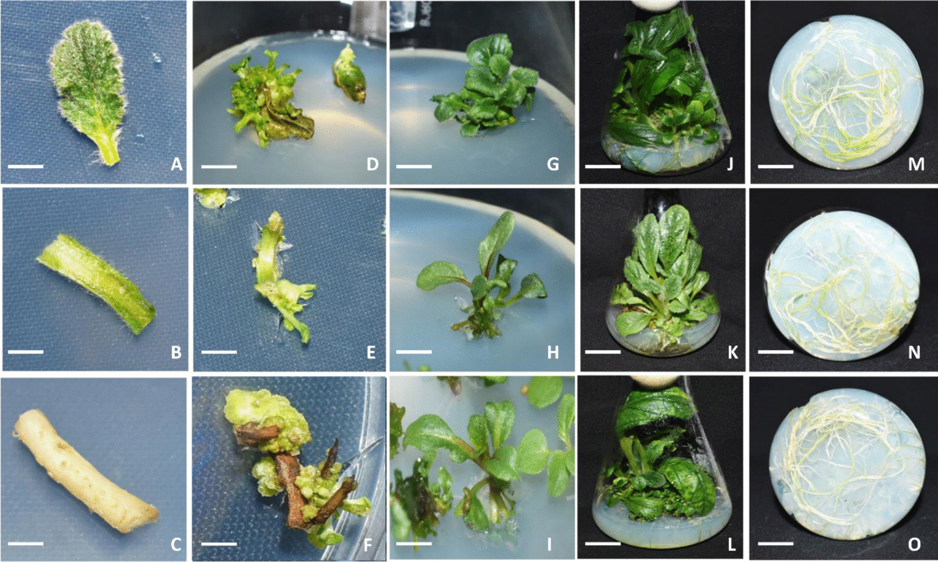
For induction and propagation of in vitro floral cultures, inflorescences from in vitro grown plants were chopped and placed on DKW #2. Floral cultures propagated in Petri dishes grew rapidly and were sub-cultured every 12 to 14 d (Fig. 1A). To obtain more compact inflorescences, GA3 was removed from the medium and DKW #2 medium with 2.0 mg L−1 ancymidol (Ancym), which is an inhibitor of GA3 biosynthesis, was used (Fig. 1B, C, D). Arabidopsis plants on such medium look stunted, compact, and phenotypically similar to Landsberg erecta ecotype (Rédei 1992). This medium was periodically used for maintenance of floral culture. Arabidopsis floral culture to some extent was different from floral culture of quinoa, described earlier (Sidorov et al. 2024). Floral cultures of Arabidopsis consisted entirely of inflorescences, while quinoa floral cultures mainly contained flowers at different stages of development.
Figure 1.
In vitro floral culture of Arabidopsis and its propagation on different media. (A) 8.5-cm Petri dish with floral culture on Driver and Kuniyuki Walnut (DKW) medium with 20.0 g L−1 sucrose, 1.0 mg L−1 Kin, 0.5 mg L−1 BA, 0.1 mg L−1 GA3, and 3.5 g L−1 agarose; pH 5.7. (B) DKW medium with 20.0 g L−1 sucrose, 1.0 mg L−1 Kin, 0.5 mg L−1 BA, and 3.5 g L−1 agarose; pH 5.7. (C, D) DKW medium with 20.0 g L−1 sucrose, 1 mg L−1 Kin, 0.5 mg L−1 BA, 2.0 mg L−1 ancymidol, and 3.5 g L−1agarose; pH 5.7. Bar = 4 mm.
On the medium for propagation, it was possible to identify abnormal development of pistils. Distinct stages of abnormal pistil development are presented in Fig. 2A, B, C. Media used for propagation could also induce shoot regeneration from different parts of the inflorescence (Fig. 2D, E). The most interesting observation is that media DKW #2 and DKW #2 without GA3 also induced multiple pistil formation (Fig. 3A, B, C). In vitro pistil culture is a unique phenomenon not described before, although abnormal development of floral organs has been demonstrated in vivo in different mutants (Okamuro et al. 1993; Ung et al. 2011). Multiple pistil cultures on DKW #2 medium or on the same medium without GA3 formed normal inflorescences. Pistil cultures were used for Agrobacterium-mediated transformation. GUS expression in stably transformed pistil cultures is shown in Fig. 3D. Since this culture is differentiated and maintained on medium without 2,4-D, relatively little if any somaclonal variation is anticipated.
Figure 2.
In vitro pistil development and regeneration from various parts of Arabidopsis inflorescence. (A, B, C) Abnormal growth of pistils on DKW medium supplemented with 1.0 mg L−1 Kin, 0.5 mg L−1 BA, and 0.1 mg L−1 GA3. Bar = 2.5 mm. (D, E) Regeneration of shoots. Bar = 6 mm. (F) Diagram of Arabidopsis flower showing pistil.
Figure 3.
Establishment, propagation, and transformation of Arabidopsis in vitro pistil culture with NPTII/GUS construct. (A, B, C) Establishment of in vitro pistil culture. (A) Bar = 2.5 mm. (B) Bar = 4 mm. (C) Propagation of pistil culture on DKW medium supplemented with 1.0 mg L−1 Kin, 0.5 mg L−1 BA, and 0.1 mg L−1 GA3; 8.5-cm plate. (D) Stable GUS expression in transformed pistil culture after 25 d of selection. Bar = 3 mm.
Transformation of Floral Cultures and Precultured SeedlingsIn each treatment, approximately 100 floral cultured clumps were inoculated with Agrobacterium (AB32 carrying pVS1 binary plasmid with nptII and uidA markers). The essential steps of transformation were the vacuum infiltration of explants in Agrobacterium-solution and centrifugation. For selection of transformants, we used the same medium as for propagation, DKW #2 medium supplemented with 50.0 mg L−1 Par. Selection was applied without delay and was stringent enough to cause bleaching and prevent growth of non-transformed tissue. Transient and stable GUS expression shows that the target for transformation was flowers and different parts of the inflorescence (Fig. 4A, B); however, we were mainly interested in isolating transformants from meristematic tissue of inflorescences. During each sub-culture, only the green viable tissue was transferred to a new selection medium. Approximately 45 d after transformation, clumps of what appeared as non-chimeric tissue were clearly visible on the selection medium (Fig. 4C); however, after staining with X-Gluc, several of these clumps were still chimeric. Regeneration of chimeric plants is always a concern in meristem explant-based transformation systems (Ye et al. 2022, 2023). To address these concerns, extended selection time (up to 3 mo) was used. After the extended selection period, 24 lines resistant to paromomycin were isolated. TAQMAN PCR analysis showed that out of 24 selected lines, 20 lines were nptII positive and 4 were negative, 12 lines had 1 copy of nptII, and 7 lines had 2 copies of nptII (Table 1). All nptII positive lines were also stained with X-Gluc and were identified as GUS positive (Fig. 4D, E).
Figure 4.
Transformation and selection of Par-resistant inflorescence shoots of Arabidopsis. Transient and stable GUS expression in plant material during selection. (A) 1-d culture after transformation. Bar = 4.2 mm. (B) 12-d culture after transformation. Bar = 3 mm. (C) 1-mo selection on medium with 50.0 mg L−1 Par. (D, E) 2-mo selection; transformed and non-transformed, bleached shoots. (D) Bar = 10 mm. (E) Bar = 3.4 mm.
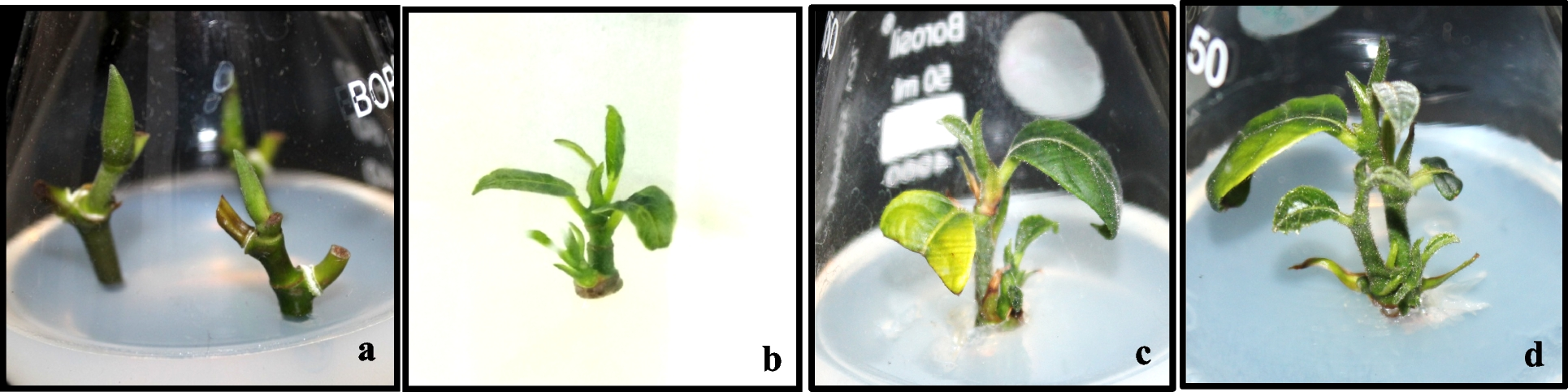
Table 1 TAQMAN PCR analysis of Arabidopsis events obtained after transformation with constructs containing nptII/GUS and aadA/Ruby as selectable and visual markersIn the current work with germinated seed transformation, seeds after sterilization were precultured in liquid medium (Fig. 5A); seedlings were blended and then transformed. The target for transformation was the shoot apical meristem, however, small pieces of seedlings were also transformed and initiated pink callus (Fig. 5B). On the medium used for selection (DKW #2, 25.0 mg L−1 Spec, 500.0 mg L−1 Carb, 200.0 mg L−1 Cef), very fast shoot germination from meristems was observed. In preliminary experiments, it is also possible to use PGR-free medium; however, the number of germinated shoots was smaller. In an experiment with approximately 150 seeds on DKW #2 selection medium, 16 pink plants, 2 green plants with pink flowers, and 6 green plants (Fig. 5D, E) were obtained. Eighteen plants expressing Ruby were analyzed by TAQMAN PCR (Table 1). All of them except one, analysis of which was failed, were aadA positive, and 80% of events had single aadA copy.
Figure 5.
Application of the RUBY visual marker in Arabidopsis transformation. (A) Germination (1 wk) Arabidopsis seeds in vented flask with liquid DKW medium, supplemented with 1.0 mg L−1 Kin, 0.5 mg L−1 BA, and 0.1 mg L−1 GA3. Single seedling with shoot apical meristem after 7-d culture presented in insertion. Bar = 4 mm. (B, C) 19-d culture after transformation of blended seedlings and selection on DKW medium, 1.0 mg L−1 Kin, 0.5 mg L−1 BA, and 0.1 mg L−1 GA3 and supplemented with 25.0 mg L−1 Spec. (C) Germination of Ruby shoots. Bar = 2.5 mm. (D, E) Selected flowering plants on 35 d after transformation. Bar = 10 mm.
Time frame for production of transgenics with the described method can be considerably short in comparison to the floral dip method. Especially Ruby expression allowed to identify the transformant very early. The described methods based on using floral culture and shoot apical meristem also have some important benefits over the widely used floral dip method. In floral dip, transgenic T1 progeny are hemizygous, and must be grown to the T2 generation to produce homozygous progeny. With currently recommended methods, some homozygous progeny are produced immediately in the T1 generation.
Production of Seeds and Analysis of T1 ProgenySeeds from transgenic inflorescences were produced in vitro on DKW #1 medium supplemented with 2.0 mg L−1 IBA. On this medium, the inflorescences produced normal flowers, which were spontaneously self-pollinated and produced siliques full of seeds (Fig. 6A). Culture conditions for seed production were the same as for growing plants (25°C, 80 μE m−2 s−1 light intensity, 16-h day length).
Figure 6.
In vitro seed production and analysis of different transgenic lines of Arabidopsis. (A) Induction and maturation seeds in vitro on DKW medium supplemented with 2.0 mg L−1 IBA. (B, C) Seed germination of lines #4 and #12 on DKW PGR-free medium with 50.0 mg L−1 Par; arrows indicate Par-sensitive seedlings. Bar = 17 mm. (D) GUS expression in seedlings of line #12.
Interestingly, seeds could not be obtained on DKW #1 (PGR-free) medium. Seeds of Arabidopsis produced in vitro were germinated on screening medium. No refrigeration was required for seed germination (there appeared to be no dormancy period). Seeds produced in vitro were placed on DKW #1 medium with 50.0 mg L−1 Par for germination and analyzed for segregation in progeny (Fig. 6B, C).
Analysis of T1 progeny from two initial proof-of-concept (POC) events (#4 and #12), which had single copy for nptII confirmed normal Mendelian inheritance of transgenes in progeny. Green seedlings germinated on PGR-free medium with 50.0 mg L−1 Par were classified as resistant (NPTII+), while yellow, smaller seedlings were designated as Par-sensitive (NPTII−). As shown in Fig. 6D, most seedlings were GUS positive. In event #4, out of 140 seeds tested, 105 seedlings were green (NPT+), 33 were yellow (NPTII−), and two failed to germinate. Similar results were obtained with event #12. Segregation of transgenes in the T1 progeny for events 4 and 12 was statistically analyzed using a chi-square test, which confirmed a statistical fit to a 3:1 segregation ratio as expected for self-pollinated progeny of a single copy T-DNA insertion. Production of seeds from RUBY transgenics was more difficult. On DKW #1 medium with 2 mg L−1 IBA, pink inflorescences were weak and produced siliques without normal seeds. Medium used for seed production also usually induced multiple roots. So, plants could be transplanted to soil for production of seeds in vivo. Unfortunately, it was not checked. Earlier, He et al. (2020) also could not show seed set in pink Arabidopsis plants, expressing Ruby in leaves and stem, growing in soil but they demonstrated production of clearly pink seeds in green normal-looking Ruby positive plants transformed with seed-specific promoter At2S3.
Protoplast Isolation and CultureDifferent Arabidopsis explants have been used for protoplast isolation, culture, and plant regeneration (see refences in “Introduction”). In this work, the possibility to isolate and culture protoplasts from established in vitro floral cultures was analyzed. Floral cultures can be easily induced and maintained in vitro. In pilot experiments, protoplasts from floral cultures divided on several media have been previously described, but the best results were obtained on Kao and Michyluk (KM) modified medium (see Sidorov et al. 2022), containing 0.2 mg L−1 NAA, 0.2 mg L−1 BA, and 1.0 mg L−1 2,4-D. Isolation of protoplasts from floral cultures was very quick and efficient, and therefore has potential as a high-throughput system for DNA-free gene editing of Arabidopsis.
Comments (0)