Blue Nevus of The Palate: A Case Image
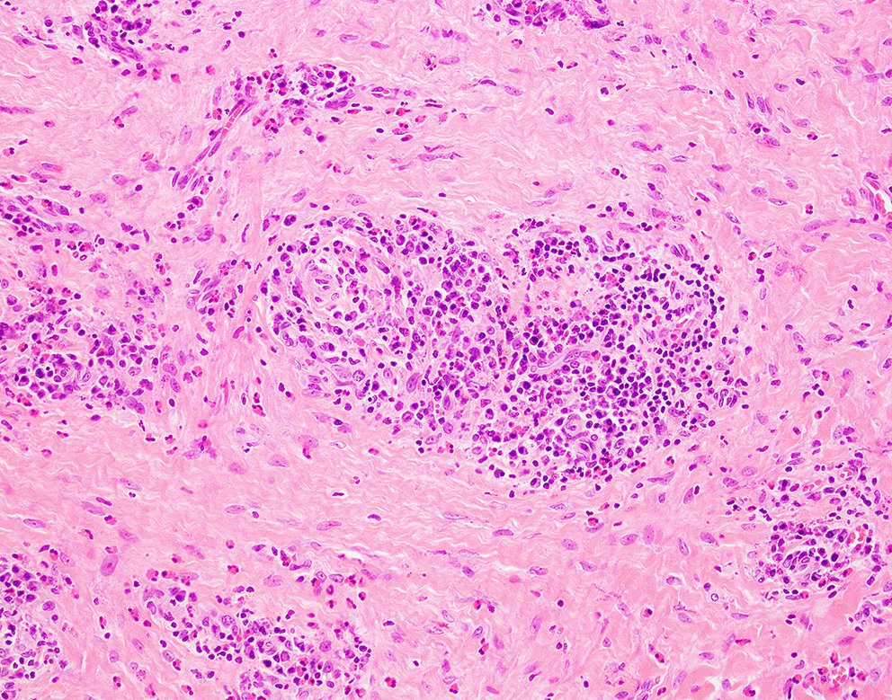
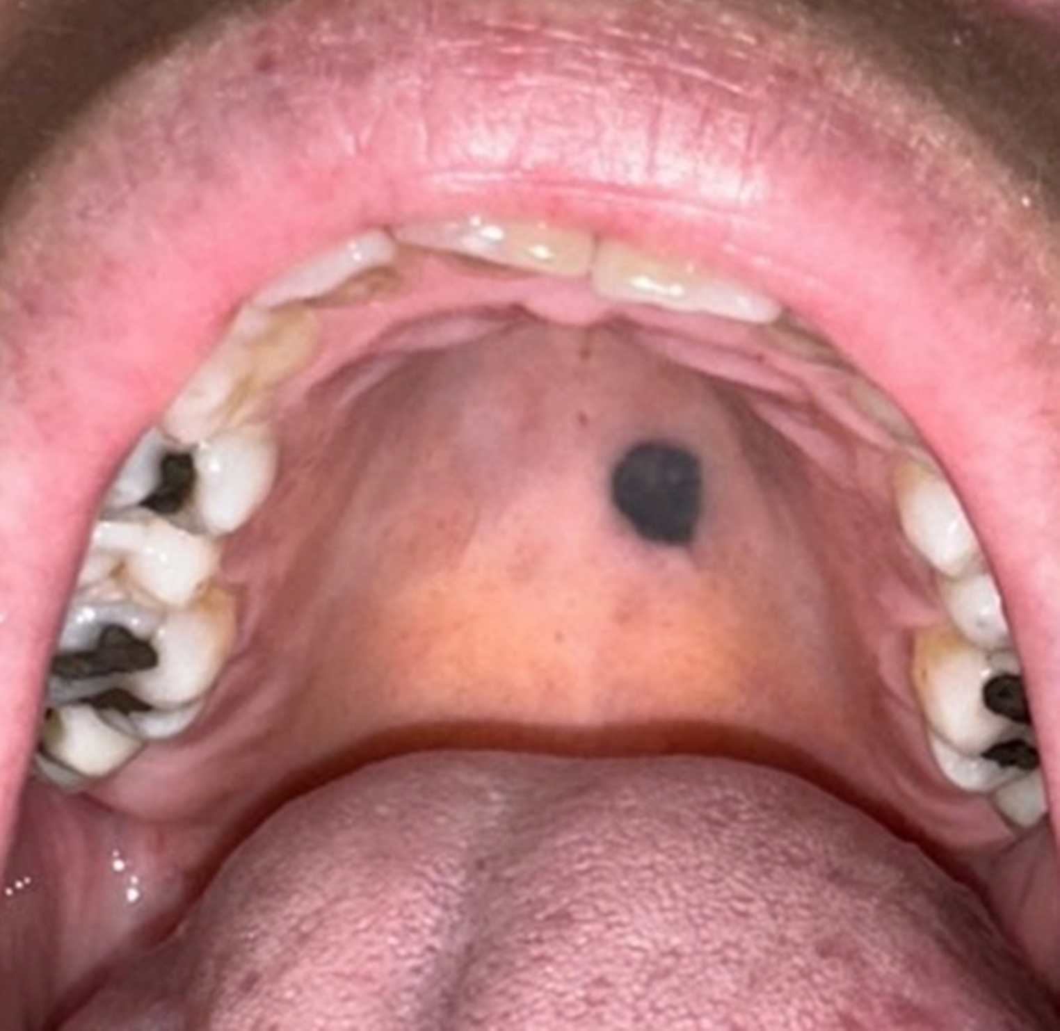
A 54-year-old female patient was referred to the oral surgery department of a public university with a complaint of a dark spot on the palate that had been present for eight months. Clinical examination revealed a well-circumscribed, darkly pigmented macule measuring approximately 1.5 cm × 1 cm located in the posterior hard palate. The lesion’s location and size raised suspicion of malignancy. An incisional biopsy was performed, and histopathological analysis showed the presence of spindle cells containing light brown pigmented granules compatible with melanin. The final diagnosis was of blue nevus. The lesion was surgically excised, with satisfactory healing, and a follow-up period of 5 months. Blue nevus is a melanocytic lesion that typically presents as a small pigmented spot and is rare in the oral cavity. This case is notable due to the lesion’s size and location and overlapping features commonly associated with malignancy, posing a diagnostic challenge, particularly for pathologists who infrequently review melanocytic lesions. This highlights the critical role of histopathological analysis in establishing a definitive diagnosis. Additionally, careful clinical follow-up is essential to monitor healing and detect any potential recurrence.


Comments (0)