
Remember me

A 63-year-old male patient presented with a one-year history of a mass located inferior to the right ear. Ultrasonography of parotid glands revealed lobulated solid lesions with cystic degenerative changes, measuring 3.4 × 3 cm on the right and 4.5 × 2.1 cm on the left. Fine-needle aspiration biopsies (FNAB) of both parotid masses were consistent with WT. The patient subsequently underwent right superficial parotidectomy.
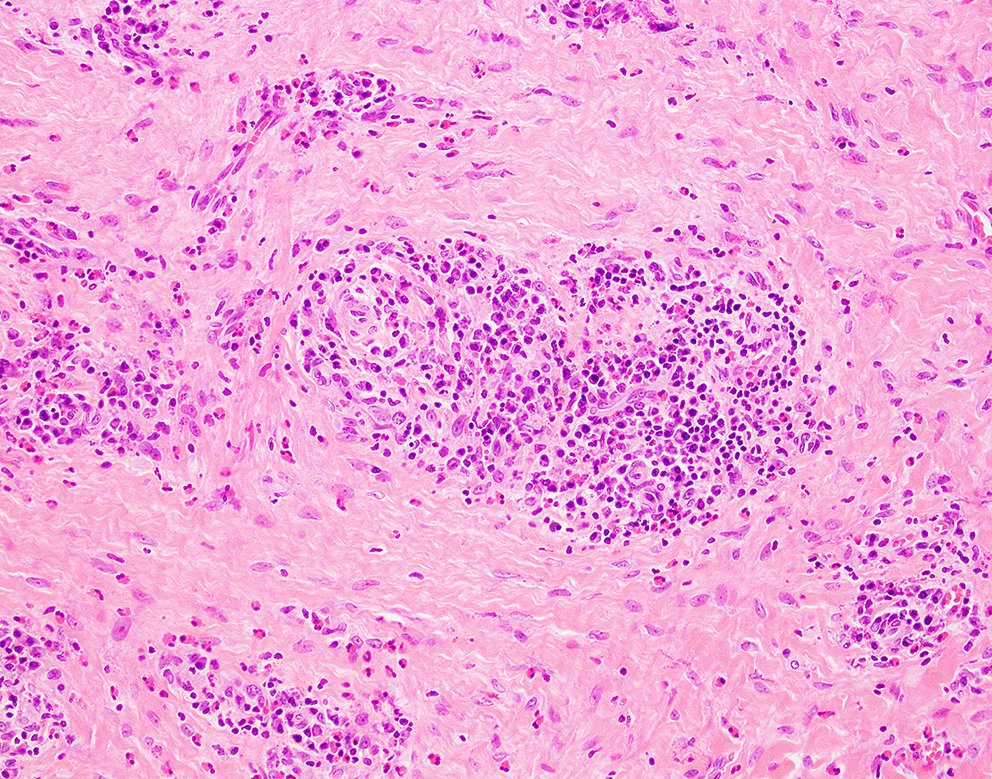
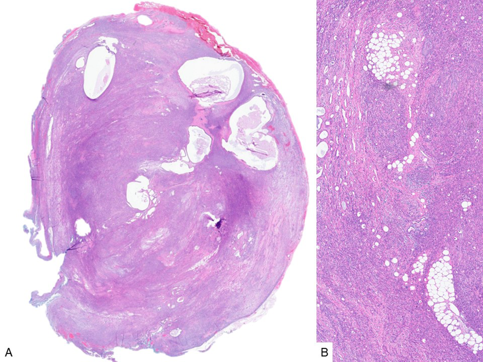
On macroscopic examination, the parotid gland measured 8.5 × 6 × 4 cm and contained a nodular, multilobulated, brown-colored mass measuring 4 × 3 × 2 cm, exhibiting hemorrhagic and cystic areas. Histopathological evaluation confirmed the diagnosis of WT, characterized by cystic spaces lined with bilayered oncocytic epithelium and accompanying lymphoid stroma (Fig. 1A). Notably, a 0.4 cm focus of mature adipocyte proliferation, consistent with lipomatous metaplasia, was identified within the tumor (Fig. 1B-E). The possibility of entrapped adipose tissue was considered; however, this was ruled out because the adipose focus was located within the tumor rather than at its periphery. Additionally, the adipose focus was surrounded by bilayered oncocytic epithelium and associated with lymphoid stroma, supporting its intratumoral origin.
Fig. 1
Histologic features of Warthin tumor with lipomatous metaplasia. Warthin tumor shows cystic spaces lined by bilayered oncocytic epithelium and prominent lymphoid stroma (A) (H&E, ×100). Foci of mature adipocyte proliferation within the tumor stroma, consistent with lipomatous metaplasia (B–E) (H&E, ×40, ×200, ×200, ×400, respectively)
Comments (0)