
Remember me

Among the 430 AAV patients, 61 patients (14%) (41 GPA, 11 MPA, 9 EGPA; 34 men (56%); mean (SD) age: 51.6 (15.4) years) had neurologic signs or symptoms at some time during the disease course. Neurologic signs or symptoms were present at disease onset in 25 (41%) patients. The remaining 36 (59%) patients developed neurologic signs or symptoms within a mean (SD) follow up of 33.4 (40.4) months after AAV diagnosis. The most common neurologic signs or symptoms attributable to CNS involvement were headache (n = 20), muscle weakness (n = 17), numbness (n = 17), and visual impairment (n = 16). At the time of the occurrence of neurologic symptoms, all patients had active disease, with a median BVAS score of 11 (IQR: 7–15)]. Additionally, 21 patients (34%) had accompanying PNS involvement.
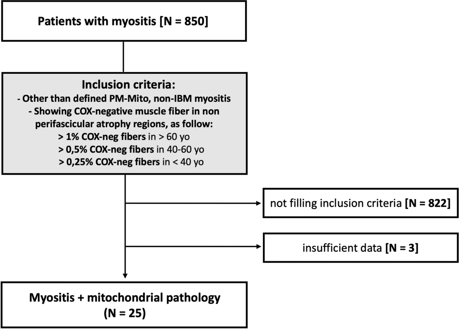
A final diagnosis of CNS involvement of AAV was established in 7 (11.4%) patients. The neurologic symptoms were associated with AAV manifestations other than CNS involvement in 30 (49.1%) patients. The causes of neurologic symptoms were drug-related adverse events or comorbidities affecting the CNS in 15 patients (24.5%). The neurologic work-up did not reveal an underlying condition in 9 patients (15%). Causes for neurologic signs or symptoms and demographic data are presented in Fig. 1 and Table 1.
Fig. 1
Causes for neurologic sign or symptoms suggesting CNS involvement among 61 AAV patients. a Sinonasal involvement in 4 patients, nasal involvement in 2 patients, nasopharyngeal mass and PNS involvement in 1 patient. b Orbital involvement in 1 patient, ocular involvement in 1 patient, retinal vasculitis in 1 patient, orbital and paranasal involvement, secondary facial paralysis and PNS in 2 patients. c Facial paralysis due to facial nerve involvement, orbital involvement and parotid gland involvement in 1 patient each. d Cardioembolic CVA due to cardiac mass and cardiac thrombus in 1 patient each. e Vertigo and nausea associated with CYC in 1 patient, subretinal fluid associated with glucocorticoid in 1 patient
Table 1 Characteristics of the 61 AAV patients and causes for neurologic signs or symptomsAAV patients with CNS involvementFinal diagnosis was CNS involvement of AAV in 7 (11.4%) patients (5 GPA, 1 MPA, 1 EGPA; 5 men). The frequency of CNS involvement for each type of AAV was 5/41 (12.1%) for GPA, 1/11 (9%) for MPA and 1/9 (11.1%) for EGPA. Mean (SD) age at the time of CNS involvement was 39.5 (12.1) years. The mean time from AAV diagnosis to the development of CNS involvement was 10 (range 0–23) months. Three patients presented with headache, muscle weakness, seizures, and numbness, two patients presented with blurred vision, one patient with hearing loss, and one with dizziness. Three patients also had PNS involvement. In two patients, CNS involvement was one of the initial manifestations of AAV. In the remaining five patients, CNS involvement occurred while they were on immunosuppressive therapy. One patient was on AZA, one patient on both RTX and CYC, one patient on both CYC and low-dose GC, one patient on CYC, and one patient on both AZA and low-dose GC.
Neurologic events due to CNS involvement were meningeal involvement in three patients. One of these patients had ischemic cerebrovascular accident (CVA) and another had both ischemic CVA and syringomyelia in addition to meningeal involvement. The neurologic events were vascular in two other patients, presenting with ischemic CVA and ischemic CVA together with hemorrhagic transformation. The remaining two patients had intracranial hypertension and cerebral venous sinus thrombosis in one patient each.
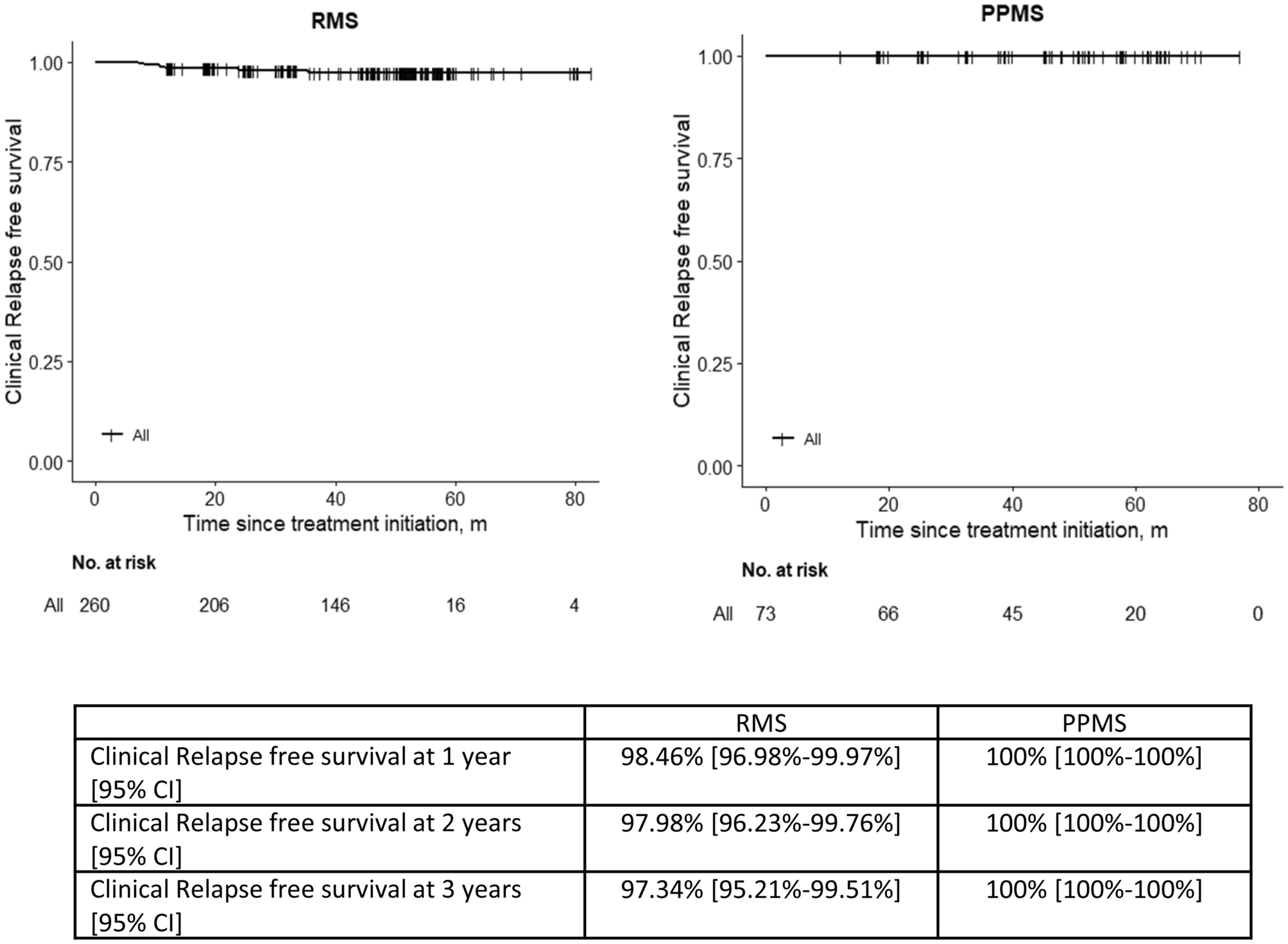
Cranial magnetic resonance imaging of these patients revealed meningeal infiltration and cortical laminar necrosis in the right parietal lobe, effacement and slight expansion in the sulci in case 1 (Fig. 2A); dural contrast enhancement in case 2; acute diffusion restriction consistent with ischemia at the cervicomedullary junction and increased T2A/FLAIR signal in this area in case 3 (Fig. 2B); cerebral ischemic lesions in white matter and left internal capsule and hemorrhagic transformation in case 4 (Fig. 2C); diffuse thrombosis in the venous sinuses in case 5 (Fig. 2D); severe stenosis of the cerebral sinuses secondary to intracranial hypertension and increased cerebrospinal fluid distance in the optic nerve sheath in case 6 (Fig. 2E); and dural thickening and hyperintense lesions in the spinal cord on spinal MRI during the initial neurological involvement in case 7 who had a relapse 6 months later, with signal changes in the cortical gray matter and white matter in the left frontotemporal region, along with pial and dural contrast enhancement at this level, granulomatous meningeal involvement-like appearance, and chronic lacunar infarcts in the inferior part of the cerebellar hemisphere. In his second relapse, which occurred 74 months later, there were increased signals in the subcortical white matter in both parietal lobes, end artery ischemia, sequelae in the vascular territory of the PICA in the left cerebellar hemisphere, and an infarct in the left frontoparietal internal carotid artery territory area. In the third relapse, 19 months later, there was dural contrast enhancement and a widespread syringomyelia cavity in the cervical and dorsal spinal cord on MRI (Fig. 2F)).
Fig. 2
Radiological images of AAV patients with CNS involvement. A Axial Flair Image demonstrates right hemisphere meningeal involvement (arrow). B DWI demonstrates hyperintense area due to ischemia in brainstem (arrow). C SWI demonstrates hemorrhagic transformation in left internal capsule (arrow). D Right internal carotid artery selective DSA examination venous phase image shows occlusion and partial recanalization of the superior sagittal sinus (white arrow) as well as right transvers sinus (black arrow). E Axial T2W image demonstrates optic nerve sheath tortuosity due to intracranial hypertension (arrow). F Axial T2 weighted image shows syringomyelia in the right half of the spinal cord (white arrow) and associated increased signal in the dorsal aspect and left half of the spinal cord (black arrow) related with former myelitis. AAV ANCA associated vasculitis, CNS Central nervous system, DSA Digital subtraction angiography, DWI Diffusion weighted imaging, MRV Magnetic Resonance Venography, SWI Susceptibility weighted imaging, T2W T2-weighted imaging
Cerebrospinal fluid (CSF) analysis was available in 2 of these patients. In Case 6, the CSF revealed elevated intracranial pressure (66 cm H2O), low glucose levels (56 mg/dL), and the presence of type 2 oligoclonal bands. In Case 5, the only abnormal finding in the CSF was a mildly elevated protein level (48 mg/dL).
Among the 7 patients, 5 patients were treated with CYC and high dose GC for induction treatment. One patient who was already on CYC treatment at the occurrence of CNS involvement, received only IVIG treatment due to suspicion of meningitis, and 1 patient with cerebral venous sinus thrombosis was treated with RTX and high dose GC for induction. Maintenance treatment of the 6 patients who did not experience a relapse included both RTX and MMF in 2 patients, RTX in 2, both RTX and AZA in 1 patient, and mepolizumab in 1 patient. The last patient (case 7) was treated with alternating oral and intravenous CYC for 23 months due to refractory AAV manifestations other than CNS involvement. The first neurologic involvement presented as hypertrophic pachymeningitis 23 months after diagnosis, and high-dose GC and CYC treatment was administered. After 6 cycles of monthly intravenous CYC, he experienced a relapse with meningeal involvement and a lacunar infarct in the cerebellar hemisphere. He received high dose GC and CYC dose was increased by 50%. Following 74 months of alternating oral and intravenous CYC treatment, he had a relapse with a frontoparietal infarct. CYC was stopped, and RTX, AZA, and high dose GC treatment were initiated. 19 months later, he had another relapse with meningeal involvement, and secondary syringomyelia was detected. IVIG and high dose GC were started. He was relapse-free with RTX, IVIG, AZA, and low dose GC for 3 years. Then, he was lost to follow-up and died of an unknown cause, 16 years after CNS involvement.
Overall, 6 patients were still alive for a mean (SD) duration of 38 (24.1) months after AAV diagnosis and for a mean (SD) duration of 27.8 (26.9) months after CNS involvement. The last patient (Case 7) who had died was the only patient who experienced relapses of CNS involvement. The other patients experienced only one episode of CNS involvement, but except for one patient, all of them recovered with sequela including epilepsy, blurred vision, muscle weakness, numbness, or hearing loss, each affecting one patient. The demographics, AAV manifestations, neurologic symptoms, management, and outcome of these 7 patients are presented in Table 2.
Table 2 Demographic and clinical characteristics of AAV patients with CNS involvementAAV manifestations mimicking CNS involvementAmong the 30 (49.1%) patients who presented with neurologic symptoms suggesting CNS involvement, neurologic work-up revealed that their symptoms were due to AAV manifestations other than CNS involvement. Table 3 shows the demographic features, type of AAV, ANCA status, symptoms or signs that led to a CNS work-up, AAV manifestations other than CNS involvement that contributed to these symptoms or signs, and the outcome. The most common AAV manifestation mimicking CNS involvement was PNS involvement and, the symptoms were numbness, muscle weakness, or headache. All of these 30 patients were treated with immunosuppressives and 23 of them recovered without sequela. 4 patients with PNS involvement recovered with some residual numbness. Blurred vision persisted in the patient with ocular involvement and in the 2 patients with orbital involvement.
Table 3 Patients with neurologic signs or symptoms caused by other AAV manifestationsDrug-related adverse events and comorbidities mimicking CNS involvementNeurologic symptoms were due to drug-related adverse events or comorbidities in 15 (24.5%) patients. Cardiovascular events were the leading secondary comorbidity (n = 8), followed by infections (n = 4), drug-related adverse events (n = 2), and posterior reversible encephalopathy syndrome (PRES) (n = 1). Among the 8 patients with cardiovascular disease, 3 recovered completely with anticoagulant and cardiac treatments, whereas 3 patients recovered with residual muscle weakness. One patient with hemorrhagic CVA secondary to hypertension and cardioembolic CVA secondary to atrial fibrillation recovered without sequelae. In the last patient with muscle weakness, symptoms regressed after treatment for heart failure.
Among the 4 patients whose neurologic symptoms were related to infections, one patient with spondylodiscitis complicated with aortic pseudoaneurysm, one with septic emboli and one with diabetic ketoacidosis complicated with pneumosepsis had died. The patient with skull base osteomyelitis recovered with a sequela of blindness. Among the 2 patients whose neurologic symptoms were drug-related adverse events, dose reduction of CYC in 1 patient and of glucocorticoid in 1 patient was sufficient to eliminate their symptoms. The patient diagnosed with PRES recovered with epilepsy sequelae.
CNS findings with unclear etiologyNeurologic work-up did not lead to an underlying condition in the remaining 9 (14.7%) patients. Neurologic symptoms were transient in four patients, and did not relapse during follow-up periods of 36, 52, 57, and 120 months. In three patients, it was not possible to clearly differentiate CNS involvement from other pathologies.
The first patient, who presented with coma, died suddenly and cranial infection or CNS involvement could not be diagnosed. The second patient, who presented with syncope and later developed sequelae of muscle weakness, had a delayed presentation and concomitant acute kidney injury, making it difficult to make a clear distinction, possibly indicating a transient ischemic attack. The third patient, who presented with muscle weakness and imbalance, recovered with sequelae of muscle weakness after receiving immunosuppressive therapy. The remaining two patients were lost to follow-up after they visited our clinic with neurologic symptoms.
Comments (0)