Chemicals
AU2–94 was kindly provided by Changzhou LeSun Pharmacuticals Ltd., China, following previously reported synthetic procedures [18]. Cisplatin, 5-fluorouracil (5-FU), and gemcitabine were purchased from MedChemExpress (New Jersey, USA), while paclitaxel was obtained from the Royal Adelaide Hospital pharmacy (Adelaide, Australia). Primary antibodies against phospho-H2AX (Ser139), RB, CDK4, CDK6, c-Myc, cyclin D1, cyclin E1/E2, p16, CDK2, thymidylate synthase (TS), cleaved PARP (c-PARP), phospho-JNK (Thr183/Tyr185), phospho-p38/MAPK (Thr180/Tyr182), caspase-3, GAPDH and β-tubulin were purchased from Cell Signaling Technology (Victoria, Australia).
Cell culture
Breast cancer cell lines were cultured in RPMI-1640, or Dulbecco’s Modified Eagle Medium (DMEM) supplemented with 10% fetal bovine serum (FBS) in a humidified incubator at 37 °C with 5% CO2. Murine BM cells were isolated from the tibiae and femurs of BALB/c mice and cultured in DMEM medium, containing 25% FBS, 5% L-glutamine (Sigma-Aldrich, Australia), and 1.5% streptomycin/penicillin solution (Sigma-Aldrich, Australia).
MTT assay
The MTT assay was employed to assess the effects of AU2–94 on BM cells and breast cancer cells. To evaluate the myeloprotective potential of AU2–94, BM cells (10 × 103) were treated with either 0.1% dimethylsulfoxide (DMSO) or AU2–94 for 24 hours, followed by exposure to cisplatin, gemcitabine, 5-FU, or paclitaxel for an additional 24 hours. Optical density (OD) was measured using a PerkinElmer plate reader (Buckinghamshire, UK), and cell viability percentages as well as GI₅₀ (Growth Inhibition 50) values were calculated using GraphPad Prism 10 (California, USA).
Propidium iodide (PI) staining assay
Cell cycle studies were conducted on breast cancer cells and murine BM cells (10–30 × 104 cells/well) following a 24-hour treatment with AU2–94. A wash-out experiment was conducted by treating BM cells with AU2–94 for 24 hours, rinsing with phosphate-buffered saline (PBS), and incubating in fresh medium for another 24 hours.
To assess the effect of AU2–94 on chemotherapy-induced cell cycle arrest, BM cells and MDA-MB-468 cells were treated with AU2–94 or 0.1% DMSO for 24 hours, followed by exposure to cisplatin or gemcitabine for an additional 24 or 48 hours. Subsequently, cells were fixed with ethanol, stained with PI solution (50 μg/ml PI, 0.1 mg/ml RNase A, 0.05% Triton X-100), and analyzed using a CytoFLEX flow cytometer (CytExpert, Beckman Coulter, California, USA).
Caspase-3/7 activity assay
BM cells (10 × 103) were seeded in a white-walled 96-well plate and treated with AU2–94 (1–4 μM) or DMSO (0.1%) for 24 hours. After removing the medium, cells were exposed to cisplatin (100 μM), 5-FU (250 μM), or paclitaxel (250 nM) for an additional 24 hours. Caspase-Glo® 3/7 reagent (Promega, Australia) was added, and luminescence was measured after a 3-hours incubation at room temperature using a PerkinElmer plate reader.
Annexin-V/PI staining assay
BM and breast cancer cells (1–3 × 10³) were treated with AU2–94, and chemotherapy drugs as previously described. Following treatment, apoptosis was assessed using an Annexin-V/PI staining assay, as described in [19]. The percentage of apoptotic cells was determined using a CytoFLEX flow cytometer (CytExpert, Beckman Coulter, California, USA).
Colony formation assay
To confirm that AU2–94 pre-treatment does not antagonize chemotherapy effects, MDA-MB-468 cells were treated with AU2–94, cisplatin, or gemcitabine for 7 days, with medium changes every 72 hours. Afterwards, cells were exposed to chemotherapy-containing media for an additional 7 days. Following treatment, colonies were stained with crystal violet solution (0.05% crystal violet, 1% formaldehyde, 1% PBS, 1% methanol). Colonies were quantified using ImageJ software (NIH, Maryland, USA).
Western blot
BM cells or MDA-MB-468 cells treated with AU2–94 and chemotherapeutic drugs, following the mentioned schedule. Cells were lysed following treatment, and the extracted proteins were subjected to western blot analysis as described in our previous study [20]. Band intensities were quantified using ImageJ software, and the data were normalized to a housekeeping protein.
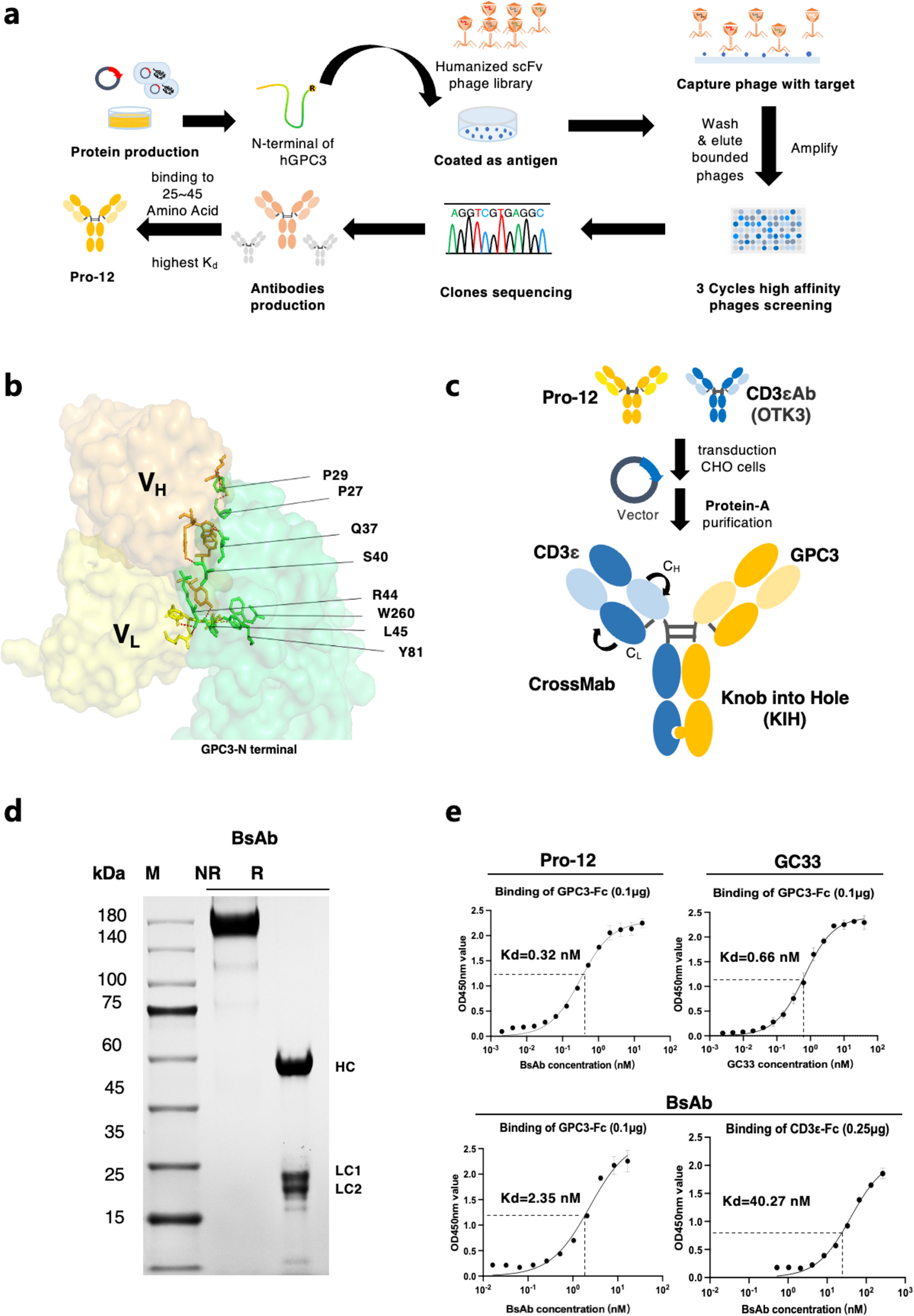
In vivo MDA-MB-468 xenograft study
All animal experiments were conducted in compliance with institutional ethical guidelines for animal care and were approved by the University of South Australia Animal Ethics Committee (Animal Ethics Numbers: U12–23). MDA-MB-468 breast cancer cells (5 × 10⁶) suspended in a 1:1 mixture of Matrigel (Corning, Australia) and FBS-free DMEM were injected into the right flank of female nude BALB/c mice. Once the tumour size reached 80–100 mm3, mice were randomly divided into five groups (n = 5) and treated for 21 days with the following regimens: (i) vehicle (distilled water PO QW), (ii) AU2–94 (100 mg/kg PO QW), (iii) cisplatin (5 mg/kg IP QW), (iv) and (v) AU2–94 (75 mg/kg PO QW and 100 mg/kg PO QW) administered 2 hours before cisplatin.
Mice were monitored daily for any signs of toxicity and tumour volume was measured every other day. Tumour growth inhibition (TGI) was calculated as described in our previous study [19]. %T/C was calculated using the formula: %T/C = (Vt/Vc) × 100, where Vt and Vc represent the tumour volumes of the compound-treated and vehicle-treated groups, respectively, at a particular time point. Data were analyzed using GraphPad Prism version 10 for Windows (GraphPad Software, USA).
In vivo assessment of myeloid-protective effect of AU2–94 against 5-FU
To assess the effect of AU2–94 pre-dosing on 5-FU-induced myelosuppression, female BALB/c mice were injected with EMT-6 cells (0.5 × 10⁶). Once tumours were established, mice were randomly assigned into four treatment groups (n = 10) and received the following treatments: (i) vehicle (distilled water PO and 0.9% saline solution, IP), (ii) a single dose of AU2–94 (200 mg/kg PO), (iii) 5-FU (100 mg/kg IP), and (iv) AU2–94 (200 mg/kg PO) administered 2 hours before 5-FU (100 mg/kg IP). To prepare each compounds for in vivo administration, AU2–94 was dissolved in distilled water. 5-FU was also dissolved in 0.9% saline solution according to previous reports [21].
Two- and six-days post-treatment, spleen and BM cells were collected from 5 mice in each treatment groups. The blood samples were randomly collected from 3 mice in each treatment group and sent to Gribbles Veterinary Pathology (Adelaide, Australia) for complete blood counts (CBC) analysis.
BM cells were isolated by flushing the femur and tibia bones with ice-cold Hank’s balanced salt solution (HBSS, Sigma-Aldrich, Australia) containing 0.5 mM ethylenediaminetetraacetic acid (EDTA, Sigma-Aldrich, Australia) and 2% FBS. Harvested cells were centrifuged, RBCs were lysed by eBioscience™ 1X RBC Lysis Buffer (Invitrogen, Australia), and single-cells were stained with Annexin-V/PI (BD Biosciences, Australia) or phycoerythrin (PE) mouse anti-Ki-67 (BD Biosciences, Australia) according to the manufacturer’s instructions. For Ki-67 staining, cells were fixed and permeabilized using BD Cytofix/Cytoperm™ Fixation/Permeabilization Solution and BD Perm/Wash™ Buffer, respectively. Cells were then exposed to PE anti-Ki-67 antibody for 1 hour at 4 °C. Flow cytometry analysis was performed using a CytoFLEX flow cytometer (CytExpert, Beckman Coulter, California, USA).
AlphaLISA® SureFire® ultra™ assay
AlphaLISA® SureFire® Ultra™ assay was employed to assess the expression levels of RB and phosphorylated RB (serine-780) in breast cancer cell lines and BM cells, respectively. BM cells (100 × 103 cells/well) were treated with AU2–94 for 24 hours. Cells were lysed using AlphaLISA Lysis Buffer (PerkinElmer, Australia) and were incubated with acceptor mix, and donor mix according to the manufacturer’s instructions. Luminescence was measured using an alpha-enabled plate reader (Victor Nivo, Multilabel plate reader).
Statistical analysis
All in vitro studies were conducted in at least three independent experiments, unless otherwise specified. Statistical analyses were performed using t-test, one-way ANOVA or two-way ANOVA where appropriate, to determine the significance of differences among means of treatment groups. Differences between means were considered significant when p ≤ 0.05. All statistical analyses were carried out using GraphPad Prism 10 (GraphPad Software, California, USA).

Comments (0)