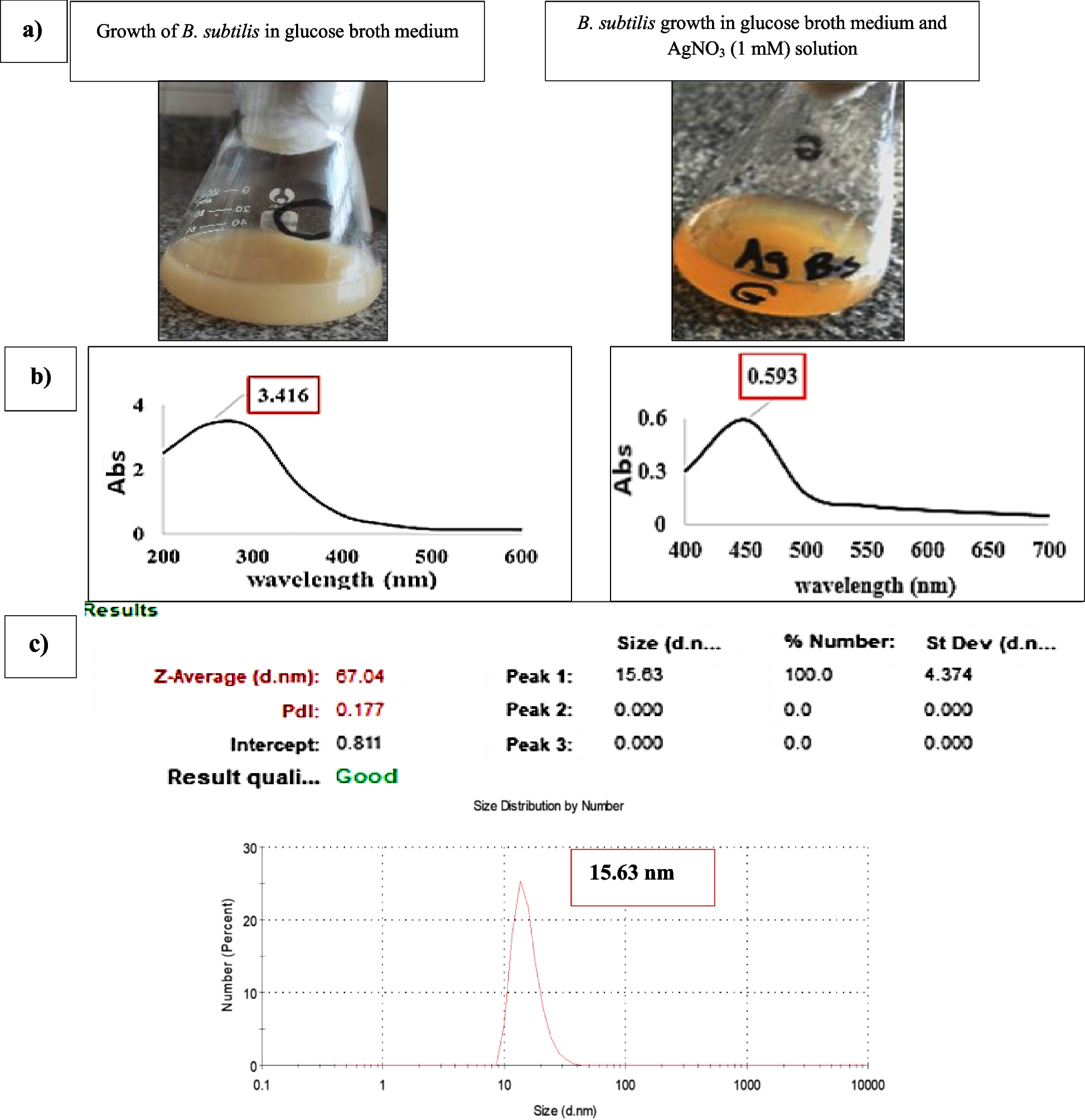
The use of nanoparticles in biomedical applications has garnered considerable attention due to their unique physical and chemical properties, which confer remarkable antimicrobial effects. The biosynthesis of NPs utilizing environmentally friendly methods, such as through the activity of microorganisms or plant extracts, has emerged as a promising avenue for producing these nanomaterials sustainably [25,26,27,28,29]. The ecologically benign aspect of the green synthesis of AgNPs utilizing B. subtilis AMD2024 makes it a unique biosynthesis strategy when compared to physical and chemical procedures. The mixture’s reaction to AgNO3 in combination with B. subtilis cell-free extract changed color, confirming the green production process of Bs-AgNPs. The production of AgNPs employing B. subtilis PP273436, in contrast, changed the hue from yellow to brown, according to Abdulrazzaq and Abas [30]. Using B. zanthoxyli GBE11 ON340757, [10] noticed a similar color shift. The findings of the UV-vis spectroscopy of the culture supernatant in comparison to the Bs-AgNPs solution indicated the culture supernatant showed an absorbance peak of 3.416 at λ max 250 nm, and at λ max 450 nm, the Bs-AgNPs reaction mixture showed an absorbance peak of 0.593. The obtained results were in close agreement with the findings of Alfryyan et al. [31] using B. cereus at 450 nm for AgNPs formation. In a study by Muchanyereyi et al. [32], the high absorbance utilizing plant extracts was approximately 420 nm. According to [33], the spectra of AgNPs produced by the bioreduction activity of the bacterial extract B. subtilis CBPPR1 revealed an absorption peak at 445 nm which is very close to the recent findings.
Proteins and enzymes like nitrate reductase in the supernatant are crucial for lowering silver ions, claims [34, 35], who investigated the impact of endophytic A. baumannii and P. aeruginosa secondary metabolites, particularly the phenolic compounds as antioxidant agents, on the production of AgNPs from silver nitrate solution, also validated this. The bioconversion of silver ions to nanoparticles in the reaction mixture was strongly indicated by the strength of the acquired SPR peak, which was scored at the optimal wavelength, thereby guaranteeing the biosynthesis process [36, 37].
Particle size of the biosynthesized AgNPs varies depending on the green synthesized procedures and the biosynthesizing conditions (temperature, shaking speed, incubation length, and metabolites for reduction of AgNO3). Using B. subtilis, the particle size of Bs-AgNPs was 15.63 nm after 48 hours at 30 °C and 150 rpm shaking flask. In the same manner, B. subtilis and E. coli produced AgNPs with an average size of 11.10 and 38.89 nm, respectively, according to Mbagwu et al. [38]. However, according to [39], the average size of AgNPs produced by B. amyloliquefaciens was 11.10 nm.
With a size reduction of 68.97% to 4.849 nm, blackstrap sugarcane molasses was the best byproduct and carbon source. With a size increase of 82.87% using Bs-AgNPs, the waste from arish cheese whey, on the other hand, had the largest particle size, measuring 91.28 nm. Abd-Elhalim et al. [21] found that blackstrap sugarcane molasses, which had a size of 80.5 nm, was the most appropriate carbon source for the formation of CuNPs using P. silesiensis strain A3, as opposed to glucose, which had a size of 87.1 nm.
Furthermore, as reported by Plaza et al. [40], brewery liquor and molasses inoculated with B. subtilis T-1cell-free supernatant were the most effective biosynthetic sources for the production of AgNPs. Similarly, Sherien et al. [41] showed that Chaetomium globosum can produce zinc nanoparticles from a range of agro-industrial wastes, such as leftover olive cake and apple, carrot, and potato peels.
Different agro-industrial wastes contain varying compositions of organic compounds, including carbohydrates, proteins, amino acids, and phytochemicals, which can influence the growth of B. subtilis leading to the biosynthesis and stabilization of the nanoparticles process. The amount of sugars, vitamins, and other growth elements that are essential for the synthesis of proteins, secondary metabolites, enzymes, and microbes may be the reason for the high quality of blackstrap sugarcane molasses [40]. In contrast, waste with a higher fiber or starch content might lead to larger aggregated nanoparticles due to inadequate stabilization.
By looking at the time course of the green-produced Bs-AgNPs, it was found that 48 hours of incubation was the best amount of time to achieve the appropriate concentration. The concentration of the biosynthesized AgNPs did not increase after this period. The reaction concluded at the eighteenth hour of incubation, according to the UV-Vis spectra absorbance for 1.0 mM silver nitrate combined with cell-free supernatant, and the creation of silver nanoparticles is unaffected by longer incubation times using B. subtilis, as demonstrated by [42]. It took a maximum of 16 hours for a combination of Lactobacillus and Bacillus species to finish fabricating AgNPs. Bacillus sp. alone can finish manufacturing in a maximum of 40 hours, while Lactobacillus sp. alone can do it in more than 40 hours [43]. According to [44], nanoparticle formation takes place during a specific period, after which the concentration and particle size stay constant due to non-agglomeration. The time course data support these findings.
Using HR-SEM, the shape and form of Bs-AgNPs were investigated. The nanosphere-shaped biosynthesized Bs-AgNPs in this work are coated with components of the free extract from B. subtilis cells. In the study of [41], sphere-shaped AgNPs were exposed using B. cereus cell-free extract. Another work by [45] used green tea leaf extract to fabricate spherical and quasi-spherical AgNPs.
According to the work headed by [37], the JCPDS cards No. 04–0783 are crucial for identifying phases in XRD analysis. Each phase has a unique card number associated with its peak positions and intensities. For AgNPs, the observed peaks in the XRD pattern should be compared against the corresponding JCPDS card to confirm the presence of silver and the absence of undesired compounds. An XRD pattern displaying peaks at 2θ values of approximately 38.12°, 44.23°, and 64.32° for silver nanoparticles suggests the presence of the (111), (200), and (220) crystallographic planes, respectively. These peaks when agreeing with the JCPDS support the identification of the synthesized nanoparticles as metallic silver. For the current results, the XRD analysis showed that the sample was not well-crystallized and free of AgNO3 impurities and that there was no total reduction of Ag+ to AgNPs. Six compounds, including Ag2CO3, Ag2O, Ag2O3, AgNO3, and FeAg, were mixed in the Bs-AgNPs’ XRD pattern, according to the results of the XRD investigation. Aswini et al. [46] demonstrated that the metallic NPs’ face-centered cubic structure’s crystalline planes were represented by the XRD peaks at 2θ values of 27.7950, 32.2943, 46.3023, 54.8490, 57.5382, 67.4964, 74.4280, and 76.7858 cm−1. One possible indication that silver ions are present in the generated AgNPs is the strongest peak, which is located at 32.2943 cm−1.
There could be many strategies to minimize the presence of additional phases such as optimization of synthesis conditions as adjusting the concentration of the silver precursor (AgNO₃) and the reducing agents can lead to a more controlled synthesis process, potentially minimizing the formation of undesired products. For instance, lower concentrations might reduce the likelihood of side reactions. Tweaking reaction parameters such as temperature and reaction time can also optimize the conditions for producing pure AgNPs. Shortening the time or controlling the temperature may prevent the formation of multiple phases.
FTIR analysis was used to examine the functional groups in the Bs-AgNPs cell-free extract, which ranged from 400 to 4000 cm−1. Peak positions ranged from 438.88 to 3306.63 cm−1. It was demonstrated that the following functional groups were active: aliphatic primary amine, alkane, carbon dioxide, alkyne, ketenimine, allene, alcohol, amine, primary alcohol, and alkene, in that order. The active functional groups were demonstrated to be N-H stretching, C-H stretching, O=C=O stretching, C≡C stretching, C=C=N bending, C=C=C bending, O-H stretching, C-N stretching, C-O, and C=C stretching. According to the results of FTIR analysis, proteins in particular were in charge of capping and reduction, whilst other components, such as fatty acids, were in charge of stabilizing utilizing Ulvophyte sp. MBIC10591 and Coelastrella aeroterrestrica (Strain BA_Chlo4), respectively [47, 48]. The present study’s functional groups (N=C=S, O-H, N-H, and C-H) are identical to those in the B. coagulans GBI-30, 6068 extract, with a few exceptions in other functional groups [49].
The XRD results suggest a strong molecular connection between Bs-AgNPs and B. subtilis development in blackstrap sugarcane molasses. Proteins like protein coating and the enzyme Ag reductase, which are responsible for the reduction and stability of AgNPs, are present when the FTIR analysis indicates bending amide and amine stretching functional groups [21].
Due to the electrostatic repulsion between charged particles, zeta potential is an essential concept for the stability of NP suspensions. Bs-AgNPs’ zeta potential analysis showed a value of −4.57 mV, according to Liu et al. [50].
In general, a zeta potential value of ±30 mV or higher is considered stable, while values below this threshold may indicate a risk of instability and agglomeration over time [2, 6]. This indicates that the biosynthesized AgNPs using B. subtilis were moderately stable and had a nonionic character in the cell-free supernatant of the capping molecules. AgNPs zeta potential analysis revealed a negative charge for Rhodococcus sp., Brevundimonas sp., and Bacillus sp., respectively, with values of 32.8, 29.6, and 28.1 mV [51].
DLS analysis gives a comprehensive characteristic of the homogeneity of the NPs in colloidal solutions by evaluating the polydispersity index (PDI) values. If the PDI score is greater or less than 0.4, the homogeneity of the NPs solution is raised; if the PDI result is equal to or greater than 1, the NPs is termed heterogeneous. According to the current findings, the biosynthesized AgNPs’ PDI score was 0.177 indicating good homogeneity [7].
Many studies have shown a wide range of IC50 values for AgNPs depending on various factors such as the synthesis method, size, shape, and biological source of the nanoparticles, as well as the type of cell lines used.
The cytotoxicity (IC50) of Bs-AgNPs on ccl-81 cells was determined to be 0.2 mg/mL (200 µg/mL). In previous studies, the cytotoxicity effect of AgNO3 and Ag-NPs on normal Vero cells (African green monkey kidney) produced by B. cereus showed IC50 values of 304.8 and 290.2 μg/mL, respectively, according to Alsharif et al. [52]. In another work, the IC50 value of AgNPs biofabricated by Juglans regia extract on normal (NOF18 cells) was 93.3 μg/mL, according to Ghabban et al. [2]. For renal normal cell lines originating from Vero cells, the bacterially produced Ag-NPs utilizing B. amyloliquefaciens had an IC50 value of 383.7±3.1 µg/mL, according to [54, 55].
The safety of nanoparticles often depends on the application. For biomedical applications, IC50 values that exceed 100 μg/mL can sometimes be considered within a “safe” range, indicating lower cytotoxicity against normal cells [2]. If the observed IC50 value from the current study aligns with or exceeds this threshold, it signifies that the AgNPs may be safer for potential therapeutic applications, such as drug delivery or antimicrobial use. The effectiveness of AgNPs is also critical in medical and environmental applications. Low cytotoxicity can be beneficial for applications like wound dressings, water purification, or as antibacterial agents in food packaging. Comparing the study’s IC50 results with other formulations helps in determining whether these nanoparticles possess a favorable balance of potency (antimicrobial activity) and safety (cell viability) for potential therapeutic and industrial applications. So it is essential to approach the use of these AgNPs with caution in therapeutic contexts. Exploring mechanisms to reduce cytotoxicity and carefully considering dosing regimens will be essential steps in optimizing their application while minimizing the risk of adverse effects. Understanding how these nanoparticles interact with biological systems will continue to be a significant area of research aimed at enhancing both safety and efficacy in biomedical applications.


Comments (0)