Plant material
Leaves of T. usneoides L. were collected in Villa de Leyva, Boyacá, Colombia and were identified by Nestor Garcia from the Herbarium of Pontificia Universidad Javeriana (voucher specimen number 30547). This research was authorized by the Contract of Access to Genetic Resources and Derived Products N° 212 (RGE 0287-6) between the Pontificia Universidad Javeriana and the Ministry of Environment and Sustainable Development of the Republic of Colombia.
Tumor cell lines and culture conditions
The 4T1, MCF-7, B16-F10, K562, U937, and 3T3 cell lines were cultured in RPMI-1640 (Eurobio, Toulouse, France) supplemented with 10% heat-inactivated fetal bovine serum (FBS), 2 mM L-glutamine, 100 U/mL penicillin, 100 µg/mL streptomycin, 0.01 M HEPES buffer, and 1 mM sodium pyruvate (Eurobio) and were incubated in a humidified environment at 37 °C and 5% CO2.
DA-3/ER-GM cells were kindly provided by Prof. Jose Arteaga (Department of Basic Science, Medical School, Universidad Industrial de Santander, Bucaramanga, Colombia). DA-3/ER-GM cells were cultured in RPMI-1640 medium supplemented as described above and also with 50 pg/mL gentamycin (Gibco, Grand Island, NY, USA).
Mice
Young (6 to 12 weeks of age) female BALB/cAnNCrl mice were housed at the animal facilities of Pontificia Universidad Javeriana (PUJ, Bogotá, Colombia) according to the established protocols of the Ethics Committee of the Faculty of Sciences and National and International Legislation for Live Animal Experimentation (Colombia Republic, Resolution 08430, 1993; National Academy of Sciences, 2010). The present study was approved by the ethics committee of the Faculty of Sciences, PUJ, on August 9, 2018. Each specific protocol was also approved by the animal experimentation committee of PUJ (FUA-104-21). Mice were maintained in polyethylene cages with food and water provided ad libitum on a 12h light/dark cycle at 20–22 °C and 40–60% humidity.
Chemicals and reagents
The solvents used in the chromatographic analyses were as follows: LC‒MS grade formic acid from Merck (Merck, Darmstadt, Germany); acetonitrile (LC‒MS and HPLC grade), methanol (LC‒MS and HPLC grade), chloroform, and analytical grade dichloromethane from J.T. Baker (J.T. Baker, NJ, USA); type I water from Milli-Q equipment (MilliPore, MA, USA); and methanol, ethyl acetate, toluene, and derivative solutions such as p-anisaldehyde and 2-aminoethyl diphenylborinate (natural product reagents) were purchased from Sigma‒Aldrich (Sigma–Aldrich, MO, USA). Silica gel 60–200 Mesh LOBAChemie and Sephadex LH-20 (GE Healthcare, WI, USA) were used as the stationary phases for column chromatography.
Extraction and fractionation
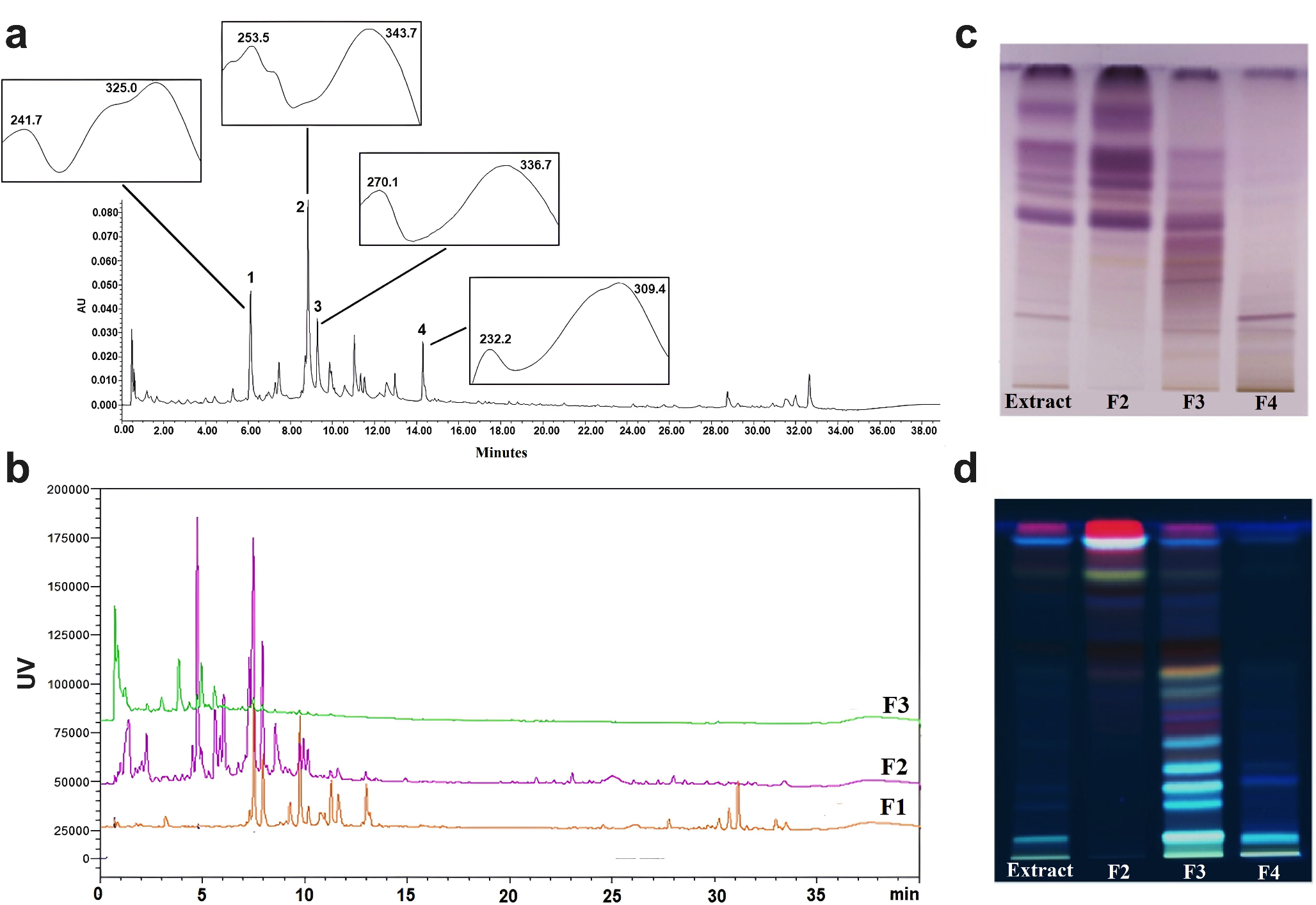
Leaves of T. usneoides L. were dried in a circulating air oven at a controlled temperature of 35 °C. The dried plant material (200 g) was macerated at room temperature with ethanol (EtOH) at a 1:30 w/v ratio for 72 h in the dark, with solvent replacement every 24 h. The obtained extract was filtered and dried in a rotary evaporator to obtain the total ethanolic extract or EtOH extract. The EtOH extract (5.5 g) was fractionated via vacuum chromatography with silica gel (60–200 mesh) using solvents of increasing polarity. The obtained fractions of hexane (F1), dichloromethane (F2), ethyl acetate (F3), and a hydroethanolic solution (F4) were also dried under reduced pressure in a rotary evaporator. The hydroethanolic fraction was subsequently lyophilized for water removal. Subsequently, the F2 fraction (500 mg) was further fractionated via silica gel on glass column chromatography and eluted with a gradient of hexane: dichloromethane (5:5; 3:7; 1:9; 0:1) and dichloromethane: methanol (9:1). The F3 fraction (300 mg) was fractionated on a Sephadex LH-20 column, with methanol used as the mobile phase. Five fractions were obtained: F3 1 (149.7 mg), F3 2 (18.5 mg), F3 3 (13.8 mg), F3 4 (1.9 mg), and F3 5 (2.1 mg). The F4 fraction was separated by centrifugal partition chromatography (CPC) on a PLC-CPC 2250 − 250 System (Gilson, USA) via UV/Vis detection. The solvent systems tested for each fraction were variations of the HEMWat system (hexane-ethyl acetate–methanol–water), according to the polarity of the analytes of interest (Neves Costa and Leitão 2010). The selected solvent system was HEMWat at a ratio of 3.5:6:3.5:6. The selected solvent system was then brought to equilibrium in a separating funnel at room temperature. Each of the phases was separated immediately before use. The lower aqueous phase was used as the stationary phase, while the upper organic phase was used as the mobile phase. The sample (500 mg) was prepared in both the upper and lower phases (1:1 v/v). For the equilibrium step, the rotor was filled with the aqueous stationary phase at 500 rpm at a flow rate of 50 mL/min. Once the stationary phase eluted, the mobile phase was pumped through the column at 1500 rpm and 5 mL/min. With the hydrostatic equilibrium reached, 5 mL of the sample solution was injected through the injection valve in ascendant mode. Detection was performed at 280 and 350 nm.
Analysis by high-performance thin-layer chromatography (HPTLC)
HPTLC analysis of the extract and fractions was performed using Merck HPTLC silica gel plates with glass supports and the fluorescence indicator F254 in CAMAG HPTLC equipment. For triterpene analysis, we used a mobile phase composed of toluene: chloroform:methanol (40:40:10 v/v) and sulfuric anisaldehyde/heat as a detection reagent. The plate was observed under visible light. For the monitoring of polyphenols and flavonoids, chloroform: acetone: formic acid (75:16.5:8.5 v/v) was used as the mobile phase, and a natural product reagent (2-aminoethyl diphenylborinate)/UV 365 nm was used as the detection reagent.
Analysis by ultra-performance liquid chromatography (UPLC-PDA and UHPLC-MS-TQ)
UPLC-PDA analysis was performed on a Waters Acquity H Class UPLC with an Acquity photodiode array detector (PDA). The data were processed using Empower 3 software. Chromatographic separation was performed on a Phenomenex Kinetex C18 column (100 × 2.1 mm, 1.7 μm) maintained at 25 °C ± 1 with an elution gradient of acetonitrile (solvent A) and 0.1% formic acid in water (solvent B) as follows: 17–70% A (0–20 min), 70–95% A (20–30 min), 95% A (30–32 min), and 95 − 17% A (32–40 min), with a run flow rate of 0.40 mL min-1 and an injection volume of 3 µL. The wavelengths used for detection were 274 nm and 350 nm. UHPLC-MS-TQ analysis was performed on a Nexera UHPLC instrument coupled to an LCMS 8060 Shimadzu Scientific Instruments (Maryland, USA) mass spectrometer. The same chromatographic conditions used in the UPLC-PDA analysis were also used here. The mass spectrometer was operated in positive and negative ionization modes, and the data were acquired within 100–2000 m/z. The capillary conditions were as follows: capillary voltage, + 3 kV; sample cone voltage, 33 kV; drying temperature, 250 °C; nebulization gas, nitrogen at 350 L/h; collision gas, argon at 50 L/h; and collection energy, 2.5 eV. The data were processed with Lab Solutions software.
Analysis by gas chromatography coupled to mass spectrometry (GC‒MS-TQ)
For gas chromatography, a GC-MS-TQ8040 (Shimadzu Scientific Instruments, Maryland, USA) was used. The column used was an HP5-MS with the following dimensions: 60 m (L) × 0.25 mm (ID) × 0.25 μm (film width) (Agilent J&W Scientific, California, USA). The reaction conditions were as follows: injection port (250 °C), split mode (20:1), injection volume (1 µL), temperature ramp: 40 °C (Tinicial), 15 °C/min up to 200 °C (held for 2 min), 4 °C/min up to 280 °C, and 5 °C/min up to 320 °C (held for 10 min), for a total run time of 50.67 min. Helium was used as the carrier gas, and the mass spectrometer was set to positive electron impact ionization at 70 eV in full scan mode over a mass range of 30 to 600 m/z. The identification criteria were based on the percentage of agreement (SI) (> 80%) with the NIST library and an abundance limit > 2.5%.
In vitro cytotoxicity assays
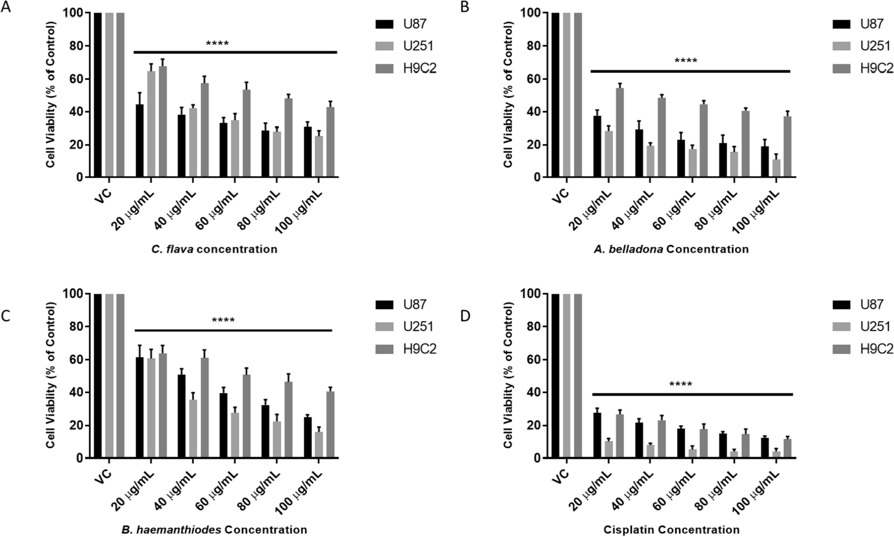
Cytotoxic effects were evaluated using the colorimetric 3-[4,5-dimethylthiazol-2-yl]-2,5-diphenyl tetrazolium (MTT; Sigma‒Aldrich, Saint Louis, MO, USA) assay (Mosmann 1983). To perform this assay, 3 × 10³ cells/well were seeded in 96-well plates and incubated for 12 h at 37 °C with 5% CO2. After incubation, serial dilutions were performed with a 1:2 factor, starting from 250 µg/mL to 1.95 µg/mL for each T. usneoides extract, fraction, and subfraction. Ethanol (EtOH) and dimethyl sulfoxide (DMSO) were used as negative controls, and the chemotherapeutic drug doxorubicin (DX) was used as a positive control at concentrations ranging from 2.5 µM to 0.019 µM. After 48 h of treatment, MTT reagent (1 mg/mL) was added, after which the cells were incubated for 4 h, and the absorbance at 540 nm was read in a Multiskan plate reader (FC, Thermo Scientific). The half-maximal inhibitory concentration (IC50) values were calculated using GraphPad Prism version 9.0 for Mac OS X statistics software (GraphPad Software, San Diego, CA). The selectivity index (SI) was calculated using the following formula: SI = IC50 no tumor cells/IC50 tumor cells.
Proliferation assay
In all, 1 × 10⁵ DA-3/ER-GM cells were seeded in 12-well plates with EtOH (extract vehicle, 0.02%) and incubated at 37 °C in a CO2 atmosphere with the IC50 of the T. usneoides extract or EtOH (extract vehicle, 0.02%) for 24, 48, and 72 h. After that time, the cells were stained with trypan blue and counted. The exponential (Malthusian) growth formula Y = Y0*exp(k*x) of the software GraphPad Prism version 9.0 for Mac OS X was used to calculate the doubling time.
ROS measurement
To evaluate reactive oxygen species (ROS) generation in response to treatment, 1.5 × 10⁵ DA-3/ER-GM cells were plated in 12-well plates and treated with the IC50 of the T. usneoides extract, doxorubicin (pro-oxidant control), DMSO (doxorubicin vehicle), or EtOH (extract vehicle, 0.02%) for 12 h and 24 h. The cells were stained with 1 µM 2′,7′ dichlorodihydrofluorescein diacetate (H2DCFDA) (Sigma Aldrich, Saint Louis MO, USA) for 40 min at 37 °C, followed by PI (Sigma‒Aldrich). Samples were acquired using a Cytek Aurora Cytometer (Cytek Biosciences, Fremont, CA, USA) and analyzed with FlowJo v10.8.1 software (BD Life Sciences). Three independent experiments were performed in triplicate. The data are presented as the means ± SEMs.
Glucose uptake assay
DA-3/ER-GM cells were cultured in 12-well plates at a density of 3 × 10⁵ cells/well and incubated for 1, 6, 12, or 24 h with T. usneoides extract at the IC50, 20 µM LY294002 (control of decreased glucose uptake), or EtOH (extract vehicle, 0.02%). After treatment, the cells were washed and resuspended in 40 µM 2-NBDG [2-(N-(7-nitrobenz-2-oxa-1,3-diazol-4-il) amino)-2-desoxiglucosa] (Invitrogen Molecular Probes). Then, the cells were incubated for 30 min at 37 °C and washed with cold 1X PBS. Live/dead cell labeling was performed with the LIVE/DEAD Fixable Aqua Dead Cell Stain Kit (Life Technologies, Thermo Scientific, Eugene, OR, USA). Data were acquired with a Cytek Aurora Cytometer (Cytek Biosciences, Fremont, CA, USA) and analyzed with FlowJo v10.8.1 software (BD Life Sciences). Two independent experiments were performed in triplicate. The data are presented as the means ± SEMs.
In vivo AML model and treatment with T. usneoides extract
For the development of the in vivo model, 5 × 10⁴ DA-3/ER-GM cells were injected into the lateral tail vein as previously described (Murillo et al. 2023). To evaluate the effect of the extract on circulating blasts and their dissemination, 3 days after the inoculation of tumor cells, the mice were treated with the extract or PBS (negative control) twice a week via intraperitoneal (i.p.) injection. The dose of T. usneoides extract used was 142.5 mg/kg body weight, which corresponds to a dose 4 times lower than the lethal dose 50 (LD50), which ensures that the concentration of the extract is not toxic to the animals (Lasso et al. 2022). Mice were monitored three times per week for changes in body weight and signs of disease. Additionally, 20 µL of peripheral blood drawn from the tail vein was collected to evaluate hematological parameters with a blood count using a Mindray BC-5150 analyzer (Mindray Bio-Medical Electronics Co., Ltd., Shenzhen, China). A Wright´s peripheral blood smear was also performed using an automated stainer (Aerospray Stainer, Wescor, Logan, Utah, USA). When warranted for animal welfare reasons, animals were euthanized by CO2 inhalation. At necropsy, the spleen, femur, and lungs were removed to evaluate the presence of blasts in these organs.
Statistical analysis
Statistical analyses were performed using the software GraphPad Prism version 9.0 for Mac OS X (GraphPad Software, La Jolla California USA, www.graphpad.com). Differences between the two groups were determined using the Mann‒Whitney U test. Differences among subject groups were evaluated using the Kruskal‒Wallis test and Dunn’s post hoc test for multiple comparisons. Values were considered statistically significant when p < 0.05.


Comments (0)