
Remember me


The aim of this study was to describe the distribution of neutrophil granulocytes by tracing NE in a porcine Escherichia coli (E. coli) model of sepsis using PET–CT and the selective NE PET-tracer [11C]NES [16, 19]. The experiment took place at the Hedenstierna laboratory, a facility for large animal intensive care research and the PET-centre at Uppsala University hospital. A timeline for the experiment is shown in Fig. 1.
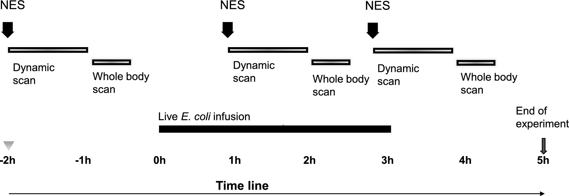
Fig. 1
A timeline of the experimental setting. All animals were anaesthetised, prepared and transported to the PET-camera. At -2 h the first scan was started with an injection of [11C]NES and a 60 min dynamic scan followed by a 20–24 min whole-body scan. At 0 h sepsis was induced by the infusion of live E. coli during three hours. At 1 h the second scan was started with an injection of [11C]NES followed by the same dynamic and static protocol. For three of the animals a third scan was performed starting at 3 h. The experiment was terminated at 5 h
The subjects and intensive care protocolSeven healthy land-breed pigs of both sexes weighing between 25 and 30 kg were used (P1–P7). They arrived at the facility in the morning and were immediately anaesthetised, tracheotomised and mechanically ventilated. The animals were handled and prepared as previously reported [19] and described in detail in Supplement 1.
InterventionsHeart rate, mean arterial pressure (MAP) and mean pulmonary arterial pressure (MPAP) were monitored continuously. Cardiac output (CO) was measured hourly through thermodilution, and airway pressures including wedge pressure were registered hourly. Cardiac index was calculated as CO/body surface area (BSA) and pulmonary vascular resistance index (PVRI) was calculated as (MPAP-wedge pressure) * 80/CI. BSA was calculated using Meeh’s formula: BSA = k * body weight (kg) (2/3) [20].
Arterial blood gases were drawn hourly and analysed for oxygen pressure, carbon dioxide pressure, lactate, glucose and haemoglobin. Intensive care interventions were made according to Supplement 2. The depth of sleep, signs of pain and shivering were continuously observed by trained personnel and when needed, the animals were given additional doses of morphine, ketamine or rocuronium bromide.
Ethical statementThe pigs were given food and drink ad libitum until one hour before arrival to the research facility where they immediately were anaesthetised. They were kept under deep anaesthesia until killed at the end of the experiment. Every effort was made to reduce suffering and the experiment was approved by the Animal Ethics Board in Uppsala, Sweden, permit number 5.8.18–08592/2019, supplementary decision made 2022–04-25. The study was designed with consideration of Minimum Quality Threshold in Pre-clinical Sepsis Studies and reported in coherence with the ARRIVE 2.0 guidelines [21, 22].
Blood sample analysisThe arterial blood gases and the venous blood or plasma samples were analysed for oxygen pressure, carbon dioxide pressure, lactate, glucose, haemoglobin blood cell count, creatinine, tumour necrosis factor- alfa (TNF-α) and NE. The analysis methods are described in detail in Supplement 3.
BacteriaA clinical isolate of E. coli was prepared as described in Supplement 4. The bacteria were given as an intravenous (i.v.) infusion of 8.3 log10 colony forming units (CFU) × hour (h)−1 for three hours. The infusion was changed every hour to assure that the bacteria remained in log-phase.
Radiochemistry[11C]NES ([11C]GW457427) was produced as earlier described and obtained with a radiochemical purity higher than 95% [16]. Usually around 2 gigabecquerel (GBq) of activity was obtained at the end of synthesis. The positron emitting radionuclide carbon-11 has a short half-life of only 20.4 min, which allows repeated administrations of [11C]NES in the same animal on the same day, as each tracer administration will have decayed by more than 98% after 2 h (6 radioactive half-lives). Up to three synthesises were made per experimental day around 2 h apart, to enable a baseline scan and one or two follow-up scans after induction of sepsis. [11C]NES passes through the neutrophil cell membrane, and thus binds to both extracellular and intracellular NE.
In vivo [11C]NES PET–CT examinations in pigsFor an overview of the PET–CT protocol, see Fig. 1. The pig was positioned in a supine position on the PET–CT scan bed. Next, an i.v. injection of [11C]NES at a dose of 12 megabecquerel (MBq) × kg−1 (around 300–360 MBq) was administered, followed by a 60-min dynamic PET-scan also functioning as a stabilisation period for tracer uptake and distribution. A whole-body CT scan was performed for attenuation correction and anatomical co-registration. Thereafter, a whole-body PET-scan lasting 20–24 min was performed. For more details see Supplement 5.
After the first PET-scan was finalised, experimental sepsis was induced by an intravenous bacterial infusion as described above. One hour after the start of the bacterial infusion, a second injection of [11C]NES was administered. At this time point, around three hours had passed from the first [11C]NES injection, and negligible radioactivity (< 0.3%) remained in the body. After 60 min of dynamic PET-scanning and stabilisation the whole-body PET and CT scans were repeated as described above.
Finally, in three of the pigs a third injection of [11C]NES was administered around 2 h after the second injection. As before, the [11C]NES injection was followed by dynamic PET-scan for stabilisation and static whole-body PET–CT scans.
Image analysisPET–CT image analysis was performed using the PMOD Base Functionality (PBAS) tool (PMOD Technologies LLC, Zurich, Switzerland). Manual segmentation and delineation of the lungs, bone marrow, liver and spleen were performed with assistance by CT images. Tracer uptake was quantified in Becquerels per cubic centimetre (Bq × (cc)−1) and subsequently converted to standardised uptake values (SUV) by correcting for the total administered dose (in kBq) and body weight (in kg). The unit is thus g/ml, but when the density as here can be approximated to 1 g/ml SUV becomes a unitless measure of PET-tracer uptake in tissues which can be compared within and between individuals.
Calculations of inflammatory volume of the lungThe PET–CT images of the lungs were further analysed in more detail. The volume of the lungs with inflammation was calculated from CT-images using Hounsfield units (HU) and from PET based on SUV. Segmentations of the lungs were applied on the CT-image; a range of HU between − 980 and − 150 to − 50, to cover the entire lung including parts affected by atelectasis. The upper limit (− 150 to − 50) was adjusted to make the delineation as similar as possible before and after induction of sepsis. Within the segmented lung tissue a cutoff was set to include SUV above1.3 or 1.6, whereupon a region was delineated and subsequently used to exclude spillover of uptake from bone marrow in the ribs and other tissues close to the lung. The total inflammatory volume (TIL) was calculated by multiplying SUVmean with the lung volume having a SUV above the selected cutoff. The selection of the SUV cutoff was set based on the background uptake in the baseline scan before sepsis was induced and was then fixed for the scans at 2 h and 4 h to exclude the tissue with atelectasis and gravitational effects from the TIL. Image processing and analysis was performed with the software Affinity 3.0.5 (Hermes medical solutions, Stockholm, Sweden).
Blocking of extracellular NE in vivoOne of the animals, P6, was administered the extracellular NE inhibitor sivelestat (400 mg; 14 mg × kg−1) as an i.v. infusion (400 mg in 50 mL DMSO, 2 mL × min−1, 10 min in total) 30 min prior to the third [11C]NES injection.
Bone marrow samplingFrom subjects P6 and P7, a bone marrow biopsy was taken from the femoral bone before (− 2h) and after (3h) sepsis induction. The object glasses with imprints and the biopsies were stained both with standard stain for cell count and with antibodies targeting MPO and NE, respectively. See Supplement 6.
Blocking of NE in purified human neutrophils in vitroNeutrophils were purified and either lysed or intact incubated with [11C]NES in the presence of three different elastase inhibitors, GW457427 (NES), GW311616 and sivelestat. The trapped radioactivity was measured in an in-house built scintillation counter of NaI type and corrected for radioactive decay. For details see Supplement 7.
StatisticsStatistica 14.0 (StatSoft, Tulsa, OK, USA) was used for most statistic calculations. Mean and standard deviations (SD) were used (unless otherwise stated) and a p-value < 0.05 was considered significant. Because of the biological variations we chose the non-parametric Wilcoxon test when testing for differences before and after sepsis induction. For the analysis of blocking of NE in vitro GraphPad Prism 10.4.0 was used for a t-test and Fig. 8.
Comments (0)