
Remember me

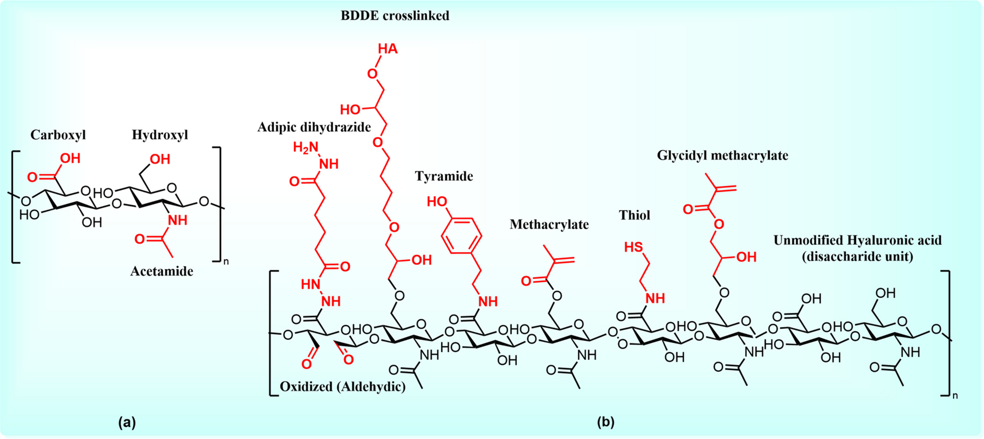

UPLVG alginate (0.180 g, 1 mmol) was dissolved in 41 mL ultrapure (type 1) water with 10% (v/v) isopropanol and degassed with nitrogen for 1 h. Following degassing, NaIO4 (7 mg, 0.0327 mmol) was added to the solution which was then covered with aluminum foil and stirred under a nitrogen atmosphere for 48–72 h. The oxidized solution was transferred to a cellulose acetate dialysis membrane with a molecular cut off of 12,000 g/mol. Dialysis was carried out in 1.0 M NaCl for 24 h and then in ultrapure water for 48 h with the bath being changed every 12 h. Dialyzed mixture was lyophilized at − 50C and a pressure of ≤ 400 mbar for 24 h. A cottony, white oxidized product (0.178 g) was recovered. This oxidized alginate was dissolved in 15 mL ultrapure water with 12% (v/v) MeOH. Once dissolved, 4-(2-aminoethyl) benzoic acid HCl (3.6 mg, 0.020 mmol) and 2-methylpyridine borane complex (21 mg, 0.2 mmol) were added to the solution and phosphate buffer (pH = 6.01) was added to adjust the pH to ~ 6. The solution was again covered in foil and stirred under N2 for 48 h. The now aminated alginate solution was again dialyzed in 1 M NaCl followed by ultrapure water as it was after the oxidation and lyophilized. Cottony benzoic acid–modified alginate (0.172 g, 96%) was recovered and analyzed via water suppression. 1H NMR characteristic aromatic signals: (400 MHz, D2O) δ 7.90 (d, J = 8.1 Hz, 2H), 7.44–7.30 (m, 2H). 1H NMR and DOSY spectra for this material were reported previously [5].
Preparation of Phenethylamine Modified Alginate (2)Oxidized alginate was prepared as described above for ABA-modified alginate (1) and dissolved in 15 mL ultrapure water with 12% (v/v) MeOH. Once dissolved, phenethylamine (3.0 mg, 0.027 mmol) dissolved in 1 mL ultrapure water and 2-methylpyridine borane complex (21 mg, 0.2 mmol) were added to the solution and phosphate buffer (pH = 6.01) was added to adjust the pH to ~ 6. Solution was again covered in foil and stirred under N2 for 48 h. The now aminated alginate solution was again dialyzed in 1 M NaCl followed by ultrapure water as it was after the oxidation and lyophilized. Cottony phenethylamine–modified alginate (0.176 g, 99%) was recovered and analyzed via water suppression. 1H NMR characteristic aromatic signals: (400 MHz, D2O) δ 7.42–7.33 (m, 2H), 7.32–7.22 (m, 3H). 1H NMR and DOSY spectra supplied in supplementary materials.
Preparation of Methyl 4-(2-Aminoethyl) Benzoate Modified Alginate (3)Oxidized alginate was prepared as described above for ABA-modified alginate (1) and dissolved in 15 mL ultrapure water with 12% v/v MeOH. Once dissolved, methyl 4-(2-aminoethyl) benzoate HCl (3.8 mg, 0.020 mmol) and 2-methylpyridine borane complex (21 mg, 0.2 mmol) were added to the solution and phosphate buffer (pH = 6.01) was added to adjust the pH to ~ 6. Solution was again covered in foil and stirred under N2 for 48 h. The now aminated alginate solution was again dialyzed in 1 M NaCl followed by ultrapure water as it was after the oxidation and lyophilized. Cottony methyl benzoate–modified alginate (0.178 g, 99%) was recovered and analyzed via water suppression 1H NMR characteristic aromatic signals: (400 MHz, D2O) δ 7.94 (d, J = 4.7 Hz, 2H), 7.44–7.31 (m, 2H). 1H NMR and DOSY spectra supplied in supplementary materials.
Preparation of 1-[4-(2-Aminoethyl) Phenyl] Ethanone–Modified Alginate (4)Oxidized alginate was prepared as described above for ABA modified alginate (1) and dissolved in 15 mL ultrapure water with 12% (v/v) MeOH. Once dissolved, 1-[4-(2-aminoethyl) phenyl] ethanone HCl (3.6 mg, 0.020 mmol) and 2-methylpyridine borane complex (21 mg, 0.2 mmol) were added to the solution and phosphate buffer (pH = 6.01) was added to adjust the pH to ~ 6. Solution was again covered in foil and stirred under N2 for 48 h. The now aminated alginate solution was again dialyzed in 1 M NaCl followed by ultrapure water as it was after the oxidation and lyophilized. Cottony acetophenone–modified alginate (0.178 g, 99%) was recovered and analyzed via water suppression. 1H NMR characteristic aromatic signals: (400 MHz, D2O) δ 8.04–7.84 (m, 2H), 7.51–7.25 (m, 2H). 1H NMR and DOSY spectra supplied in supplementary materials.
1D NMR Quantitative Characterization ExperimentalThe water suppression proton nuclear magnetic resonance (1H NMR) spectra were obtained using a Bruker Ascend 400 MHz spectrometer operating at 400.1 MHz [6]. 1H NMR spectra were referenced to the residual proton or carbon signals of the respective deuterated solvents. Chemical shifts were reported in parts per million (δ) relative to tetramethylsilane (TMS) or to residual resonances of the deuterated solvents: deuterium oxide (D2O). Spin multiplicities were indicated by the following symbols: s (singlet), d (doublet), t (triplet), q (quartet), dd (double doublet), and m (multiplet). Deuterated solvents were purchased from Cambridge Isotope Laboratories.
Maleic acid (11.6 mg, 0.1 mmol) was dissolved in 2 mL deuterium oxide NMR solvent to give a 50 mM solution. Ten milligrams of the modified alginate product was added to an NMR tube with 250 μL of the maleic acid solution and 750 μL D2O. Sonication was used to fully dissolve the solid and the sample was analyzed via water suppression 1H NMR. Aromatic peaks in the spectrum were integrated along with the maleic acid peak at 6.25 ppm. Maleic acid integration was normalized to 1 and the corresponding integration of one aromatic peak (corresponding to 2H) was used to derive the percentage of alginate units modified.
DOSY ExperimentalDOSY spectra were obtained using a Bruker Ascend 400 MHz magnet operating at 400.1 MHz. Pertinent parameters are shown in Table 1.
Table 1 Pertinent DOSY acquisition parametersPreparation of Simulated Gastric Fluid and Simulated Intestinal FluidThese solutions were prepared as previously described [1]. Briefly, the simulated gastric fluid (SGF) was prepared by mixing 2 g/L of NaCl with DiH2O and adjusting the pH to 2.0. The simulated intestinal fluid (SIF) was prepared by mixing 6.8 g/L monobasic KH2PO4 with DiH2O and adjusting the pH to 6.8 or 7.4.
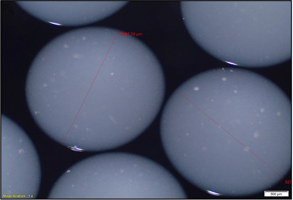
In Vitro Testing of Alginate Material Hydrogels Stability and DisintegrationUnmodified and modified alginates were dissolved in Hanks balanced salt solution (HBSS) at a concentration of 1.5% (w/v) overnight. Ten hydrogel beads were made by manually extruding the alginate solution through an 18-guage blunt needle into 3 ml of 100 mM CaCl2 cross-linking solution in a 6-well plate, the diameter of the hydrogel beads ranged from 2.6 to 3.27 mm with average diameter size = 2.97 mm and standard deviation = 0.19 mm (Fig. 1). The beads were allowed to cross-link for 10–15 min before being incubated in either SGF for 6–24 h or SIF for 3 h while being shaken at 60 RPM at 37 °C. At corresponding time points, the beads were visually counted by two individuals to determine the number of intact beads remaining in each respective medium. The term intact describes beads that have a completely intact structure (Fig. 2A), beads that are partially dissolved (Fig. 2B) or beads that have lost the shell but still preserve the opaque core (Fig. 2C). The arrows in 2B and 2C indicate beads counted as intact.
Fig. 1
Representative sizes of alginate hydrogel microbeads
Fig. 2
Different stages of bead disintegration as intact beads were assessed. Beads with intact structure (A), beads that are partially dissolved (B), or beads that have lost the shell but still preserve the opaque core (C). The arrows in B and C indicate beads counted as intact
Determination of Protection of Encapsulated Protein from Acidic Destruction0.5 ml of HBSS was mixed with 0.5 ml of 2 mg/ml bovine serum albumin (BSA) (Thermo Scientific #23,209), and the mixture was added to 15 mg of unmodified UPLVG to reach a final concentration of 1 mg/ml BSA in a 1.5%w/v alginate. As described earlier, 10 hydrogel beads were made manually by extruding the alginate solution containing BSA through an 18-guage blunt needle into 3 ml of 100 mM CaCl2 cross-linking solution in two 6-well plates. In the first plate (control group), the beads were left to cross-link for 10 min before the CaCl2 was replaced by 5 ml of Dulbecco phosphate-buffered saline (DPBS) (Thermo Scientific #14,190,144) to dissolve the beads. In the second plate (test group), the beads were left to cross-link for 10 min before the CaCl2 was replaced by 3 ml of SGF (pH = 2). After 2 h, the SGF was replaced by 5 ml of DPBS. After 3 h, beads from both groups were completely dissolved in the DPBS medium, and this medium from each group was placed in a 15-ml tube and centrifuged at 1000 rpm for 5 min to precipitate any remnant particles of the dissolved beads. Then the BCA assay (Thermo Scientific # 23,225) was performed on clear samples from both groups to quantify the concentration of albumin in each group.
Comments (0)