
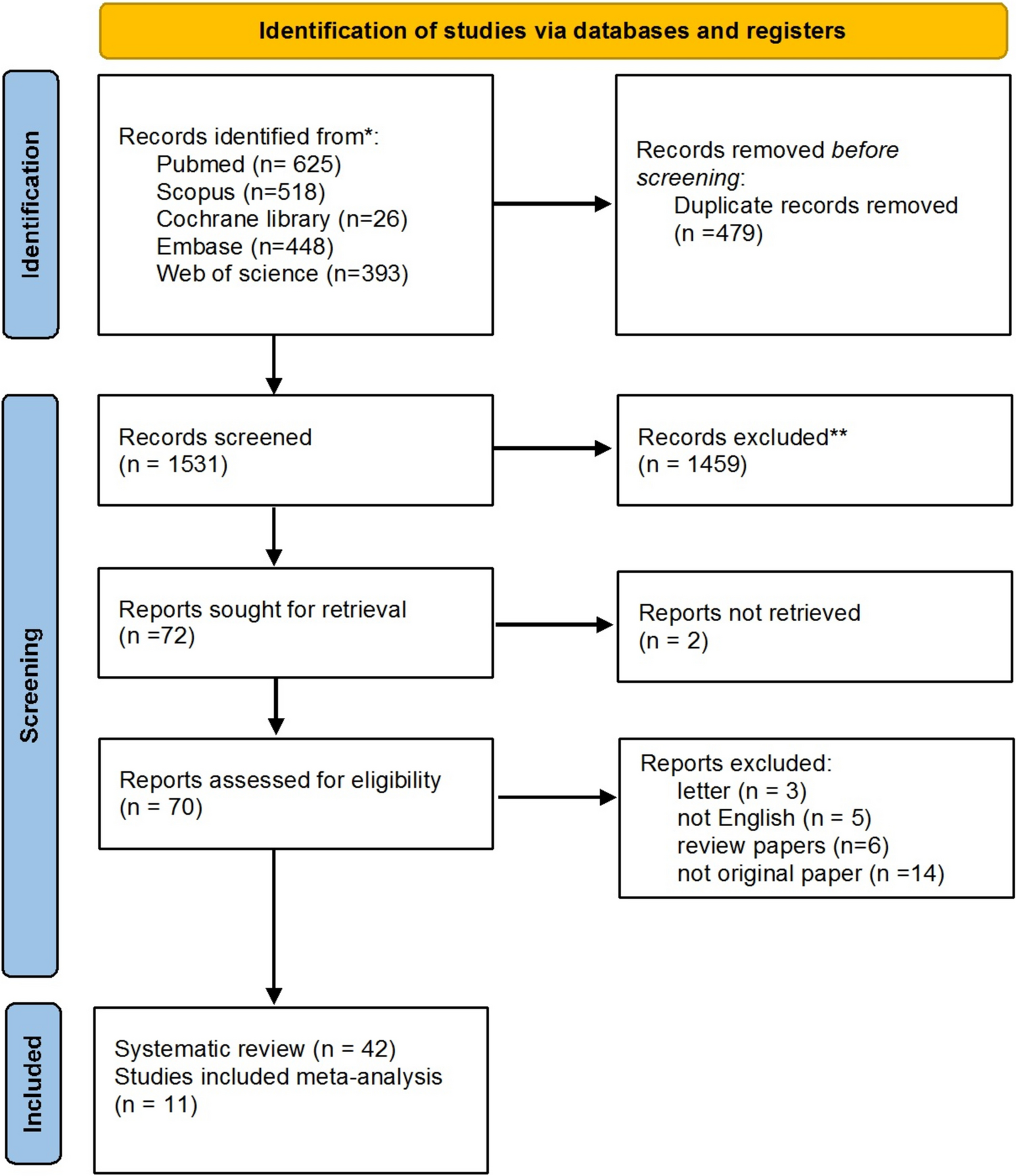
Remember me
Vertebral artery stenosis is a condition characterized by the compression and narrowing of the vertebral artery lumen, which is often asymptomatic [2, 12]; symptomatic vertebral artery stenosis may manifest as dizziness (47%), unilateral limb weakness (41%), dysarthria (31%), headache (28%), and nausea or vomiting (27%) [13, 14]. The V1 segment of the vertebral artery at its origin is more prone to stenosis compared to other segments [15,16,17].
Evidence suggests that simultaneous surgery on the vertebral and carotid arteries may lead to increased mortality and stroke rates within 30 days postoperatively (8-33%), and the occurrence of transient or permanent Horner’s syndrome (2-21%), thus concurrent carotid and vertebral artery surgery is not recommended for patients with combined carotid and vertebral artery disease [2]. This study posits that for patients requiring the removal of carotid plaques simultaneously, the basic condition of the patient should be combined with an assessment of the surgical timing and tolerance. Preoperative imaging examinations should be completed to accurately measure the degree and length of carotid stenosis, and the anastomotic site should be preselected. After a thorough evaluation of the surgical risk by anesthesiologists and other relevant department physicians, the surgery should commence. During the surgical process, it is recommended to fully dissect and expose the vertebral artery first, perform the carotid endarterectomy, and after the intraoperative plaque removal and confirmation of unobstructed carotid blood flow, proceed with the bypass surgery based on the preselected anastomotic site.
The summary of studies related to microsurgical treatment for partial proximal vertebral artery stenosis is presented in Table 5. The compiled data indicates that transposition surgery has a significant proportion within microsurgical procedures, and is associated with a relatively high incidence of postoperative complications. Pure vertebral artery endarterectomy has fewer complications compared to transposition surgery, yet the sample size is small and further validation is required through multicenter studies with larger cohorts. Postoperative complications of microsurgical treatment primarily include Horner’s syndrome, surgical site hematoma, cranial nerve injuries, chylous leakage, and stroke. As depicted in Fig. 3, among the related studies, Horner’s syndrome accounts for the highest proportion of transient postoperative complications.
Table 5 Microsurgical treatment of proximal vertebral artery stenosis: a review of related studiesFig. 3
The proportion of postoperative complications across different studies
Horner’s syndromeHorner’s syndrome is a clinical syndrome resulting from the interruption of any part of the ocular sympathetic efferent pathway, characterized by ptosis, miosis, and anhidrosis on the ipsilateral side of the face. Surgical injury to the cervical sympathetic trunk may involve thermal or anatomical damage. Thermal injury is often due to the heat generated by energy devices such as electric or ultrasonic scalpels, leading to injury of the cervical sympathetic trunk. Mild thermal injuries are generally reversible due to the integrity of the nerve. Anatomical damage usually involves the transection of the preganglionic fibers of the cervical sympathetic nerve. The local nerve ganglion may be palpated similarly to lymph nodes, and inexperienced operators may mistakenly excise the nerve ganglion along with lymph nodes. Therefore, when palpating enlarged lymph nodes posterior to the carotid artery, it is important to distinguish them from nerve ganglia, especially when the lymph nodes are adherent to surrounding tissues.
Postoperative surgical site hematomaIn the event of a postoperative surgical site hematoma, attention should be paid to whether the patient’s trachea has shifted, and whether there are symptoms such as difficulty swallowing or respiratory distress, as asphyxiation is the greatest risk associated with wound hematoma. If the patient presents with symptoms of asphyxiation or stridor, consider the possibility of rupture at the site of vascular anastomosis. Perform tracheal intubation, promptly open the incision, remove the blood clot, and achieve hemostasis. If the patient has no symptoms other than the surgical site hematoma, consider the possibility of inadequate hemostasis during surgery; if the swelling continues to increase, it may also be necessary to reopen the incision for hemostasis. Layer-by-layer dissection during surgery and timely separation and ligation of blood vessels are crucial to prevent postoperative bleeding, especially the superficial vascular structures within the fat pad, which should not be overlooked.
Cranial nerve injuryThe incidence of cranial nerve injury following carotid endarterectomy is as high as 8-10% [18], but there are no study reports on its occurrence in the treatment of vertebral artery stenosis. Intraoperative injury to the vagus or recurrent laryngeal nerve may lead to unilateral vocal cord paralysis. Damage to the unilateral hypoglossal nerve may cause difficulties in speaking, chewing, and swallowing, while bilateral damage can lead to upper airway obstruction. During surgery, traction on the nerves should be minimized, and once symptoms of nerve injury appear, timely administration of neurotrophic drug therapy should be initiated. In most cases, symptoms will gradually alleviate if the nerves were not damaged during surgery. However, it should be noted that unilateral hypoglossal nerve paralysis is a contraindication for contralateral endarterectomy, and the subsequent treatment should be deferred until recovery on that side is achieved.
Postoperative hoarseness and chylous leakHoarseness, if present postoperatively, should prompt consideration of whether the surgical field involved the superior laryngeal nerve or the recurrent laryngeal nerve. However, the most common cause is laryngeal edema, which typically resolves as the swelling subsides, leading to the disappearance of the patient’s symptoms.
Chylous leaks are more prevalent in patients undergoing surgery on the left vertebral artery. During surgery, careful management of the thoracic duct is essential. A significant leakage of chyle can lead to electrolyte imbalances, hypoproteinemia, infection, and even bleeding. It may also result in chylothorax or chyloperitoneum, causing respiratory distress and thoracoabdominal infections, with severe cases potentially life-threatening. Patients with milder symptoms are treated with a high-protein, low-fat diet and local compression bandaging. If the patient has a large volume of chyle drainage or shows no significant improvement after three days of compression therapy, surgical exploration may be considered.
In this study, 14 patients with proximal vertebral artery stenosis had previously undergone failed interventions due to vascular reasons, and 4 patients experienced in-stent restenosis following stent therapy. The vertebral artery has more developed elastin and smooth muscle compared to the coronary artery, which can make stent placement more challenging. Additionally, the origin of the vertebral artery is more flexible, increasing the inflammatory response to stent placement and potentially leading to in-stent restenosis [19]. A meta-analysis of non-randomized studies reported no difference in technical success and procedural complications between drug-eluting stents (DES) and bare-metal stents (BMS), but patients treated with BMS had a higher incidence of recurrent symptoms and postoperative reintervention compared to those treated with DES [20]. A multicenter study involving 420 patients who underwent endovascular stent placement reported an average in-stent restenosis rate of 26% at 12 months [21]. In a single-center series, the stent fracture rate was 5%, 15%, and 30% at one, three, and five years, respectively, with most being asymptomatic [21, 22]. Compared to endovascular treatment, microsurgical treatment has a relatively lower rate of restenosis.
In these 34 cases, 94% of the patients received active antiplatelet therapy and/or anticoagulant therapy. Some patients had previously undergone endovascular stent treatment, but their clinical symptoms did not show significant improvement. For all patients who underwent microsurgical treatment, we confirmed that their symptoms were alleviated or improved postoperatively, and successful revascularization was achieved. The modified Rankin Scale (mRS) scores before and after surgery showed statistical significance with a paired t-test P < 0.01, as seen in Fig. 4. Moreover, compared with other studies as shown in Table 5, vertebral artery endarterectomy alone is associated with relatively fewer complications than surgical treatment involving more than two vessels. Compared to arterial transposition surgery, when the supplying vessel has severe stenosis or unstable atherosclerotic plaques, performing vertebral artery endarterectomy alone does not involve dealing with other vessels, nor does it require a second surgery, resulting in less trauma and preserving the original physiological and anatomical structure of the vertebral artery [15]. Under permissible conditions, vertebral artery endarterectomy alone may be a better choice in the microsurgical treatment of proximal vertebral artery stenosis.
Fig. 4
Comparison of preoperative and postoperative modified Rankin Scale (mRS) outcomes
Overall, the prognosis and follow-up results suggest that microsurgical revascularization surgery is highly feasible, with good safety and clinical outcomes in the treatment of proximal vertebral artery stenosis.
Comments (0)