FDG PET/CT in Hantavirus Hemorrhagic Fever With Renal Syndrome
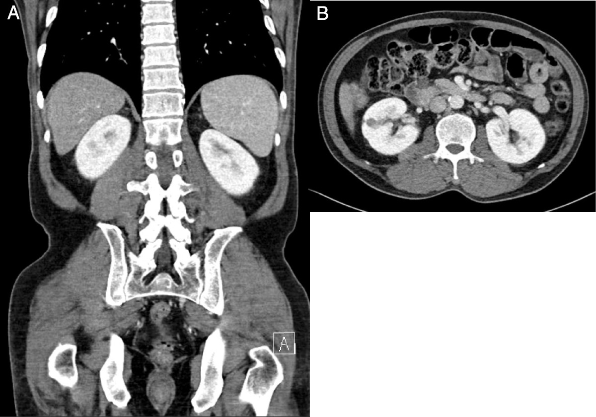
Because of his prolonged fever and gradual increase in blood leukocytes, 18F-FDG PET/CT was performed at 5 days after abdominal CT. Surprisingly, bilateral kidneys were diffusely enlarged for 5 days, with the renal parenchyma showing intense hypermetabolism. The maximal diameters of the right kidney in axial and coronal views increased from 6.9 to 7.8 cm and from 9.8 cm to 10.8 cm, respectively (A, fusion coronal image; B, axial images). Bilateral nephritis was highly suspected. Hemorrhagic fever with renal syndrome was diagnosed with typical clinical symptoms and Hantaan virus antibody (indirect immunofluorescence antibody test; immunoglobulin M 1:512, immunoglobulin G 1:4096). Hantaan virus is one species of the genus Hantavirus. Supportive care such as intravenous hydration and control of blood pressure and serum glucose was provided. Follow-up FDG PET/CT after 1 month of recovery showed normal size and usual metabolism of both kidneys (C, fusion coronal image; D, axial images). Hemorrhagic fever with renal syndrome is a potentially fatal infectious disease with a worldwide distribution.
1–3 It is caused by Hantavirus when humans inhale aerosolized excrements of infected rodents. Its clinical hallmarks include fever, thrombocytopenia, acute renal insufficiency, and pathologically typical features of acute interstitial nephritis. Most patients go through a series of stages called acute febrile, hypotensive, oliguric, and diuretic phases, which may take several weeks to recover. Because there are no specific antiviral agents for Hantavirus, supportive care is the main strategy of treatment. Hemodialysis can be performed if it progresses to renal failure. FDG PET/CT has been proven useful for finding the focus of fever in various febrile diseases. Its use has expanded to the nononcologic field.
4–8Hantavirus-induced acute interstitial nephritis is characterized by a lymphocyte-predominant infiltrate with varying degrees of eosinophils and macrophages.
9 These metabolically active inflammatory cells are thought to contribute to intense FDG uptake. This case shows that FDG PET/CT could demonstrate the physiology of Hantavirus invading kidneys and causing nephritis in a short period. Furthermore, it could also be applied to evaluate treatment response by determining whether the kidney has returned to normal size with usual metabolism. FDG PET/CT could be the choice of image modality for diagnosis and treatment evaluation in patients suspected of having hemorrhagic fever with renal syndrome. To the best of our knowledge, this is the first report of FDG PET/CT image for patients with HFRS.


Comments (0)