
Remember me


Overall, there were 41 cases in which incidental findings were observed and parental testing was recommended. Results of parental testing were obtained in 13 of the 41 cases, and all results confirmed the original PGT-A predictions. In cases where the parental reports were not received, it is unknown if testing was ordered. If so the outcome of any potential testing was not reported to us by the referring clinician. In 3 cases, a balanced translocation carrier status was confirmed by conventional karyotyping, supporting IVF-derived embryo-based PGT-A incidental findings (Figs. 1 and 2; Table 1).
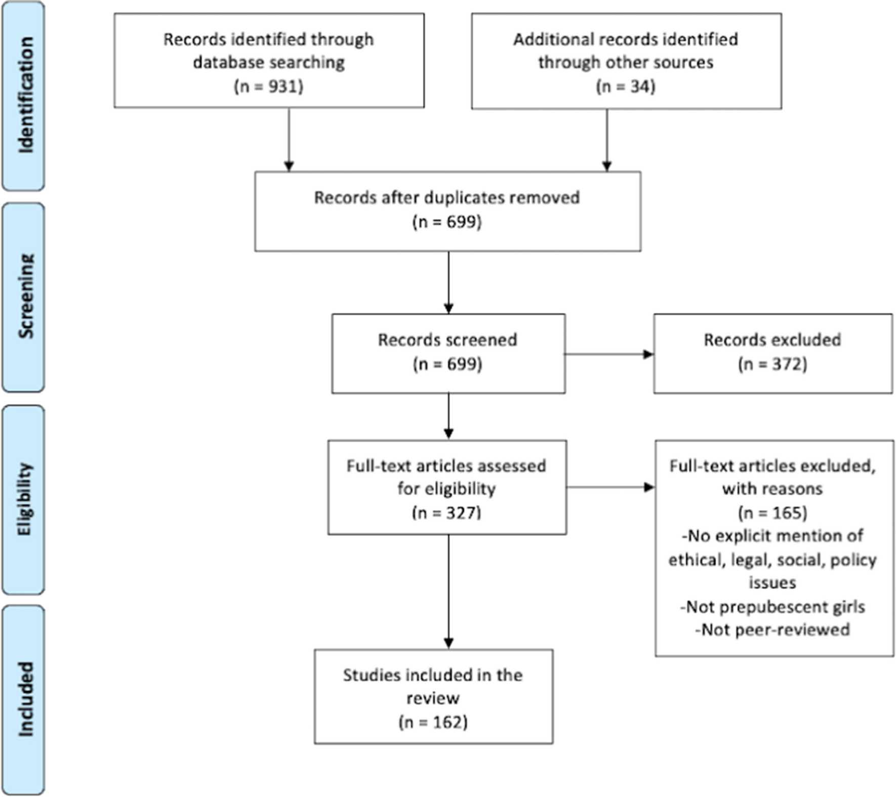
Fig. 1
Tree diagram illustrating identification and follow-up on cases with incidental findings for possible translocation or CNV
Fig. 2
Examples of SNP array PGT-A results for 3 cases leading to identification of parental carriers of a balanced translocation. In each case, two chromosomes displayed clear copy number and allele ratio imbalances (highlighted in red). Although each case displays results observed in only one embryo, multiple embryos possessed imbalances on the same chromosomes
Case 1The patient was 44 years old at the time of egg retrieval. Five trophectoderm biopsy samples were received for PGT-A. After SNP microarray analysis, 4 embryos were predicted to be aneuploid. Two of the embryos had consistent segmental imbalances on chromosomes 3 and 9. A conventional karyotype of the parents was ordered and confirmed a maternal balanced translocation, 46,XX,t(3;9)(q13.2;q21). Once a balanced translocation was documented on conventional karyotype, the PGT-A results were reinterpreted using a previously established PGT-SR approach. After re-analysis, one embryo was predicted to be a balanced translocation carrier, 2 embryos were unbalanced, and the remaining two embryos were balanced carriers but had additional chromosome abnormalities unrelated to the maternal translocation, and therefore were predicted aneuploid.
Case 2The patient was 34 years old at the time of egg retrieval. Nine trophectoderm biopsy samples were received for PGT-A. After SNP microarray analysis, 6 embryos were predicted to be aneuploid. Three of the embryos had consistent segmental imbalances on chromosomes 11 and 12. A conventional karyotype of the parents was ordered and confirmed a maternal balanced translocation, 46,XX,t(11;12)(q21;p13.3). Once a balanced translocation was documented on conventional karyotype, the PGT-A results were reinterpreted using a previously established PGT-SR analysis protocol. After re-analysis, three embryos were predicted to be balanced translocation carriers, 2 embryos were predicted to be unbalanced, but due to additional chromosome abnormalities unrelated to the maternal translocation were predicted to be aneuploid. The remaining 5 embryos were identified as aneuploid due to chromosome abnormalities unrelated to the maternal translocation.
Case 3The patient was 27 years old at the time of egg retrieval. Six trophectoderm biopsy samples were received for PGT-A. After SNP microarray analysis, 4 embryos were predicted to be aneuploid. Three of the embryos had consistent segmental imbalances on chromosomes 18 and 21. A conventional karyotype of the parents was ordered and confirmed a paternal balanced translocation, 46,XY,t(18;21)(p10;p10). Once a balanced translocation was documented on conventional karyotype, the PGT-A results were reinterpreted using a previously established PGT-SR analysis protocol. After re-analysis, two embryos were predicted to be euploid, and 2 embryos were predicted to be unbalanced. One embryo was predicted to be unbalanced but was identified as aneuploid due to additional chromosome abnormalities unrelated to the paternal translocation. The remaining embryo was identified as aneuploid due to chromosome abnormalities unrelated to the paternal translocation.
Identification of pathogenic CNV carriersCNV carrier status was confirmed in all 10 cases where parental microarray or karyotype data was obtained (Table 1). In 4 of the cases, parental studies indicated a benign CNV and the embryo PGT-A results were reinterpreted accordingly. In 4 of the cases, parental studies indicated a CNV classified as a variant of uncertain significance and the embryo.
PGT-A results were reinterpreted accordingly. In 2 of the cases, parental studies indicated a pathogenic variant with clinical implications for the associated embryos (Fig. 3) and are described in detail below.
Fig. 3
Examples of SNP array PGT-A results from 2 cases leading to the identification of parental carriers of a pathogenic CNV. Each case displays results from 3 embryos where a small imbalance was clearly seen (highlighted in red). The del 3p26.3:p26.1 was 3 Mb, and the dup 15q11.2:11.2 was 6.5 Mb
Case 1The patient was 41 years old at the time of egg retrieval. Five trophectoderm biopsy samples were received for PGT-A studies. After SNP microarray analysis, all 5 of the embryos were predicted to be aneuploid. Three of the embryos had a recurrent segmental imbalance on chromosome 15. A conventional karyotype of the parents was ordered and confirmed an abnormal female karyotype with a duplication within 15q11.2. When maternally inherited, this duplication is associated with autism spectrum disorders and had clinical implications for the patient and associated embryos. PGT-M studies were recommended but not completed.
Based on the preliminary results and maternal karyotype. all the embryos were aneuploid.
Case 2The patient was 27 years old at the time of egg retrieval. Four trophectoderm biopsy samples were received for PGT-A studies. After SNP microarray analysis, all 4 of the embryos were predicted to be aneuploid. Three of the embryos had a recurrent segmental imbalance on chromosome 3. A chromosome microarray of the parents was ordered and confirmed that the patient was heterozygous positive for an interstitial deletion of 3p26.1:p26.3, which is classified as pathogenic and associated with 3p deletion syndrome having clinical implications for the patient and associated embryos. PGT-M studies were recommended but not completed. Based on the preliminary results and maternal karyotype, all the embryos were aneuploid.
Comments (0)