
Remember me

Human life is full of decisions and actions that are taken to be totally voluntary. In everyday life, our decisions could, for example, concern the choice of food to eat, or of means of transport from home to work, which is highly dependent on societal/environmental and intrinsic information. In order to be able to unravel the mysteries of conscious (free) will, it is essential to understand which and how neural structures are encoding information during intentional control.
Different views exist on the temporal and causal relationship between intentions and decisions, and to what degree intentions are conscious (see e.g. Haggard 2008, 2019; Mele 2009, 2019; Block 2021), but the difference partly depends on the context. For example, experimental situations are usually quite different from ecological ones. Here, we adhere to the view that intentions, which may be unconscious or conscious to various degree, in general precede decisions, which typically would be conscious. This view links closely to that of Freeman (1999), who based his view primarily on physiological and behavioral studies of animals.
The process of intentionality may comprise the operations of predicting, planning, and learning actions, while intentions could imply the creation and projection of alternative future states, desired, avoided, or feared. Such hypotheses could be constructed in attractor dynamics by extrapolation from past experience, controlling choices and directions of actions in the present (Freeman 1999; Liljenström, 2018).
In principle, there could be many intentions within our brain-mind system, conscious or unconscious, and possibly competing, but one (or a few) of them may lead to a decision to act. Intentions have been classified as distal, proximal, or motor intention, depending on their temporal relation to a decision to act (see e.g. Mele 2019, and Parés-Pujolràs and Haggard 2021). There could also be different causes for the emergence of intentions, for example genetic, or physiological, but sometimes also emotional and cognitive. Hence, intentions may originate in different parts of the brain, including the limbic system.
Decisions, on the other hand, are cognitive in nature and are primarily associated with higher cortical systems/processes. Hence, many animals may have intentions to do this or that, but never really make a decision to do so. Humans, too, while still largely influenced by our limbic system, may have unconscious intentions, perhaps leading to actions without decisions. However, some of our intentions could be cognitively and perhaps also consciously formed in neocortex, e.g. in prefrontal cortex (Haggard and Parés-Pujolràs, 2021). Typically, a distal intention may last for a longer period of time, and would precede a decision to act. Proximal intentions are usually quite close in time for an action. In some cases, when immediate actions are called for by environmental conditions, intentions and decisions could be more or less simultaneous.
Intention can be viewed as a precursor to conscious (free) will, as an “urge” or “desire” to act in a certain direction, to attain a certain goal. Voluntary movement, or more generally, behavior, is based on perception and past experience (memory), which are required for prediction of (inter)actions. Attention may provide information about the internal and external worlds, but intention guides our actions (Liljenström, 2011).
During the past decades, there has been a debate in the literature regarding some experiments apparently showing that free will is just an illusion. Seemingly, the brain knows, at least half a second in advance what “you” decide to do (Libet et al. 1982, 1983). Lately, this time window has been extended to incredible ten seconds, based on more recent experiments (Soon et al. 2008; Haynes 2011). The common interpretations of these studies have, however, been criticized (see e.g. Mele 2009, 2019; van Inwagen 2017), and alternative interpretations of the experiments can be given (see e.g. Liljenström, 2022).
A series of famous EEG experiments (Kornhuber and Deecke 1965; Libet et al. 1982, 1983; Keller and Heckhausen 1990; Haggard and Eimer 1999; see Libet (2004) for a review) are often quoted as evidence for free will being an illusion. The EEG readiness potential (RP), apparent only when averaged over a large number of trials, appears to precede the conscious will for spontaneous voluntary movements. However, in contrast to Libet type of experiments where arbitrary and purposeless choices are studied, more ecological experiments, where deliberate choices are made, no RP is observed (Maoz et al. 2019).
Voluntary actions can be considered to be endogenously self-initiated, and based on internal decisions and motivations (Brass and Haggard 2007; Haggard 2008), in contrast to involuntary movements. According to Haggard (2008), volition is associated with the concept of a goal-directed activity, based on both motivation for pursuing a goal and the instrumental knowledge for controlling the motor action. In this regard, the causal relation between the action and its outcome, as well as reason-action associations are major concerns for the study of goal-directed volitional behavior. Therefore, we may be able to take into consideration context-action and action-outcome associations in the presence of endogenous signal(s) as features of voluntary actions (Fried et al. 2017). These aspects of volitional actions can be addressed with the involvements of the hierarchically higher neural regions (Friedman and Robbins 2022). The feature of goal-directedness is strongly related to the concept of cognitive control, which is different from habitual behaviors (Petrides 2005). Cognitive control is a neural-based process developed based on the adaptive function of the brain. This process is about the maintenance of neural patterns, correlating goals and potential actions (Miller and Cohen 2001).
There are certain cortical areas believed to be central for cognitive control and attentional functions (Corbetta and Shulman 2002). The lateral prefrontal cortex (LPFC) and anterior cingulate cortex (ACC) are two key neural structures involved in bottom-up and top-down attention (Rossi et al. 2009; Aarts and Roelofs 2011). The intentional processes hierarchically influence the dynamics of internally/externally stimulated neural structures towards stability for framing decisions. Accordingly, a major focus in this work is modelling the functionalities of LPFC and ACC, as well as their interactions during intention, as the preparatory phase of volitional decision making.
Undoubtedly, many different structures are directly and indirectly involved in the exertion of voluntary actions. In addition to LPFC and ACC, areas such as the orbitofrontal cortex (OFC) and amygdala are structures related to emotional processes, which were included in our previous model of decision making (Hassannejad Nazir and Liljenström, 2015). One could also consider modelling the pre-SMA and SMA, where the RP has been detected, but for simplicity, in this work we do not consider activity in OFC, amygdala, or SMA. Instead, we confine our work to the major areas supposed to be directly involved in the emergence of the early intentional process, preparing for a decision to act, and preceding the activity in SMA. We do not consider direct motor intentions in this work.
Executive functions associated with voluntary action controlDecision making processThe decision-making process (DM) is described in a simple way by Doya (2008) in five steps: (1) presentation of external/internal stimuli, (2) predicting values of the associated potential decisions, (3) selection of an option, (4) actual outcome evaluation, and finally, (5) learning, based on the difference between the predicted and actual values of the potential options. According to Doya and others (Doya 2008; Fobbs and Mizumori 2014), maximizing the utility of the decisions is considered the main goal of all humans decision-making, where different types of contextual learning contribute to the knowledge-base for reaching a decision (Gariépy et al. 2014; Lee and Harris 2013).
Working memoryWorking memory and attentional control are two strongly associated cognitive functions (Unsworth and Spillers 2010; Oberauer 2019; Kang et al. 2020). The commonly accepted features of working memory (temporarily holding, monitoring, and manipulating information) make this process an essential cognitive aspect of goal-directed voluntary actions. To accomplish voluntary actions, the selection of relevant stimuli and suppression of irrelevant ones is resulting from various procedural aspects of working memory affecting excitatory and inhibitory neurons at the site of termination (Baddeley 2012; Adams et al. 2018). Brodmann areas BA 9 and BA 46, which are laterally and highly connected subregions of LPFC, appear to be involved in working memory, where cytoarchitectural differences between these two areas bring about different functional aspects of working memory. The neural activity of these areas may represent different if–then rules, including reason-action and action-outcome associations.
Neural structures associated with volitional controlLateral prefrontal cortexIn order to be able to make decisions, while being bombarded by numerous internal and external stimuli, our brains need to be equipped with functional specifications. Keeping on hold different received environmental information, loaded memories, projected interoceptive signals and goals are essential for controlling such a great deal of information. Moreover, shifting between these signals and managing the competition between them is indispensable. In this regard, the role of the most essential neural regions must be considered.
The extrinsic and intrinsic connectivity of neural parts located in the prefrontal lobe of cortex provide a broad contribution to cognitive controlling processes and executive functions. The cytoarchitecture and laminar organization of prefrontal neural areas seem to provide a basis for structure-based hierarchical information processing required for cognitive functions (Bludau et al. 2014). The executive functionality of the cortical laminar structure of LPFC was demonstrated by Pribram and others (Pribram et al 1952). The intra-cortical connectivity of LPFC serves as an early sensory stimuli processor for subsequent cognitive reasoning (Mushiake et al. 2006; Figner et al. 2010). Cognitive flexibility, working memory and inhibitory control are major aspects of executive functions involved in the intentional controlling process, including vetoing and self-control, as well as externally driven actions.
The LPFC, located in the frontal pole of the brain, has been recognized as one of the most important structures involved in cognitive processes (Tanji and Hoshi 2008). Functional neuroimaging of LPFC illustrates its contribution, not as an integrator of external information from associative sensory structures (i.e., early sensory stimuli), but as the recipient of internal afferents from other laminar cortical structures, limbic and subcortical areas. In this regard, the intrinsic connections within its different subregions, as well as its extrinsic connectivity with some other brain areas, demonstrate its versatile role in controlling goal-directed and stimulus driven behavior.
The functionality of LPFC is highly attributed to its neural organisation. Taking into account the Brodmann structural classification, LPFC consists of different subregions with different laminar organisation including Brodmann areas BA 9, 45, 46 and 47. Some studies also consider BA 10 as part of LPFC. Among these subregions, the architectonic organisation of BA 9, 10, and 46 are similar. These three areas are composed of six granular layers including well-defined layer IV (Petrides 2005), however, with different cellular distribution, bringing about different functional capacities (Petrides 2005).
Brodmann area 9As shown in Fig. 1, BA 9 is subjected to early sensory information capable of encoding the environmental contextual information. This area also has strong connections with ACC, encoding endogenous stimuli. The high capacity of BA 9 in processing afferent endogenous signals and integrating them with environmental signals makes this structure one of the most potential neural candidates during the preparatory process of intention in the presence of endogenous signals.
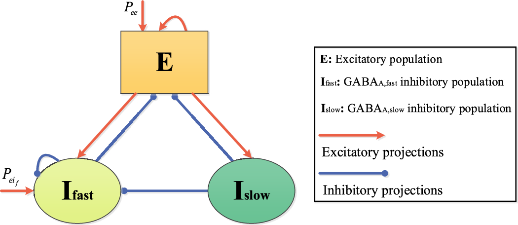
Fig. 1
Exciting and inhibiting pathways projected from ACC to BA 9. BA 9 is composed of pyramidal neurons and various inhibitory interneurons. This area is exposed to environmental early sensory stimuli and afferent endogenous signal from ACC. The integration of these signal would bring about the unconscious selective attentional control. Providing that the internal and external stimuli are correlated, ACC excites BA 9 pyramidal neurons (Ex) through projecting directly pyramidal neurons and indirectly by exciting calretinin (CR) inhibitory interneurons resulting in disinhibition of calbindin (CB) interneurons, (i.e. pathway (a)). To suppress the activity of external stimulus given that the efferent signals to BA 9 are uncorrelated, the ACC excites directly the CB inhibitory neurons (i.e. pathway (b))
This controlling process of BA 9 can be mediated through expression of calcium-binding proteins in the interneurons of this area. The abundance of calcium-binding proteins as calretinin (CR) and calbindin (CB) among the GABAergic interneurons enables BA 9 to control the environmental information considering the projected internal signal via ACC. The expression of these proteins in the interneurons activates the gating process during attentional control. Generally, CR interneurons are connected to CB interneurons, while the target of CB interneurons is excitatory (Ex) pyramidal cells. Hence, the expression of CR interneurons disinhibits the excitability of pyramidal neurons (Barinka and Druga 2010).
Brodmann areas 46 and 10BA 46 and 10 are structurally similar. Both consist of six well-developed layers with the same cellular composition. However, their different cellular intensities result in distinct functionalities. BA 46 is a well-developed six laminar structure, bordered by BA 9, BA 10. The supragranular layers of this sub-region are densely organized with medium size cells (Bludau et al. 2014; Petrides 2005). This LPFC subregion is involved in executive processes such as attentional control and working memory, as well as DM (Barbey et al. 2013; Blakemore and Frith 2000).
BA 10 is the largest area in the frontal lobe of the brain, with a wide range of connections to various neural areas. This cortical area is known as a supramodal structure widely contributing to various cognitive functions. The adjacent regions of BA 10 are BA 9, BA 46 (as parts of LPFC), ACC and OFC, indicating the key involvement of this neural part in attentional control and DM. In addition, it has extensive connections with the pre-supplementary motor area (pre-SMA), thalamus, hypothalamus, and basal ganglia (Peng et al. 2018).
BA 10 has a lower cellular density compared to the cell density distribution of BA 46. Generally, structures with lower cellular density have greater neuropil space, and subsequently larger space for higher neural connections have been observed (Hrvoj-Mihic and Semendeferi 2019). Therefore, considerable higher neuropil fraction is likely more strongly contributing to complex functions while higher level of controlling, here referred to as hyper-controlling, compared to other LPFC subregions is required (Okuda 2007; Peng et al 2018). According to Chahine et al. (2015) BA 10 is involved in branching activations in a complex solving task strongly dependent on resolving goal-tree sequences. This process also increases complexity of working memory.
Considering the above-mentioned observations, Medalla and Barbas (2010) have suggested that the cellular density difference between BA 10 and BA 46 makes them play different roles in the controlling process. Therefore, BA 10 is likely to be more involved in cognitive functions with higher-order processing (Ramnani and Owen 2004).
Anterior cingulate cortexConsidering the unique position of ACC, this structure is connected to the cortico-cortical and cortico-limbic pathways. This structure also receives dopamine, innervated by the ventro-tegmental area encoding error related negativity. The bidirectional connections of ACC with amygdala and OFC facilitate the flow of endogenous information among these structures projecting LPFC (Bush et al. 2000; Allman et al. 2001; Palomero-Gallagher et al. 2008; Medalla and Barbas 2012; Apps et al. 2016). ACC plays an important role in modulating the neurodynamics of the LPFC through projecting its subregions during different attentional control phases. The modulatory action of this neural area is highly attributed to its laminar pattern (Palomero-Gallagher et al. 2008). Hierarchical signal transmission, determining the type of projection, is strongly related to the development of granular layer IV in cortical structures. The origin of ascending (feedforward) signals is the granular neural structure with well-developed layer IV, while the descending (feedback) signals with modulatory properties are generally projected by agranular structures which lack layer IV (Braver et al. 2001). The laminar organization of ACC, with a large densely packed layer V merged with layer VI and no layer IV, provides the structural basis of selective attention function. The controlling role of ACC during selective attention could be the result of response conflict monitoring (Shipp 2005).
ACC receives afferent signals from OFC, which has long been known as a structure involved in modulating emotional arousal (Jenison 2014; Stalnaker et al. 2015). The recorded oscillatory activity of OFC represents the expected value of the associated outcome of the afferent stimulus, which is referred to as the “expectancy signal”. Therefore, ACC is capable of projecting this expectancy signal to its connected structures. According to the hierarchical control required to manage the projected signal process, it is expected that ACC evokes recognizably different neurodynamics in each of the LPFC subregions. That is why ACC targets different neural cell types in each BA areas, resulting in the emergence of different frequencies and neural pattern behaviors.
Pre-supplementary motor area (pre-SMA)The supplementary motor complex (SMC) is functionally parcellated into two areas: the supplementary motor area (SMA) and the pre-supplementary motor area (pre-SMA) (Nachev et al. 2008). The variety of observed functional activities and the outflow/inflow of information from these areas are bases for neural segregation. These two subregions of SMC, categorized as agranular structures, lack cortical layer IV. Pre-SMA has been well known as a neural structure with the function of predictive coding (Akkal et al. 2007). With regard to the neural areas adjacent to these two SMC subregions, SMA is mainly involved in movement generation, while pre-SMA provides the basis for movement initiation. Therefore, pre-SMA is considered to be strongly coupled to higher order cognitive functions (Nachev et al. 2007). Pre-SMA is characteristically well known for its oscillatory readout, and primarily the early readiness potential (RP), as a neural signature of volition. The emergence of this signal is characterized with two components (i.e. early and late RP, respectively), which supposedly are administrated by different projections from various neural structures (Seghezzi and Zapparoli 2020). The slow negative build up of the oscillatory activity consitituting the early phase of the RP, which emerges around 1500–400 ms before the movement onset, and represents the intentional preparatory process, whereas the late RP occures 400–0 ms prior to action performance (Schurger et al. 2021). The pre-SMA is exposed to afferent pathways, conveying goal-directed and motivational signals originated from different neural areas, bringing about different components of the RP. The early RP, as measured in pre-SMA, is presumed to result from input signals from LPFC subregions, while the emergence of the late RP component is the result of primary motor cortex projections (Schurger et al. 2021).
The early RP in the pre-SMA may evolve as a result of the projections of associative signals, i.e. context-action and action-outcome associations encoded by oscillatory activities of the neural attractors correlated to potential actions/goals. Hence, this cortical area mediates the required preparatory processes of DM. Subsequently, the late RP, and eventually voluntary action, would appear through bidirectional interactions between the pre-SMA and SMA, as well as some other subcortical areas, such as basal ganglia (Nachev et al. 2008).
Focus and objectivesAlthough there have been many advances in the past decades in understanding the role of the brain in decision-making and volition, there are still many unanswered fundamental questions targeting the intentional control process, preparing for a decision to act. In order to shed light on these neural-mental relations, some fundamental questions should be addressed. We propose that a mathematical representation of the neurodynamics related to cognition may demonstrate transitions between different degrees of order and disorder in the brain (Freeman
Comments (0)