
Remember me


A 58-year-old, right-handed woman underwent living-donor liver transplantation for decompensated alcoholic liver cirrhosis. She had performed DSA, was desensitized with rituximab, and had undergone plasma exchange before transplantation. The patient received methylprednisolone (4 mg/day), tacrolimus (3 mg/day), and everolimus (1.5 mg/day).
Nine months after liver transplantation, she was admitted to our hospital with self-limited focal-to-bilateral tonic-clonic seizures. On admission, a neurological examination revealed mild acalculia and agraphia. She had no history of headache, fever, infection, or vaccination. CD4-positive and CD8-positive T cell counts were 185/μL and 1383/μL, respectively.
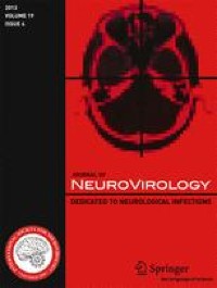
T2-weighted cranial magnetic resonance imaging (MRI) showed asymmetric, cortex-sparing white matter lesions in the left frontal, left parietal, and right parieto-occipital lobes, with weekly enlargements. Gadolinium-enhanced T1-weighted image showed partial enhancement in these brain lesions (Fig. 1A–C). Electroencephalography revealed repetitive spikes localized in the right parietal region (P4 max). Cerebrospinal fluid (CSF) examination showed no cells, a protein concentration of 33 mg/dL, an IgG index of 0.40, and positive oligoclonal IgG bands. JCV DNA was detected in the CSF using quantitative polymerase chain reaction (PCR) (166 copies/mL) at the National Institute of Infectious Diseases (Tokyo, Japan). Epstein–Barr virus, herpes simplex virus, cytomegalovirus, and varicella zoster virus were not detected by PCR. Serum and cerebrospinal fluid tests for Mycobacterium tuberculosis, Cryptococcus, syphilis, and HIV were also negative.
Fig. 1
Magnetic resonance imaging of the head. T2WI indicates T2-weigted image; T1-Gd, gadolinium-enhanced T1-weighted image. T2-weighted image at the onset of progressive multifocal encephalopathy (PML) shows asymmetric, cortex-sparing white matter lesions in the left frontal and right parieto-occipital lobes (A). Gadolinium-enhanced T1-weighted image shows partial enhancement in these brain lesions (B). The lesions enlarge weekly, with a confluent pattern (C, 1 week after initial imaging; D, 3 weeks after initial imaging)
The patient fulfilled the diagnostic criteria for definite PML (Berger et al. 2013). Immune reconstitution was initiated before confirming the PCR findings. Tacrolimus was discontinued, and the methylprednisolone and everolimus doses were gradually tapered. There was no neurological deterioration, and the JCV DNA titer was reduced from 166 to 18 copies/mL 1 week after initiating immune reconstitution. As the lesions enlarged, the treatment was switched from prednisolone and everolimus to cyclosporine alone (50 mg/day) (Fig. 1D). Mirtazapine (15 mg/day) and mefloquine (275 mg/week) were administered after approval from the institutional ethics committee. Three weeks after initiating immune reconstitution, lesion expansion ceased. Monthly tests for JCV DNA in the CSF were also negative. There was no evidence of immune reconstitution inflammatory syndrome.
Two months after initiating immune reconstitution, aspartate transaminase (AST) and alanine transaminase (ALT) levels increased to 40–60 IU/L. Liver biopsy revealed portal inflammation with lymphocytes and eosinophils, bile duct damage, and endothelialitis with bile ductular proliferation. C4d immunohistochemical staining was positive in the portal veins. Acute cellular and antibody-mediated rejection was suspected. Her immunosuppressant dose was gradually increased to 2 mg/day methylprednisolone, 1.5 mg/day everolimus, and 75 mg/day cyclosporine, with close monitoring of neurological symptoms and MRI. Subsequently, her AST and ALT levels normalized with no PML relapse.
Forty-eight months after PML onset, she lived at home with an Expanded Disability Status Scale score of 1.5. MRI performed every 6 months showed no lesion enlargement. She remained negative on PCR for CSF JCV for over 3 years and 7 months (16 PCR tests). Graft dysfunction was not observed.
Comments (0)