
Remember me

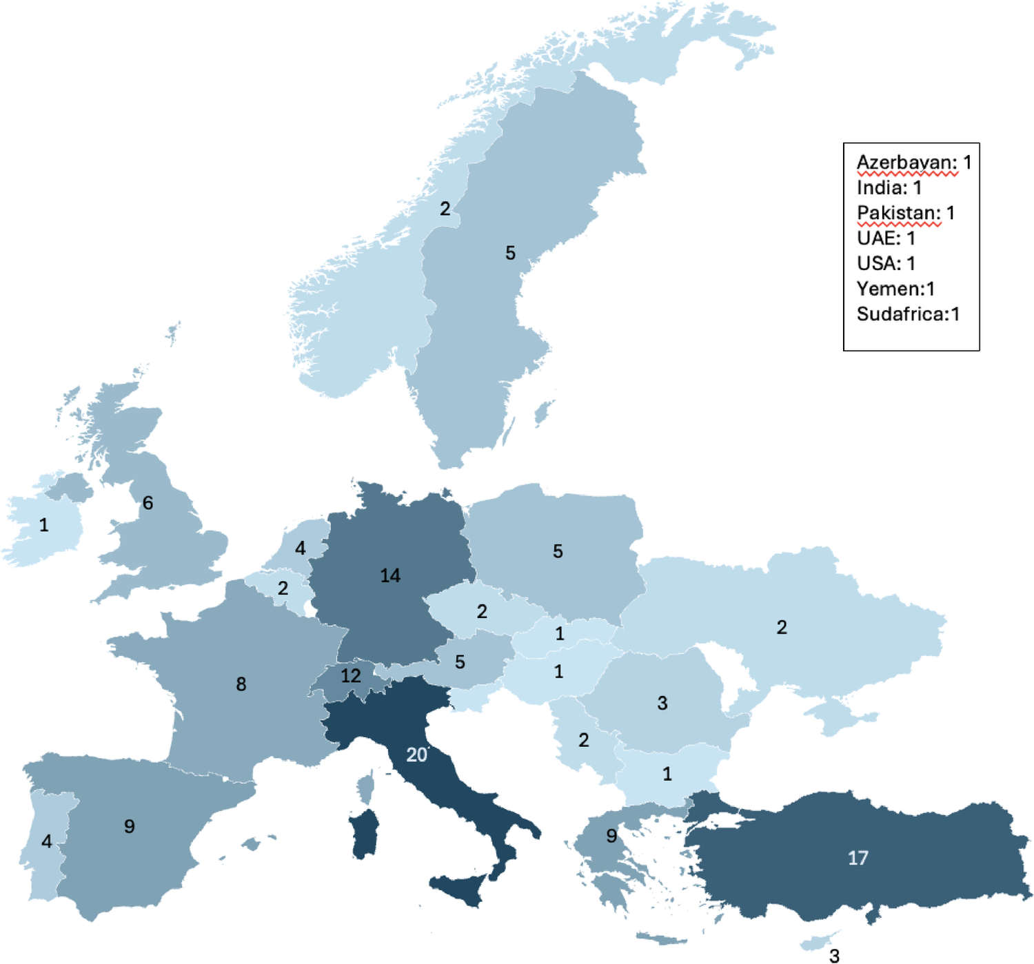
In April 2022 fluorescence-guided laparoscopic biopsy was introduced at our division. All FGLLB procedures (n = 42) from then until September 2025 were recorded in a prospective database. Patient details, including sex, age, previous abdominal surgery, and comorbidities, were collected. A control group of 55 LLB patients who were operated on from 2006 to 2021 before the use of fluorescence was introduced. Patient selection was based on the following inclusion criteria: (1) Age ≥ 18 years; (2) Absence of superficial pathological lymph nodes amenable to surgical biopsy; and (3) A clear indication for abdominal lymph node sampling necessary for diagnosing or confirming a lymphoproliferative disorder.Patients were excluded if they presented with known iodide allergies, coagulopathy, or pregnancy. For preoperative staging, all subjects received a PET/CT scan. This imaging was utilized to identify pathological lymph nodes, define their specific anatomical location, and evaluate the degree of standardized uptake value (SUV). The same senior surgeon (MC) performed all FGLLB and LLB procedures. The primary outcome of the procedures was the diagnostic yield, defined as whether the collected tissue corresponded to lymphatic tissue in sufficient quantity to allow for a definitive diagnosis. To comprehensively assess the technique’s outcome, we evaluated the following secondary performance parameters: surgical time, estimated blood loss, rate of surgical conversion, need for an additional trocar, abdominal drain placement, length of hospital stay, and postoperative morbidity. Informed consent for the procedure and the use of the dye was obtained from each patient in the study. The study was carried out according to the relevant guidelines and regulation (Declaration of Helsinki).
Surgical techniqueFor all patients, the surgical procedure consisted of a laparoscopic biopsy of lymph node tissue, which could be an entire lymph node, a fragment of a lymph node, or a lymphomatous plaque in cases of pathological extranodal lymphatic tissue. In FGLLB cases, a dedicated endoscopic system (Visera Elite II, Olympus Medical Systems Corp., Tokyo, Japan) was used to illuminate the regional lymph nodes with an infrared (IR) light source (Figs. 1 and 2).
Fig. 1
Fluorescent spot at the inner inguinal ring under near-infrared view.
Fig. 2
A pathologic lymph node is dissected and retrieved.
The surgical technique for FGLLB has been described in detail elsewhere [12]. A solution was prepared by diluting a 25 mg vial of indocyanine powder with 10 ml of sterile water. The site and route of indocyanine green (ICG) administration varied depending on the anatomical location of the target lymph nodes. In sub-mesocolic lymph nodes (i.e., para-aortic and para-iliac chains below the mesocolon) ICG was injected both intradermally into the patient’s inguinal regions and, if possible, intra-nodally. This was performed approximately four hours before surgery. In supra-mesocolic lymph nodes (i.e., periaortic lymph nodes of the celiac trunk, including its hepatic, gastric, and splenic arterial axes, as well as the retroportal and pancreatic axes) a 2 ml injection of the solution was administered intraoperatively into the peritoneum near the pathological lymph nodes cluster. During surgery, if fluorescence was absent or unsatisfactory at the pathological lymph node site, an additional 1–2 cc intravenous injection of the ICG solution was administered, and the resulting fluorescence was then assessed.
Statistical analysisThe results are expressed as mean, standard deviation (SD), median, counts, and percentages. The Shapiro-Wilk test was used to asses normal distribution of continuous variables and to choose the appropriate test for group comparisons. The nonparametric Mann-Whitney U test for continuous variables and the χ2 test for binary variables were used to compare the groups. A logistic regression model was used to identify variables potentially associated with surgical time and hospital stay in the univariate analysis. A two-tailed P value less than 0.05 was considered statistically significant. Statistical analysis was performed using the SPSS software (Version 13.0, SPSS Inc, Chicago, IL, USA). A post-hoc (retrospective) power analysis for the primary outcomes that demonstrated a strong trend toward benefit (p < 0.10) was conducted.
Comments (0)