This study aimed to evaluate the effects of LPT on muscle cells subjected to oxidative stress by assessing cell viability and the expression of genes associated with muscle regeneration, as well as a pro-inflammatory cytokine. To date, no studies in the literature have investigated this specific protocol of LPT in myoblasts under oxidative stress in vitro. However, several in vivo studies have examined muscle tissue regeneration following injury, demonstrating reductions in inflammatory markers and oxidative stress, particularly in relation to physical activity.
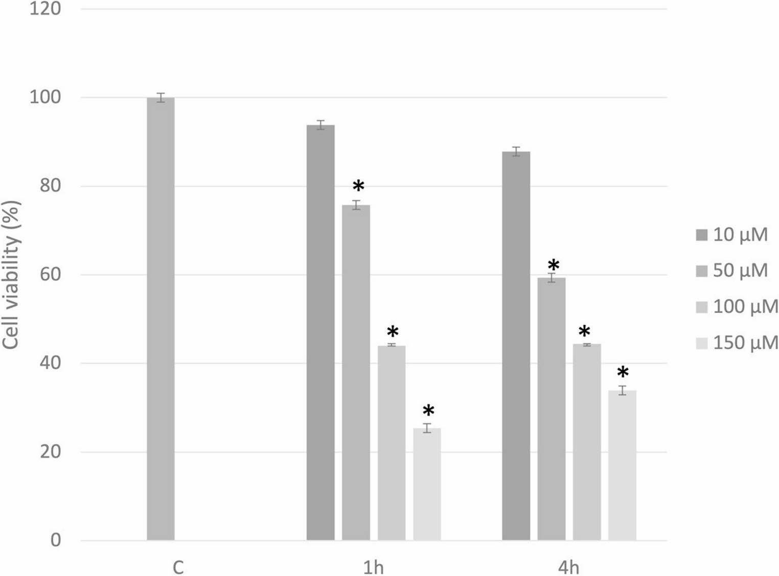
In our study, oxidative stress led to a significant reduction in cell viability after 1 h of treatment with 50 µM H2O2. Higher concentrations of H2O2 induced reductions exceeding 50% in cell viability, highlighting their detrimental effects on muscle cells.
The adopted protocol allowed the evaluation of myoblast behavior when irradiated with LPT either before or after exposure to oxidative stress. Myoblasts treated with LPT at 660 nm with an energy delivered of 3 J, and at 808 nm with energies of 3 and 10 J, prior to oxidative stress induction, exhibited attenuation of the reduction in cell viability. Except for groups irradiated at 660 nm with 10 J after oxidative stress, and at 808 nm with 5 J and 10 J before oxidative stress, all other treatments showed cell viability levels comparable to the control group, indicating that LPT mitigated the deleterious effects of oxidative stress.
One study [18] also induced oxidative stress using a different protocol, evaluating the effects of LPT on C2C12 myoblasts cultured in various concentrations of M1 phenotype macrophage-conditioned medium (MCM1) derived from J774 cells. In that study, irradiation was performed 2 h after the oxidative stress protocol. The authors reported that LPT effectively modulated the increase in viability and proliferation of myoblasts when irradiated with low-level laser at a wavelength of 780 nm and an energy delivered of 1 J. In our findings, LPT applied after oxidative stress also influenced cell viability, although at higher energies delivered − 3 and 5 J at 660 nm, and 3, 5, and 10 J at 808 nm - since the viability of these groups was comparable to that of the control group.
Studies analyzing myoblasts have also reported favorable outcomes regarding cell viability. Trajano et al. [19] investigated C2C12 myoblast cultures exposed to low-level infrared laser (808 nm, 100 mW) at fluences of 10, 35, and 70 J/cm2, with evaluations at 24, 48, and 72 h. Their findings showed that laser exposure enhanced cell viability after 48 h with a fluence of 10 J/cm2, whereas no significant increase was observed at higher fluences. These results are consistent with our study, in which LPT improved myoblast viability even under oxidative stress, particularly with the energy delivered of 10 J at 808 nm.
Similarly, using the infrared spectrum, Mesquita-Ferrari et al. [20] irradiated C2C12 cells with a GaAlAs diode laser at a wavelength of 780 nm and an energy delivered of 5 J. Cell viability and the expression of myogenic regulatory factors were assessed at 24, 48, and 72 h post-irradiation using the MTT assay and quantitative RT-qPCR, respectively. Their results showed increased cell viability in the laser-treated group compared to the control at all time points. In our study, which included oxidative stress, application of 5 J at 660 nm either before or after oxidative stress did not result in significant differences compared with the control and OS groups. These findings suggest that the presence of oxidative stress may alter the cellular response to LPT, as the energy delivered of 5 J at 660 nm did not reproduce the viability gains observed in previous studies, underscoring the importance of experimental conditions when evaluating the biological effects of laser therapy.
Regarding the gene expression results of the present study, increased levels of MyoD and myogenin were observed in the groups irradiated with 10 J at both wavelengths in the PRE-OS condition, with MyoD showing the most pronounced expression at 10 J with 808 nm. In addition, IL-6 expression was detected at the energy delivered of 5 J, with the 660 nm wavelength showing expression in the PRE and the 808 nm wavelength in the POST-OS condition. However, in the study by Mesquita-Ferrari et al. [20] using an energy delivered of 5 J, a trend toward increased myogenin messenger RNA (mRNA) was observed in the laser-treated group. Conversely, and in agreement with our findings, Trajano et al. [13] reported an increase in MyoD expression in C2C12 myoblasts treated with a low-level infrared laser at an energy delivered of 10 J.
In vivo studies have also been conducted using both animal and human models. For instance, Alves et al. [9]. demonstrated that low-level laser therapy (LLLT) administered at 780 nm and an energy delivered of 10 J following muscle injury modulated the expression of MyoD and myogenin during the repair process. Similarly, Rodrigues [21], using LPT at 660 nm with energies delivered of 10 and 50 J, observed upregulation of MyoD at 50 J and increased myogenin expression at 10 J. In contrast, our study showed expression of MyoD and myogenin in groups irradiated prior to the oxidative stress protocol, differing from these studies in which LPT was applied after the injury.
In the present study, the best results were obtained with the enegy delivered of 10 J at 808 nm in the PRE-OS protocol. This group showed a significant increase in cell viability compared to the controls, as well as a higher relative expression of the MyoD gene, indicating enhanced cell regeneration. These findings are consistent with clinical studies. For example, de Oliveira et al. [22], applied LPT at 10 J and 810 nm prior to a fatigue protocol and analyzed inflammatory markers related to fatigue and oxidative stress at 1 min, 1 h, 24 h, 48 h, 72 h, and 96 h post-treatment. They observed a reduction in these markers, which favored muscle performance. Similarly, Vanin et al. [23] investigated LPT administered before an eccentric exercise protocol using energies delivered of 10, 30, and 50 J at 810 nm on the quadriceps, with evaluations conducted at 1, 24, 48, 72, and 96 h. Their results demonstrated that laser treatment enhanced maximum voluntary contraction from 24 to 96 h post-application at enegies delivered of 10 J and 50 J, thereby improving performance and favorably modulating biological markers such as CK and IL-6.
It is well established that the effectiveness of LPT on target tissues depends on a combination of parameters, including wavelength, energy density, duration, and frequency of application. Among these, wavelength plays a pivotal role in laser-tissue interactions by modulating absorption and scattering properties [24]. Given the wide range of laser parameters and the comprehensive protocol employed in this study, we were unable to evaluate additional doses. Further studies are needed to investigate the effects of LPT under oxidative stress, optimize its application for improving muscle function, and clarify the mechanisms underlying its protective effects, which may contribute to the development of therapeutic strategies for muscle injuries and disorders associated with oxidative stress.


Comments (0)