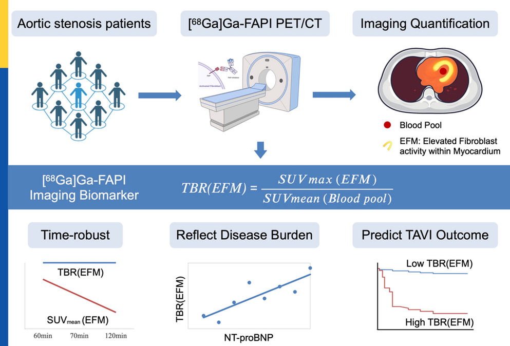
This study in patients with severe aortic stenosis undergoing TAVI yielded three principal findings with important implications for imaging protocol design for FAPI uptake, pathophysiological understanding, and patient management. First, the TBR(EFM) was markedly stable between 60- and 120-minutes p.i., even as absolute myocardial SUV declined over the same interval. Second, TBR(EFM) showed robust associations with NT-proBNP across imaging times, indicating that ratio-based quantification better reflects the biology of fibroblast activation under pressure overload. Third, higher baseline (prior to TAVI) TBR(EFM) was associated with limited clinical improvement one year after TAVI, suggesting that pre-procedural [68Ga]Ga-FAPI uptake predisposes the feasibility of reverse remodeling and has a significant prognostic value. Collectively, these findings support a streamlined and standardized workflow for myocardial [68Ga]Ga-FAPI-PET quantification in AS, consisting of a single static acquisition approximately 60 min post-injection, combined with LV masking and a central blood-pool reference.
The time stability of TBR(EFM) after approximately 60 min p.i. is consistent with rapid blood clearance and early, specific binding of [68Ga]Ga-FAPI tracers to activated fibroblasts, yielding proportional washout from myocardium and blood pool beyond the first hour. This behavior supports standardizing acquisition at a single early time point—60 min p.i.—without sacrificing quantitative fidelity. In contrast, the observed 25–30% decline in SUVmean between 60 and 120 min underscores the time dependency of absolute uptake [15] and cautions against inter-patient or inter-study comparisons based on raw SUVs when uptake times differ, even modestly. Although acceptable image quality may be achievable somewhat earlier given the fast kinetics of [68Ga]Ga-FAPI, a 60-minute standard provides a pragmatic balance between contrast and harmonization.
When interpreted in this context, the superiority of TBR(EFM) over SUVmean(EFM) is mechanistically intuitive. By normalizing to blood-pool activity, TBR(EFM) functions as an internal control that mitigates variability in delivery, clearance, and sampling time, thereby isolating a signal that more faithfully reflects fibroblast biology. The tight correlation between TBR(EFM) and NT-proBNP—an established marker of myocardial wall stress and heart-failure severity—supports construct validity, whereas the absence of a meaningful relationship between SUVmean and NT-proBNP illustrates how absolute measures can obscure clinically relevant associations [16]. Using whole-LV segmentation, elevated TBR(EFM) values substantially above unity indicates global pathological fibroblast activation; volume-based “fibrosis burden” metrics can complement this by localizing disease but require thresholding, whereas TBR(EFM) provides a threshold-free, continuous gauge of global activity.
A practical strength of this work is the self-developed, semi-automatic threshold-based segmentation and quantification pipeline used for myocardial analysis. By combining consistent anatomical masking with reproducible, threshold-driven measurements, the approach reduces operator dependence, shortens processing time, and facilitates quality control. Importantly, it aligns naturally with TBR(EFM)-based reporting, making the method easy to implement at scale and well suited for multi-center harmonization and longitudinal studies where standardization and throughput are essential.
The prognostic signal of baseline TBR(EFM) integrates naturally with these quantitative advantages. Patients exhibiting higher pre-TAVI TBR(EFM) demonstrated a lower likelihood of symptomatic or biochemical improvement at one year, supporting the concept that once diffuse, active myocardial remodeling is established, functional recovery following afterload reduction is constrained. Reports of short-term improvements in LVEF after valve intervention in some high-uptake cases can be reconciled by recognizing that acute contractile reserve does not necessarily translate into durable relief from diastolic dysfunction or neurohormonal activation. Clinically, pre-TAVI TBR(EFM) may guide patient counseling, inform the intensity of post-procedural monitoring, and support consideration of adjunctive therapies targeting myocardial fibrosis or heart failure. Recent studies in high-risk post-TAVI patients have shown that sodium–glucose cotransporter 2 (SGLT2) inhibitors, such as dapagliflozin, significantly reduce all-cause mortality and heart failure events compared with standard care [17]. Whether these benefits are linked to changes in myocardial FAPI uptake and modulation of fibrosis after TAVI remains unclear and warrants further investigation. Beyond baseline risk stratification, serial [68Ga]Ga-FAPI PET likely be a marker for both persistent adverse and reverse remodeling.
Our observations demonstrate that myocardial FAPI-PET may provide complementary information to established imaging modalities. Cardiac magnetic resonance (CMR) with LGE images and T1/ECV mapping quantifies established collagen crosslinking, deposition, and replacement fibrosis, which are structural hallmarks that may lag behind biological activity, whereas [⁶⁸Ga]Ga-FAPI PET highlights ongoing fibroblast activation and pro-fibrotic signaling [18], providing a more immediate measure of fibrogenic activity, as demonstrated in recent translational studies of cardiac injury (our MS : https://pubmed.ncbi.nlm.nih.gov/40430477/) Echocardiographic strain provides a functional readout that is sensitive to loading conditions; [68Ga]Ga-FAPI offers a molecular readout less affected by hemodynamics at the imaging time. A combined approach may help distinguish active from fixed fibrosis and refine the timing of intervention.
The underlying tracer kinetics help explain both the time stability and the prognostic value of TBR(EFM). [68Ga]Ga-FAPI tracers bind to fibroblast activation protein with high specificity; by about one-hour p.i., circulating tracer is low, and tissue signal largely reflects bound or retained tracer, with subsequent proportional clearance from tissue and blood. In hearts with high densities of activated fibroblasts, sustained signaling and matrix deposition create a substrate that is stiffer and less likely to reverse, aligning the molecular readout with clinical trajectories. This framework reinforces the importance of timely intervention in AS and positions [68Ga]Ga-FAPI PET as a means to estimate where an individual lies on the spectrum from reversible to entrenched remodeling.
Several limitations temper these conclusions. The sample size—particularly for one-year outcomes (n = 11)—limits power and precludes multivariable modeling, so results should be considered hypothesis-generating. Two patients lacked 120-minute scans, modestly reducing precision for time-point comparisons. The composite responder definition, while clinically meaningful, includes subjective elements; harder endpoints were too infrequent to analyze. Histopathologic validation was not feasible. Absence of uniform valve hemodynamic data at follow-up may confound associations between FAPI signal and clinical recovery. Future work should prospectively standardize acquisition at 60 min p.i., report TBR(EFM) as the primary quantitative metric, and predefine clinically actionable cut-points (e.g., ROC-guided thresholds). Multicenter studies powered for incremental prognostic testing over NT-proBNP, LVEF, strain, and CMR fibrosis markers are needed to establish generalizability. Longitudinal imaging before and after TAVI can determine whether dynamic changes in TBR(EFM) track remodeling and outcomes, and interventional trials should assess whether therapies that reduce TBR(EFM) improve clinical endpoints. If confirmed, [68Ga]Ga-FAPI PET would serve not only as a risk-stratification tool but also as a pharmacodynamics biomarker for antifibrotic strategies in AS.

Comments (0)