In this study, we investigated the clinical presentation, radiographic features, and genotypes of a small cohort of rare disease MCDS caused by COL10A1 gene mutations. We also analyzed genotype–phenotype relationships in our and previously reported MCDS cases through a careful literature review.
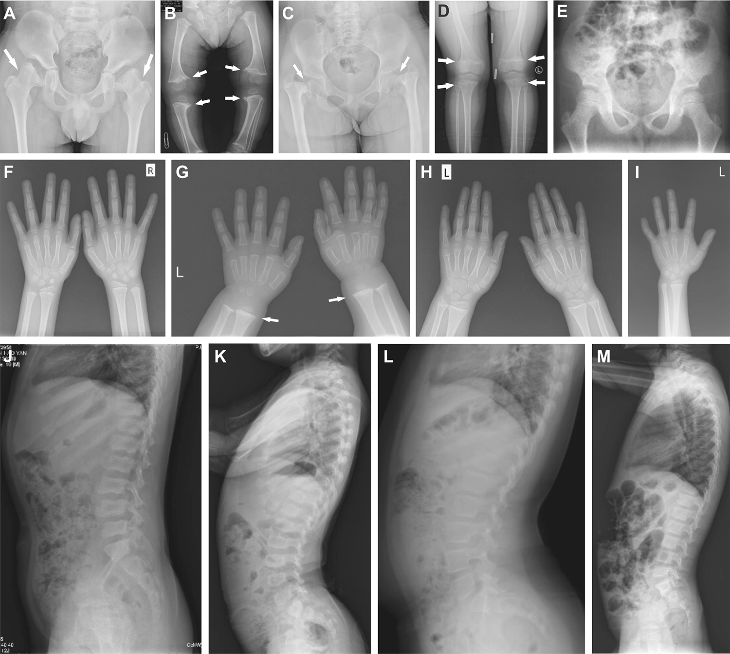
In our patients, the phenotypic profile mainly included waddling gait, bowed legs, short stature, and flattening or blurred vertebrae and femoral epiphysis widening in radiography. Patients usually had normal BMD and bone turnover biomarkers, accompanied by vitamin D insufficiency or deficiency. Calcium, vitamin D analogues, or GH therapy showed limited efficacy in improving the disease phenotype. COL10A1 mutations in our patients were all heterozygous, mainly affecting the NC1 domain. Two novel COL10A1 missense mutations, c.1925T > A (p.Ile642Asn) and c.1903C > G (p.Gln635Glu), were identified. One patient harbored a de novo NC1 domain truncating mutation and a missense mutation in the helix domain, the former possibly being mosaic, which was the first reported case of MCDS that carries mutations in both NC1 and helix domains, thereby broadening the mutational spectrum of MCDS.
MCDS is a rare disease with complex phenotypes and significant heterogeneity in clinical and radiographic manifestations. Short stature was the most common initial sign of MCDS, which persisted throughout the course of the disease. However, 40% of patients did not suffer short stature at first, suggesting the importance of other manifestations in recognizing MCDS, including shortened limbs, waddling gait, as well as short femoral neck, bowed femurs or tibiae, and metaphyseal irregularities at femurs, tibiae, and fibulae displayed on radiography. We found that 56.3% of cases also had metaphyseal irregularities at the distal radius or ulna, similar to the previous finding that wrist involvement occurred in 61.5% of cases [2]. A significant proportion of patients were initially misdiagnosed with different types of rickets, possibly due to the overlapping manifestations between the two diseases, such as bowed legs, genu varum or genu valgum, metaphyseal enlargement, fraying, and splaying [8,9,10]. However, MCDS usually lacks blurred trabecular bone and maintains normal serum levels of Ca, P, 25OHD, β-CTX, and P1NP, which help differentiate the two disorders [8, 19].
Type X collagen, a short-chain non-fibrillar collagen, is specifically secreted by hypertrophic chondrocytes in the growth plate into their surrounding extracellular matrix (ECM) [20, 21]. As a major component of the growth plate hypertrophic zone, it plays an important role in calcification and vascular invasion during endochondral ossification by assembling into hexamers [22]. Four domains comprise type X collagen, of which the carboxy-terminal NC1 domain is essential for the assembly of the α1(X) homotrimer, where MCDS mutations occur most frequently [16, 22, 23].
Of note, we identified an intricate relationship between the mutation type of COL10A1 and the degree of short stature in MCDS patients. At the first examination, no significant difference in height Z-scores was found between patients with NC1 mutations and non-NC1 mutations. However, at the last evaluation, patients with non-NC1 mutations showed significantly lower height Z-scores. This is presumably because most patients with non-NC1 mutations came from the same family carrying homozygous mutations in the NC2 domain, who showed a late onset of short stature until the age of 6, with severely low height Z-scores in adulthood (− 4.8 to − 7.5) [24]. Mechanistically, in vitro functional studies have shown that mutations in the signal peptide also considerably impair collagen X assembly, leading to a reduction of normal collagen X secretion [25]. However, the influence of NC2 mutations on collagen X expression has not been explored. This finding remains to be validated with larger samples and in vitro studies in patients with MCDS.
Intriguingly, NC1-missense patients had a significantly lower height Z-score initially than patients with NC1-truncating mutations, but the opposite was true at the last assessment, indicating that short stature may progress with age in the presence of NC1-truncating variants. It has been shown that mutant COL10A1 mRNA containing nonsense or frameshift mutations would be degraded via nonsense-mediated decay [26, 27]. In contrast, this process was not observed in COL10A1 mRNA with a missense variant in growth plate cartilage from a patient with MCDS [28], and a trace amount of mutant/normal α1(Ⅹ) chain heterotrimer assembly was detected in vitro [29]. Thus, haploinsufficiency due to NC1-truncating mutations may partially explain this phenomenon [18, 30].
The above mechanism may also explain the reduction in penetrance. The penetrance of the COL10A1 mutation has been reported as nearly 100% [1]. However, asymptomatic carrier parents exist in both the two families in our study and 4 unrelated families described in the literature, who showed normal or slightly short stature (Z-score > − 2.0) without other clinical or radiographical manifestations of MCDS [17, 31, 32]. Notably, most of them harbored missense but not truncating mutations in the NC1 domain. This further indicates that NC1-missense mutations exert a less pronounced impact on the skeletal phenotypes over time compared to NC1-truncating mutations. Consistently, previous studies have observed that certain radiographical manifestations resolve with age, including enlarged femoral head, platyspondyly, and metaphyseal cupping of phalanges and metacarpals [17, 33, 34]. Most of the cases also harbored missense mutations in COL10A1.
Furthermore, we found that the proportion of metaphyseal involvement at the distal radius or ulna was significantly higher in patients with missense or NC1 missense. No studies have yet identified an association between a specific COL10A1 genotype and a particular epiphyseal involvement. Additional cases are required to verify this finding.
To date, no medications have been approved for the treatment of MCDS. Most of the patients with skeletal deformities, such as significant lower limb curvature, received orthopedic surgery, but the height Z-score did not improve greatly after treatment. Previous studies showed that rhGH therapy modestly enhanced the height Z-score of MCDS patients during 0.5–2.25 years [16, 35]. However, our study showed that GH treatment did not significantly increase the height of patients and could not induce catch-up growth, possibly because GH therapy is unable to reverse the effects of COL10A1 mutations on bone growth [16, 35].
To find therapeutic targets for MCDS, the pathogenesis has been partially revealed. Mutant collagen is retained intracellularly, triggering endoplasmic reticulum (ER) stress and unfolded protein response, which in turn may compromise chondrocyte differentiation and polarity, leading to disorganized cell stacking and ultimately growth plate irregularities [21, 36, 37]. Interestingly, carbamazepine (CBZ), an anti-epileptic drug, has been recently repurposed to treat in vitro and in vivo models of MCDS due to its inhibitory effects on ER stress by enhancing autophagy and proteosomal degradation [21, 36, 38, 39]. In 3-week-old MCDS mice with Col10a1 p.N617K mutation, 3-week CBZ treatment (250 mg/kg/d) significantly reduced the width of the hypertrophic zone, increased femur and tibia growth, and alleviated hip dysplasia [21]. The same effects of CBZ were also observed in mice harboring Col10a1 p.Y632X mutation and medaka model with a Col10a1 frameshift at amino acid 633 [36, 39]. Mechanistically, CBZ treatment inhibited ER stress-related gene expression, including Bip, Atf4, and attenuated disruption of type X collagen expression [21, 39]. Meanwhile, CBZ restored the polarity of hypertrophic chondrocytes and the coordinated expression of their differentiation markers, Opn and Mmp13 [21, 36]. These may ameliorate the damaged growth plate, thereby restoring normal bone growth. More recently, phase 1 and phase 2/3 clinical trials of CBZ treatment on 27 children and adolescents with MCDS are ongoing in Europe [38, 40]. The participants received CBZ at a maximum dose of 20 mg/kg/day for at least 12 months [40]. Periodic reporting claims that CBZ treatment is tolerated in children with MCDS and is associated with reduced bone pain, accelerated growth, and decreased progression of deformity in the lower extremities [41]. However, long-term use of CBZ increases the risk of osteoporosis and fractures through inducing CYP24, a catabolic enzyme for 1,25(OH)2D, which accelerates 1,25(OH)2D inactivation [42, 43]. Therefore, the safety of CBZ for long-term treatment in MCDS patients remains to be evaluated.
However, our study has some limitations. First, some of the reported cases did not provide clinical manifestations in detail or undergo molecular testing, inevitably impairing the solidity of the results. Secondly, cartilage specimens were not obtained from patients to analyze the ratio of normal to mutant collagen X. Functional validation of the novel variants was not performed to rationalize the genotype–phenotype correlation. Additionally, we were unable to obtain the radiography of the unaffected carriers in families 1 and 2 to adequately explain the asymptomatic condition.
In conclusion, our findings expand the mutational and phenotypic spectrum of MCDS and shed new light on the relationships between COL10A1 mutation and the severity of MCDS, emphasizing the role of molecular typing in individualized assessment of MCDS patients. This study is of great significance for understanding the key role of collagen X in bone growth and development. Further studies will utilize larger-scale cases, combined with in vitro and in vivo experiments, to demonstrate the genotype–phenotype association and to develop safe and effective therapeutic approaches for patients with MCDS.


Comments (0)